Ophthalmological ultrasonography scanning apparatus

a scanning apparatus and ultrasonic technology, applied in the field of medical ultrasound equipment, can solve the problems of not providing the degree of resolution required for biometry of the corneal surface, and the reconstruction of a series of fan-shaped b-scan planes, so as to facilitate precise arcuate motion of an ultrasonic transducer, maintain normality of ultrasound beam, and maintain the effect of focal distance from ey

Inactive Publication Date: 2005-05-03
ARCSCAN INC
View PDF12 Cites 28 Cited by
  • Summary
  • Abstract
  • Description
  • Claims
  • Application Information

AI Technical Summary

Benefits of technology

[0008]In one aspect of the invention, an apparatus for ultrasound scanning of the eye is provided comprising a virtual center translocation mechanism that facilitates precise arcuate motion of an ultrasonic transducer to maintain focal distance from the eye and to maintain normality of the ultrasound beam with surfaces of the eye. The arcuate movement of the transducer focal path may closely approximate the surface of the cornea. Some embodimen

Problems solved by technology

The structure of the eye, particularly the cornea, presents special problems for optimal ultrasonographic B-scan imaging.
Similarly, volumetric 3-dimensional scanning by reconstruction of a series of

Method used


Image

  • Ophthalmological ultrasonography scanning apparatus
  • Ophthalmological ultrasonography scanning apparatus
  • Ophthalmological ultrasonography scanning apparatus

Examples


Embodiment Construction

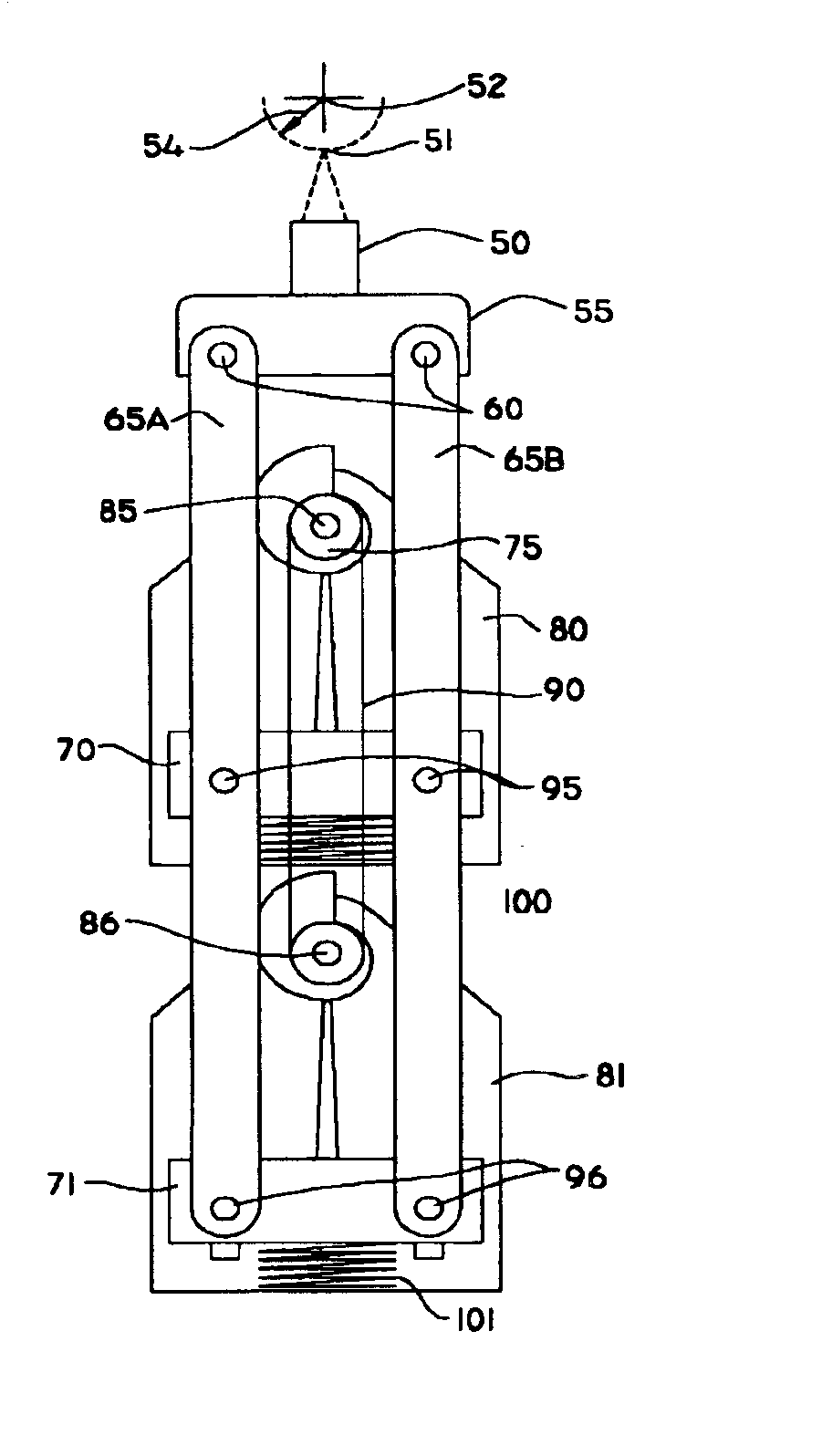
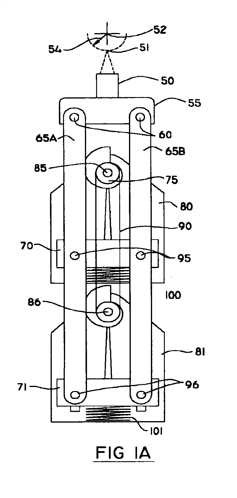
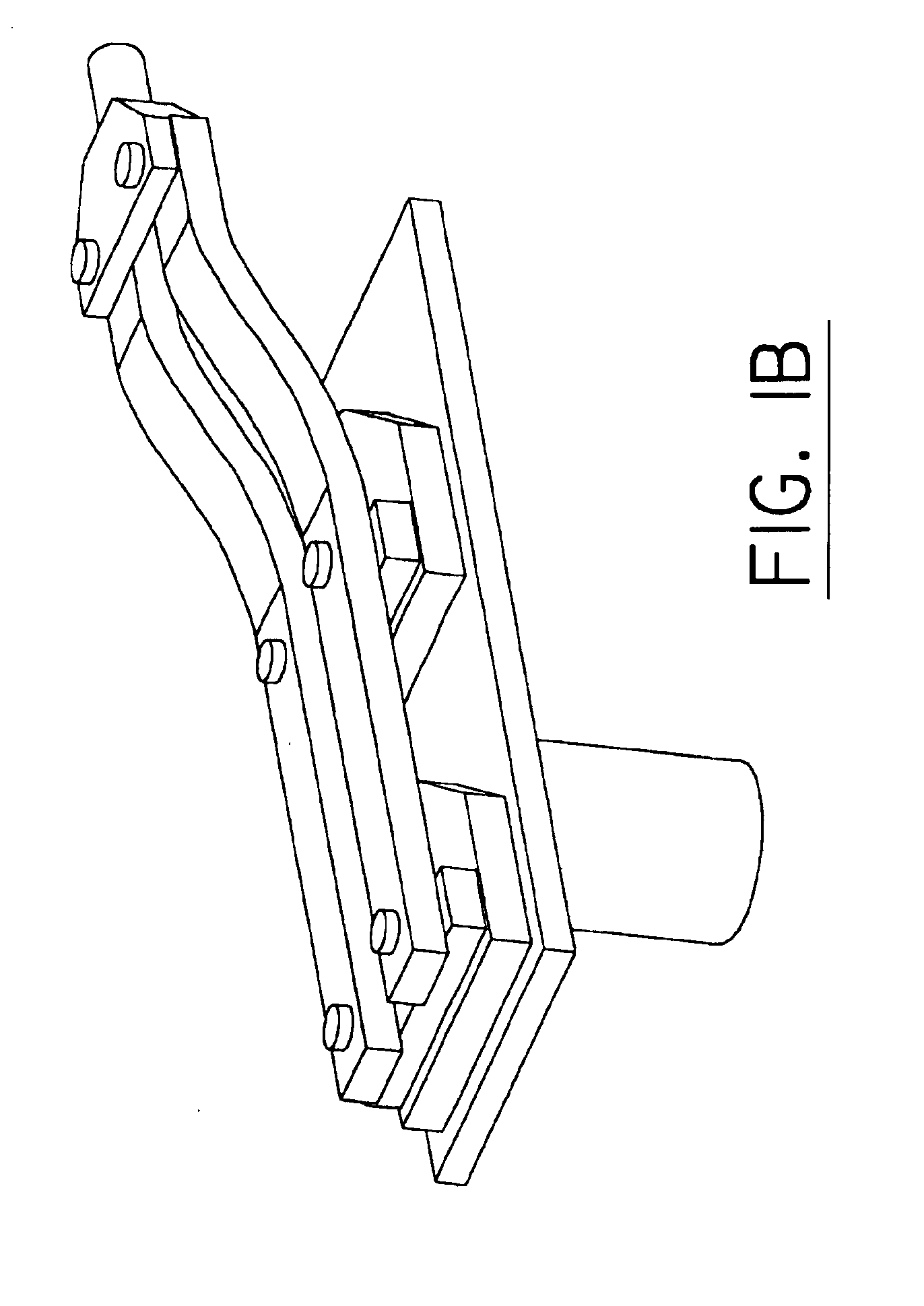
[0028]In one aspect, the invention provides an ultrasound transducer support comprising a transducer mount adapted to accommodate an ultrasound transducer, and a virtual centre mechanism. FIG. 1A illustrates an embodiment of a virtual center mechanism. First and second arm linkages 65A and 65B are each connected via three pivots to moving parts of the mechanism. Rear swinging pivots 96 connect first and second arm linkages 65A and 65B to rear radius adjust slider 71, and rear radius adjust slider 71 is attached to rear swinging linkage 81. Similarly, front swinging pivots 95 connect arm linkages 65A, 65B to front radius adjust slider 70, and front radius adjust slider 70 is attached to front swinging linkage 80. The front ends of the arm linkages 65A, 65B are connected by transducer pivots 60 to transducer mount 55, and transducer mount 55 is adapted to accommodate ultrasonic transducer 50. Front pivot 85 and rear pivot 86 are stationary relative to the swinging motion of front swin...


PUM


Abstract

An apparatus for ultrasound scanning of the eye is provided comprising a virtual center translocation mechanism that facilitates precise arcuate motion of an ultrasonic transducer to maintain focal distance from the eye and to maintain normality of the ultrasound beam with surfaces of the eye. The invention also provides a radius adjust mechanism for changing the radius of ultrasound scanning to facilitate positioning of the transducer focal point on selected surfaces of the eye. Centration optics are also provided, for aligning the ultrasound transducer with the Purkinje (or other optical or geometric) axis of a patient's eye. In addition the invention provides a hygiene barrier eyeseal for protection of the patient from disease propagation.

Description

[0001]This application is a continuation-in-part of U.S. application Ser. No. 09 / 914,924 filed Jan. 15, 2002 now U.S. Pat. No. 6,491,637, which is a national stage PCT / CA01 / 00008 filed Jan. 15, 2001.FIELD OF THE INVENTION[0002]The invention is in the field of medical ultrasound apparatus, particularly apparatus for use in ultrasonography of the eye.BACKGROUND OF THE INVENTION[0003]Ultrasound may be used in a variety of medical applications, including diagnostic ultrasonography of the eye. Diagnostic information is typically provided by an ultrasound pulse from a piezoelectric transducer, which is directed into a tissue. Reflected acoustic energy is detected (as ‘echoes’), so that the amplitude of the received energy may be correlated with the time delay in receipt of the echo. The amplitude of the echo signal is proportional to the scattering strength of the refractors in the tissue, and the time delay is proportional to the range of the refractors from the transducer. A variety of ...

Claims


Application Information

Patent Timeline
IPC IPC(8): A61F5/37A61B8/10
CPCA61B3/0083A61B8/10A61F5/3707A61B8/4461A61B8/4281
Inventor PHILLIPS, SCOTT HOWARDDENNY, CHRISTOPHER GRANT
Owner ARCSCAN INC
Why Eureka
Try Eureka