
Surgical clip applicator device

a technology of applicator device and surgical clip, which is applied in the field of surgical clip, can solve the problems of affecting the peristalsis of the sutured area, and common post-operative complications

Inactive Publication Date: 2005-05-24
NITI SURGICAL SOLUTIONS
View PDF15 Cites 303 Cited by
  • Summary
  • Abstract
  • Description
  • Claims
  • Application Information

AI Technical Summary

Benefits of technology

[0033]retaining the relative adjacent positions of the portions of the gastrointestinal tract and the clip in relation thereto, while raising the temperature of at least the intermediate portion of the clip to a temperature above an upper phase transition temperature, whereat the intermediate portion is in an elastic state, thereby causing the first and second lengths of wire to attain side-by-side registration, thereby to apply a compressive force to the walls located therebetween.

Problems solved by technology

Another disadvantage is that post-operative complications are common.
Further, suturing an organ results in lack of smoothness of the tissue therein, which, when the sutured organ is part of the gastrointestinal tract, hampers peristalsis in the sutured area.
Finally, suturing is both labor and time consuming.
However, due to the facts that such staples are not reusable and that a great many types and sizes are required, the price of staples is high.
Also, after healing, metal staples remain in place along the perimeter of the suture, which reduces elasticity of the junction and adversely affects peristalsis when the sutured organ is part of the gastrointestinal tract.
Plastic rings are cumbersome and expensive.
Such a device is limited in its uses, such as for clamping blood vessels, and is not suitable for joining portions of the gastrointestinal tract.
Such a device is disadvantageous in that its use requires a plurality of clips to be properly positioned simultaneously.
Also, there is no assurance that the resulting junction will be smooth, due to the plurality of sites of the digestive tract joined by the plurality of clips.
A major disadvantage of known memory alloy clips is that they permit compression of only approximately 80-85% of the junction perimeter, thus requiring additional manual sutures, which reduce the seal of the junction during the healing period and its elasticity during the post-operative period.
Also, this additional suturing is problematic inasmuch as it has to be carried out across a joint which includes a portion of the clip, thereby rendering difficult the sealing and anastomosis of the organ portions.

Method used


Image

  • Surgical clip applicator device
  • Surgical clip applicator device
  • Surgical clip applicator device

Examples


Embodiment Construction

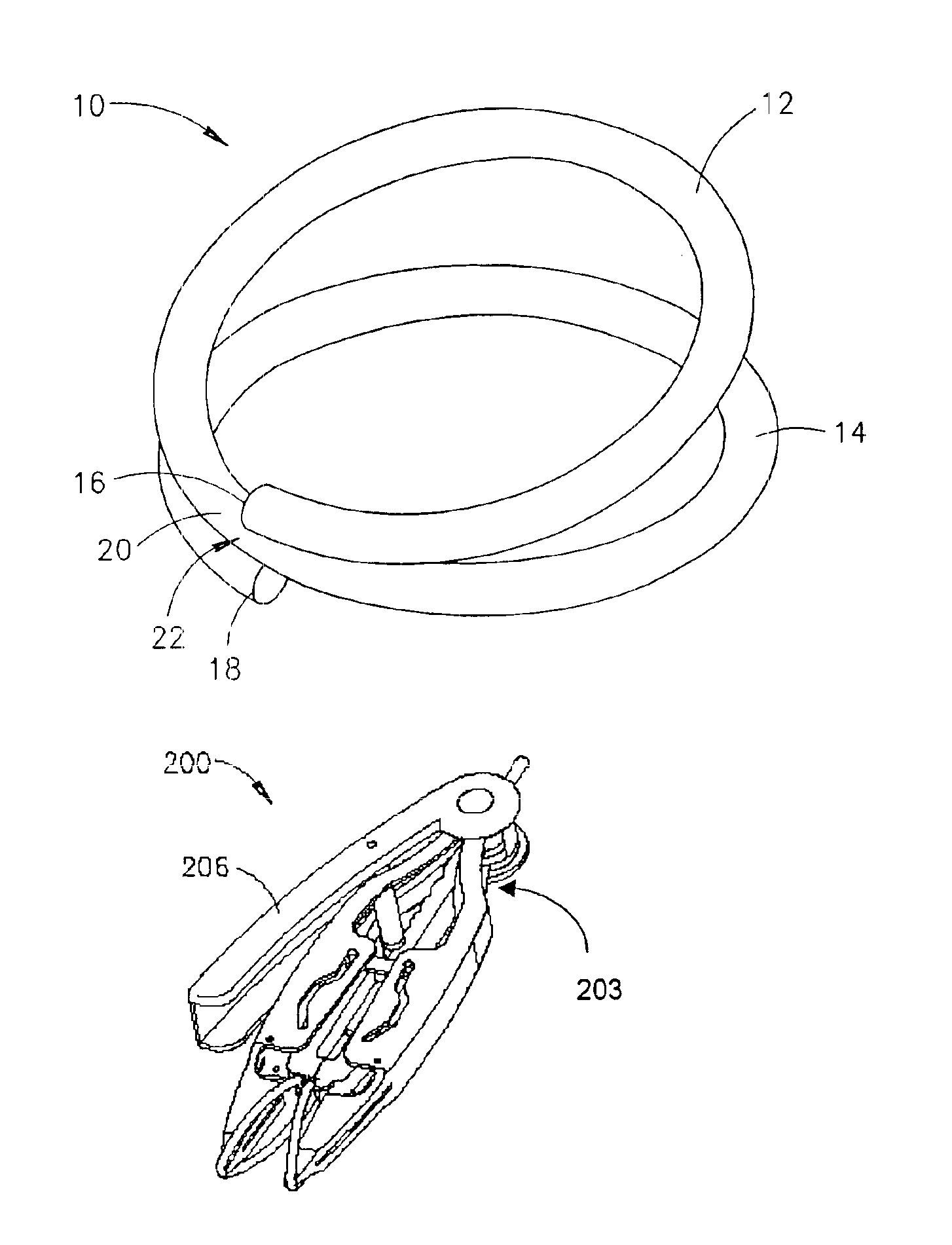
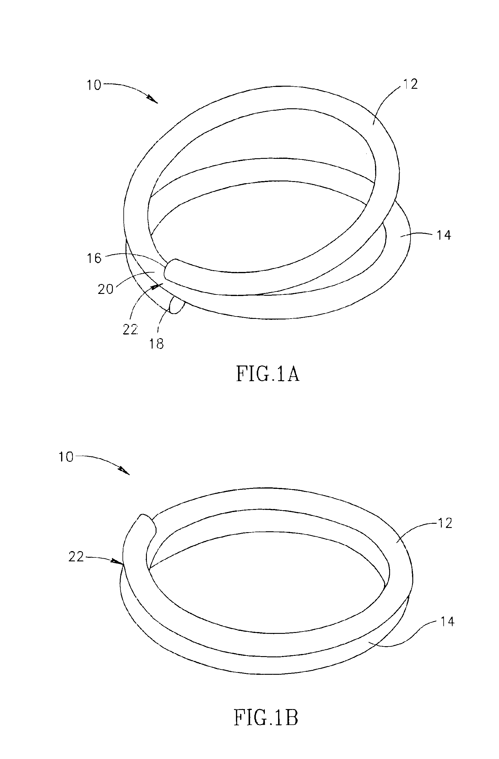
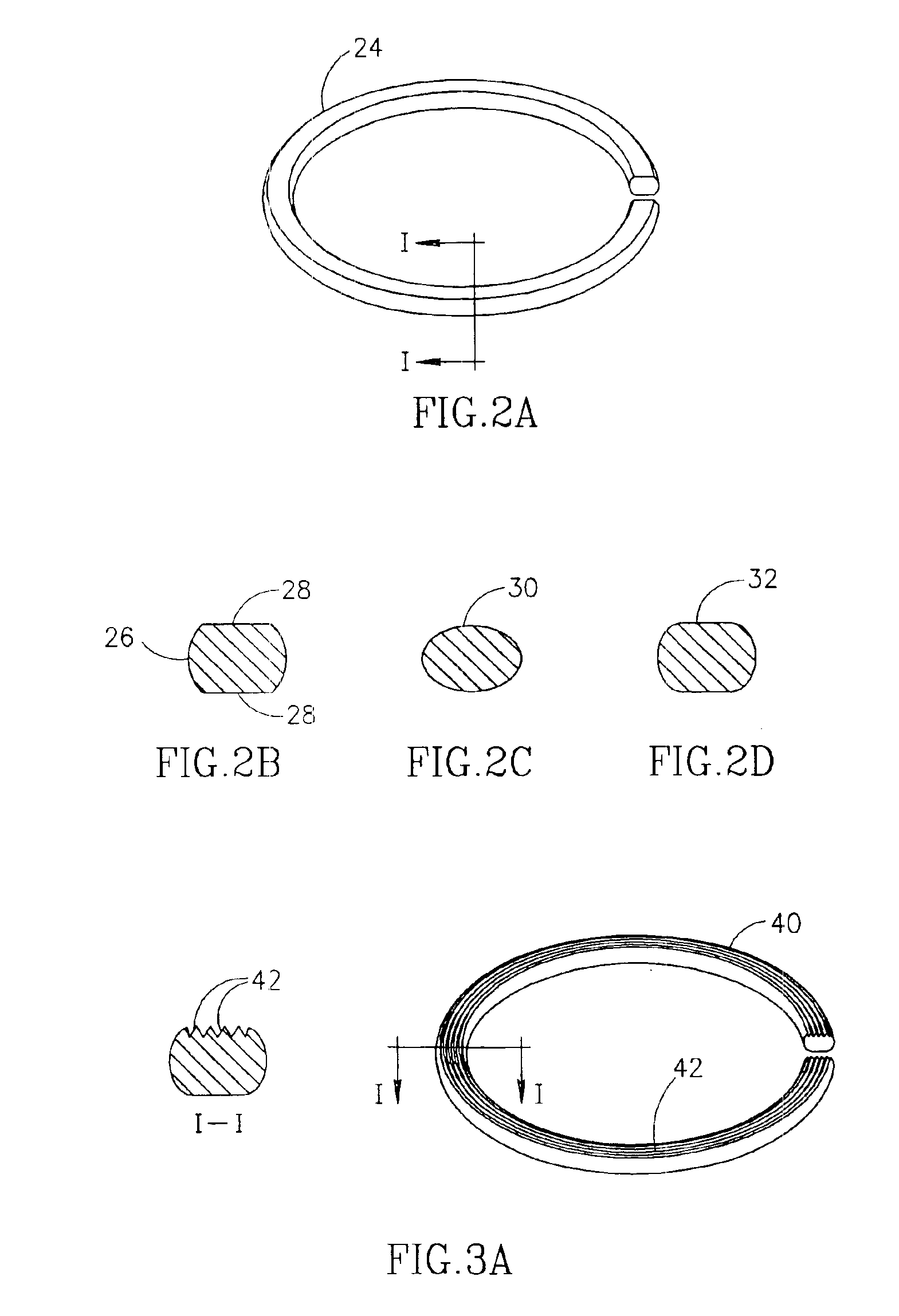
[0066]The present invention seeks to provide a surgical clip, substantially as described in the Applicant's co-pending U.S. application Ser. No. 09 / 592,518 for “SURGICAL CLIP.” The clip is at least partially formed of a shape memory alloy, such as is known in the art, and which provides organ tissue compression along the entire periphery of the clip, thereby to ensure satisfactory joining or anastomosis of portions of an organ. The present invention further seeks to provide apparatus for positioning and applying the clip and, also, for perforating tissue portions held within the applied clip, whereby initial patency of the gastrointestinal tract is created. In addition, the present invention provides a method and system for performing anastomosis of organ portions, such as those of the gastrointestinal tract. The method employs the clip as well as apparatus for positioning and applying the clip and, also, for perforating a portion of tissue held within the clip, whereby initial pate...


PUM


Abstract

The present invention, discloses an anastomosis clip applicator device for applying a surgical clip, formed at least partly of a shape memory alloy, to press together adjacent wall portions of adjacent hollow organ portions so as to effect anastomosis therebetween. The applicator device includes: gripping apparatus for gripping a surgical clip, a release mechanism, associated with the gripping apparatus, and tissue cutting apparatus, operatively associated with the gripping apparatus. There is also apparatus for activating the gripping apparatus, the release mechanism and the cutting apparatus, so as to introduce and apply the surgical clip into adjacent hollow organ portions, such that the surgical clip compresses together the adjacent walls of the hollow organ portions, and thereafter causes the cutting apparatus to perforate the adjacent pressed together organ walls to provide patency through the joined portions of the hollow organ.

Description

REFERENCE TO CO-PENDING APPLICATIONS[0001]The present application is a continuation-in-part of U.S. application Ser. No. 09 / 592,518 filed on Jun. 12, 2000 for “SURGICAL CLIP”, now U.S. Pat. No. 6,402,765, the contents of which are incorporated herein, by reference.FIELD OF THE INVENTION[0002]The present invention relates to the field of surgical clips, in general, and in particular, to the field of surgical clip applicator devices.GLOSSARY[0003]ANASTOMOSIS: The union of parts or branches (as of hollow organs or blood vessels) so as to intercommunicate therebetween.BACKGROUND OF THE INVENTION[0004]It is known in the art to provide methods for joining portions of hollow organs, such as the gastrointestinal tract. These include threads for manual suturing, staplers for mechanical suturing, and compression rings and clips.[0005]While manual suturing is universally known and relatively inexpensive, the degree of success depends considerably on the skill of the surgeon. Another disadvanta...

Claims


Application Information

Patent Timeline
Patent Type & Authority Patents(United States)
IPC IPC(8): A61B17/11A61B17/03A61B17/064A61B17/08A61B17/122A61B17/28A61B17/32A61B17/12A61B17/00
CPCA61B17/064A61B17/083A61B17/11A61B17/1114A61B17/128A61B2017/1225A61B17/0644A61B17/1227A61B17/2812A61B17/32A61B2017/00867A61B2017/0649A61B2017/1107A61B2017/1125A61B2017/1139A61B17/0643
Inventor MONASSEVITCH, LEONIDLELCUK, SHLOMOARAD, MICHAELHARARI, BOAZNEEMAN, RONEN
Owner NITI SURGICAL SOLUTIONS
Learn More