Three-dimensional ultrasound imaging method and apparatus using lateral distance correlation function

a three-dimensional ultrasound and correlation function technology, applied in the field of ultrasound imaging, can solve the problems of variation of the movement speed of the probe, and distortion of the three-dimensional image constructed from the two-dimensional frame, so as to reduce image distortion and accurately estimate the distance

Active Publication Date: 2006-01-24
MEDISON CO LTD
View PDF7 Cites 18 Cited by
  • Summary
  • Abstract
  • Description
  • Claims
  • Application Information

AI Technical Summary

Benefits of technology

[0011]Therefore, an objective of the present invention is to provide a three-dimensional ultrasound imaging method and apparatus for reducing image distortions on three-dimensional ultrasound imaging by accurately estimating a distance between consecutive two-dimensional frames that are obtained through a manual scanning by means of a lateral distance correlation function.
[0013]Also, in accordance with the present invention, a three-dimensional ultrasound imaging method is provided, comprising the steps of: a) transmitting ultrasound signals to a target object and receiving echo signals reflected from the target object; b) receive-focusing the echo signals to generate a plurality of consecutive two-dimensional ultrasound image frames; c) converting the consecutive two-dimensional ultrasound image frames into a three-dimensional ultrasound image by aligning the consecutive two-dimensional ultrasound image frames with no offset to each other in a three-dimensional space and interpolating a distance between two adjacent frames among the consecutive two-dimensional frames, to thereby make an inter-frame distance uniform; and d) displaying the three-dimensional ultrasound image.

Problems solved by technology

However, where the distance between stacked consecutive two-dimensional frames is not uniform, the three-dimensional ultrasound images constructed from the two-dimensional frames may be distorted.
For example, if a target object is an organ in a human body, then the constructed three-dimensional organ may appear distorted from its actual shape due to the non-uniformity of the distance between consecutive two-dimensional frames that represent the organ.
Such non-uniformity typically results from the variation of the movement speed of the probe.
As the variation of the movement speed of the probe increases, the non-uniformity worsens.
However, the aforementioned means have disadvantages.
However, since probes must be mechanically fixed or provided with additional devices, patients and operators feel uncomfortable.
Further, the associated manufacturing costs of ultrasound imaging apparatuses are increased.
In particular, using a three-dimensional probe requires more ultrasound sensor arrays than a two-dimensional probe, and thereby increases the manufacturing cost of the ultrasound imaging apparatus and the size of probe.
If the size of probe is increased, an operator may not easily handle the probe when diagnosing a patient.
However, since the non-mechanical means employs a fixed elevation distance correlation function, without taking into account the non-stationary characteristics of ultrasound images, obtaining an accurate distance between consecutive two-dimensional frames is very difficult and results in lower reliability of the measured ultrasound images.

Method used


Image

  • Three-dimensional ultrasound imaging method and apparatus using lateral distance correlation function
  • Three-dimensional ultrasound imaging method and apparatus using lateral distance correlation function
  • Three-dimensional ultrasound imaging method and apparatus using lateral distance correlation function

Examples


Embodiment Construction

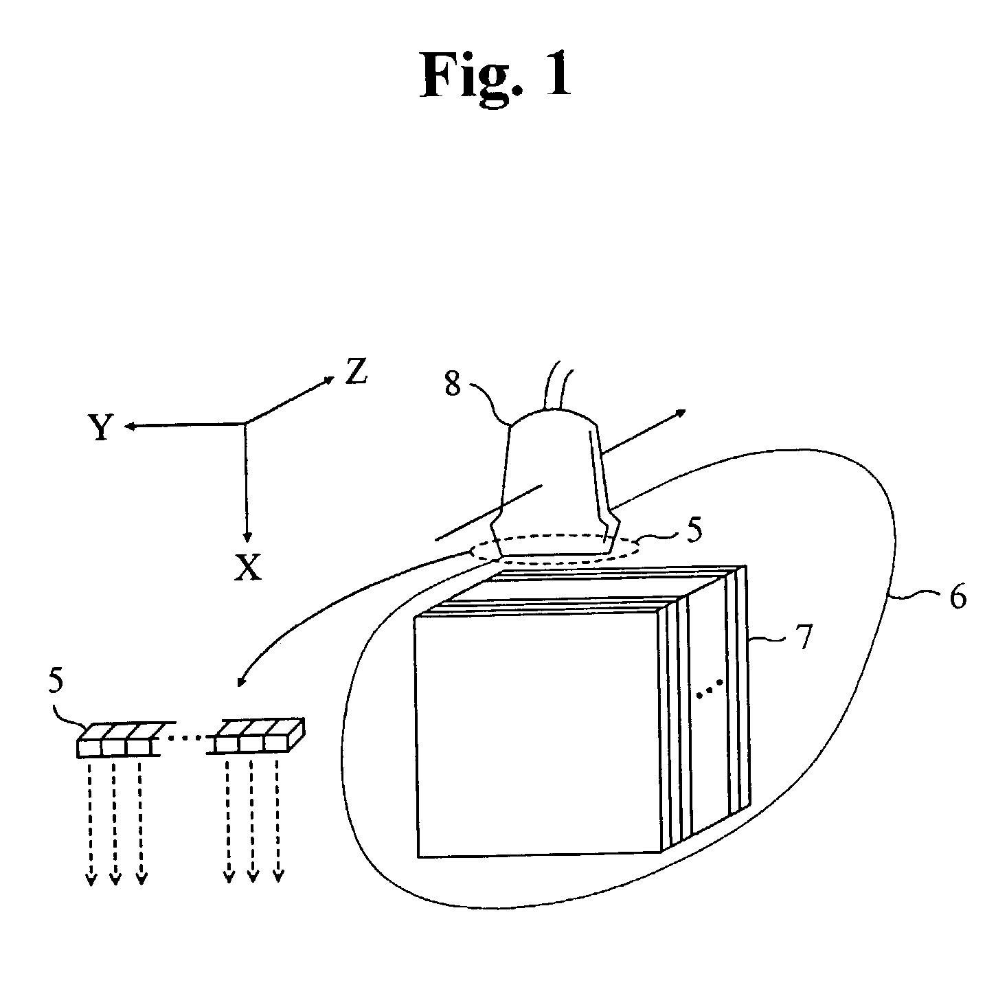
[0024]Referring to FIG. 1, which illustrates obtaining a plurality of consecutive two-dimensional (2D) frames by using a probe in a three-dimensional (3D) space. 3D space is expressed in terms of an orthogonal coordinate system of depth-lateral-elevation (X-Y-Z). Assuming that transducer array 5 is arranged in a lateral direction along the Y-axis, all frames 7 for target object 6 are located in the X-Y plane, and the interface of 2D probe 8 with target object 6 is always perpendicular to the X-axis. However, where a plurality of frame sequences are obtained by using 2D probe 8, 2D probe 8 moves non-linearly according to the shape of the curved surface of target object 6 so that the 3D positions of frames 7 are offset.

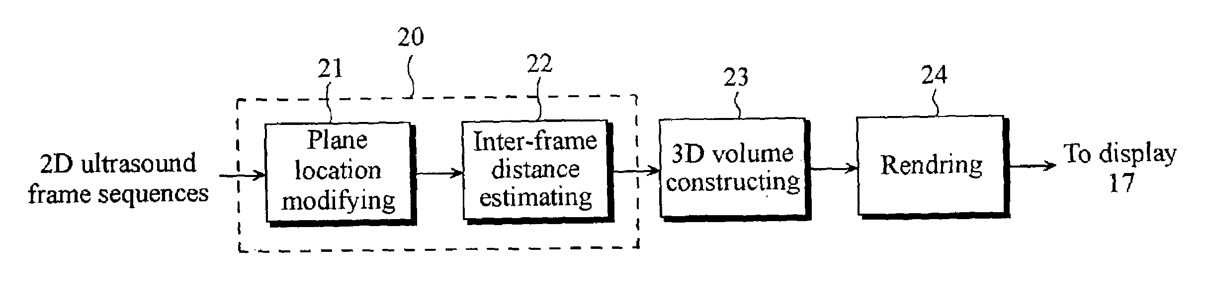
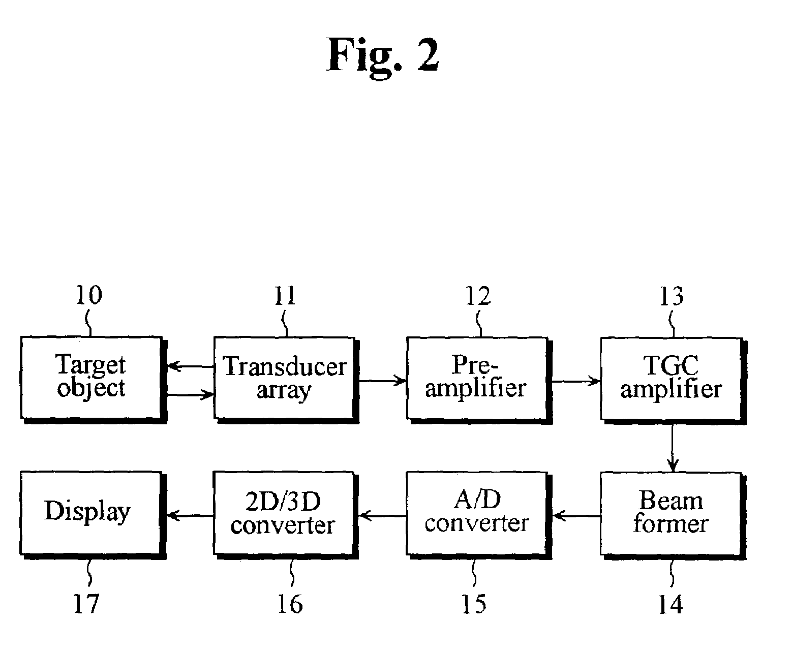
[0025]Referring to FIG. 2, which shows a block diagram of a 3D ultrasound imaging apparatus according to the present invention. Transducer array 11 transmits ultrasound signals to target object 10 and receives echo signals reflected from target object 10. The echo signa...


PUM

PropertyMeasurementUnit
lateral distance correlationaaaaaaaaaa
lateral distanceaaaaaaaaaa
distanceaaaaaaaaaa

Abstract

Disclosed is a three-dimensional ultrasound image method and apparatus for reducing distortions of ultrasound images by precisely estimating a distance between consecutive two-dimensional frames obtained by a manual scanning. The apparatus comprises a transducer array for transmitting ultrasound signals to a target object and receiving echo signals reflected from the target object; a receive-focusing unit for receive-focusing the echo signals to generate a plurality of consecutive two-dimensional ultrasound image frames; a converter for converting the consecutive two-dimensional ultrasound image frames into a three-dimensional ultrasound image; and a display for displaying the three-dimensional ultrasound image. The method comprises the steps of, a) transmitting and receiving ultrasound signals; b) receive-focusing echo signals to generate a plurality of consecutive two-dimensional ultrasound image frames; c) converting the consecutive two-dimensional ultrasound image frames into a three-dimensional ultrasound image; and d) displaying the three-dimensional ultrasound image.

Description

FIELD OF THE INVENTION[0001]The present invention relates generally to ultrasound imaging, and more particularly, to a three-dimensional ultrasound imaging method and apparatus for reducing distortions in ultrasound images, which constructs three-dimensional ultrasound images by precisely estimating the distance between consecutive two-dimensional frames obtained by manual scanning using a lateral distance correlation function.BACKGROUND OF THE INVENTION[0002]Conventionally, three-dimensional ultrasound images are obtained by rendering a three-dimensional volume consisting of consecutive two-dimensional frames stacked one-by-one. However, where the distance between stacked consecutive two-dimensional frames is not uniform, the three-dimensional ultrasound images constructed from the two-dimensional frames may be distorted. For example, if a target object is an organ in a human body, then the constructed three-dimensional organ may appear distorted from its actual shape due to the no...

Claims


Application Information

Patent Timeline
Patent Type & Authority Patents(United States)
IPC IPC(8): A61B8/00A61B8/14G01N29/44G01S7/52G01S15/89
CPCA61B8/483G01S15/8977G01S15/8993A61B8/14Y10S128/916G01S7/52034G06T19/00G06T13/40
Inventor KIM, NAM CHULSO, HYUN JOOKIM, SANG HYUNLEE, JUN HO
Owner MEDISON CO LTD
Why Eureka
Try Eureka