Method and apparatus for quantifying tissue fat content

Inactive Publication Date: 2006-02-14
GE MEDICAL SYST GLOBAL TECH CO LLC
View PDF21 Cites 60 Cited by
  • Summary
  • Abstract
  • Description
  • Claims
  • Application Information

AI Technical Summary

Problems solved by technology

Namely, a wide x-ray photon energy spectrum from the x-ray source and a lack of energy resolution from CT detection systems preclude energy discrimination CT.
Some beam-hardening artifacts can be easily corrected, but others may be more difficult to correct.
However, beam hardening from materials other than water and bone, such as metals and contrast agents, may be difficult to correct.
In addition, even with the above described correction methods, conventional CT does not provide quantitative ima

Method used


Image

  • Method and apparatus for quantifying tissue fat content
  • Method and apparatus for quantifying tissue fat content
  • Method and apparatus for quantifying tissue fat content

Examples


Embodiment Construction

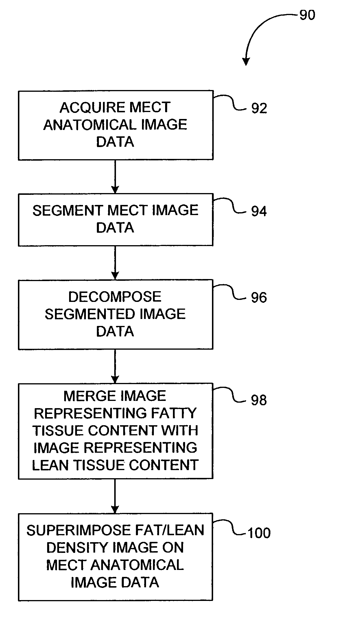
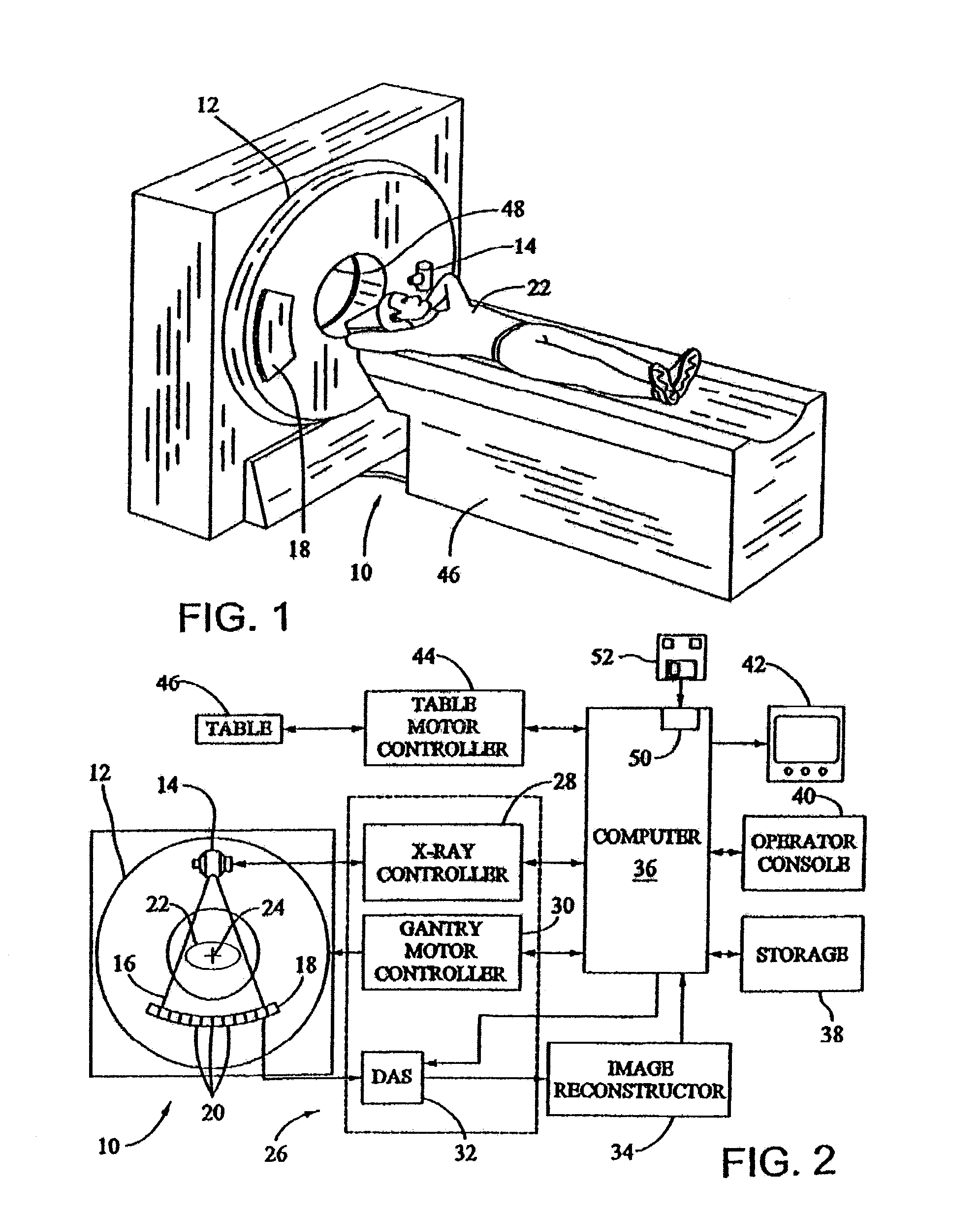
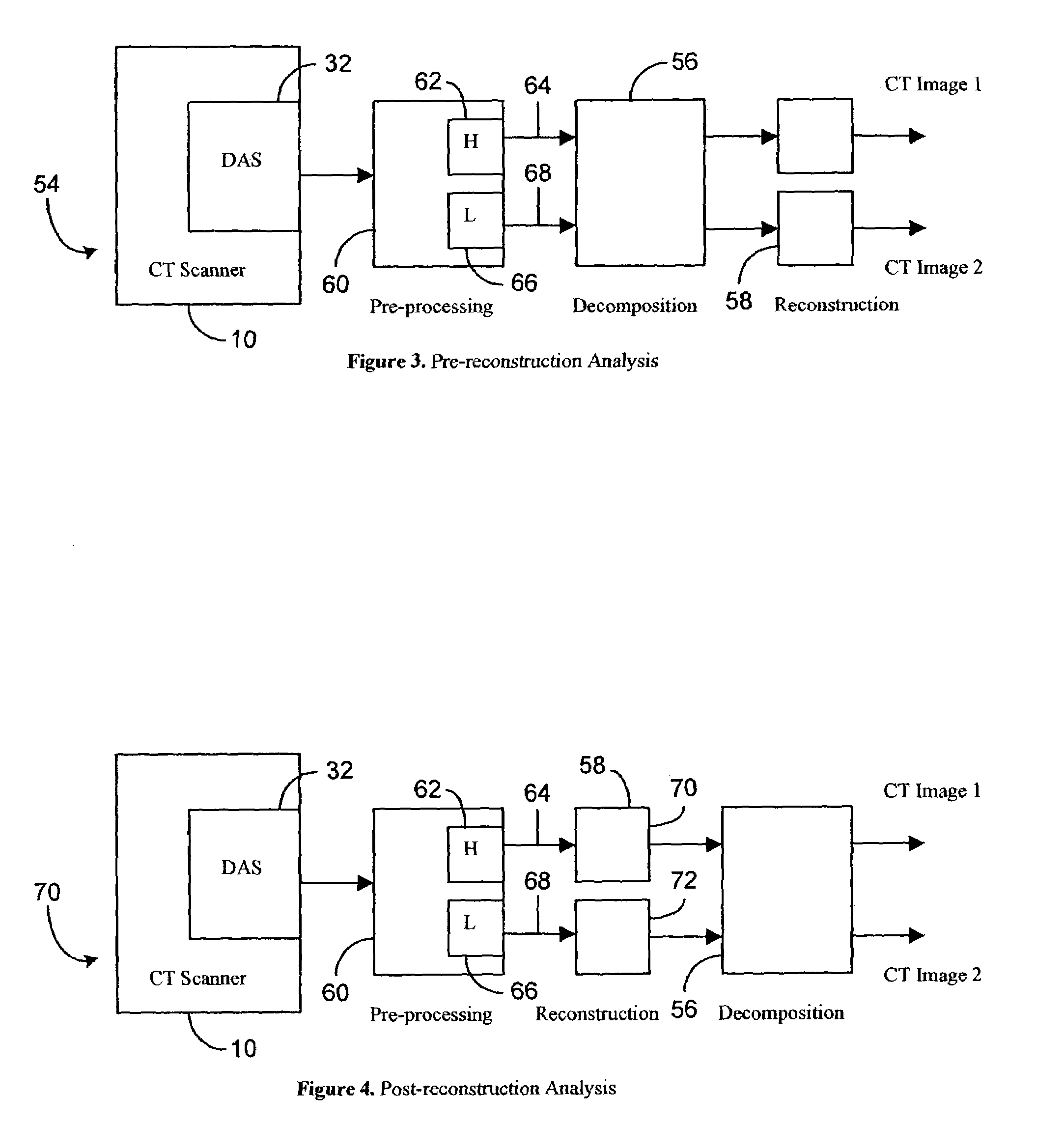
[0019]The methods and apparatus described herein facilitate accurate, non-invasive, inexpensive, and a definitive diagnosis of a fatty liver, and facilitate quantifying the tissue fat content in other organs and regions of a body. The methods and systems described herein may be used to determine the fat content, or relative fat content of any tissue or organ, in any animal, tissue specimen, or human. Additionally, the methods described herein include novel approaches to make use of the basic properties of the x-ray and material interaction. For example, for each ray trajectory, multiple measurements with different mean x-ray energies are acquired. When BMD and / or Compton and photoelectric decomposition are performed on these measurements, additional information is obtained that may facilitate improved accuracy and characterization. For example, one such characterization is determination of fat content for each voxel of acquired data. Local determination of tissue fat content facilit...


PUM


Abstract

A method for obtaining data includes quantifying tissue fat content using a multi-energy computed tomography (MECT) system.

Description

BACKGROUND OF THE INVENTION[0001]This invention relates generally to medical imaging systems, and more specifically to a method and apparatus for quantifying tissue fat content using a medical imaging system.[0002]In spite of recent advancements in computed tomography (CT) technology, such as faster scanning speed, larger coverage with multiple detector rows, and thinner slices, energy resolution is still a missing piece. Namely, a wide x-ray photon energy spectrum from the x-ray source and a lack of energy resolution from CT detection systems preclude energy discrimination CT.[0003]X-ray attenuation through a given object is not a constant. Rather, x-ray attenuation is strongly dependent on the x-ray photon energy. This physical phenomenon manifests itself in an image as a beam-hardening artifact, such as non-uniformity, shading, and streaks. Some beam-hardening artifacts can be easily corrected, but others may be more difficult to correct. In general, known methods to correct beam...

Claims


Application Information

Patent Timeline
IPC IPC(8): A61B6/00G01N23/087A61B6/03
CPCA61B5/4872A61B6/032A61B6/482A61B6/405A61B6/4241A61B6/4035
Inventor SABOL, JOHN MICHAELWALKER, MATTHEW JOSEPHAVINASH, GOPAL B.JABRI, KADRI NIZAR
Owner GE MEDICAL SYST GLOBAL TECH CO LLC
Why Eureka
Try Eureka