Medical Digital X-Ray Imaging Apparatus and Medical Digital X-Ray Sensor

a digital x-ray imaging and sensor technology, applied in the field of medical x-ray imaging apparatus and medical digital x-ray sensor, can solve the problems of unnecessarily increasing the exposure to x-ray radiation, wasting time therefor, and transferring excess data, so as to optimize the data acquisition time. , the effect of reducing the image data capacity

Active Publication Date: 2009-07-02
MORITA MFG CO LTD
View PDF11 Cites 84 Cited by
  • Summary
  • Abstract
  • Description
  • Claims
  • Application Information

AI Technical Summary

Benefits of technology

[0012]It is an advantage of the invention that an area from which image data in the digital X-ray sensor are read is changed for each imaging mode so that image data of an optimum data capacity can be acquired in an optimum time.
[0013]It is another advantage of the invention that the image data can be acquired without excess time for each imaging mode by optimizing data acquisition time.
[0014]It is a still another advantage of the invention that image data capacity can be decreased by combining signals of adjacent imaging elements to be dealt as a pixel.
[0015]It is a further advantage of the invention that the frame rate can be changed during imaging so that the amount of image data is controlled to become just enough if necessary.
[0016]It is a still further advantage of the invention that an optimum data capacity and an optimum data acquisition time can be realized for various imaging modes such as computed tomography scanning mode, panoramic imaging mode, cephalometric imaging mode, cross tomography imaging mode and dental imaging mode, so that a dose of exposure to radiation of a patient can be decreased and diagnosis efficiency can be improved.

Problems solved by technology

Therefore, if the same digital X-ray sensor is used both for computed tomography imaging and for panoramic imaging, data are read out from pixel not needed to be read, so that excess data are transferred and stored, and a time therefor is wasted.
Further, exposure to X-ray radiation is increased unnecessarily in proportion to the area in the two-dimensional detection plane.

Method used


Image

  • Medical Digital X-Ray Imaging Apparatus and Medical Digital X-Ray Sensor
  • Medical Digital X-Ray Imaging Apparatus and Medical Digital X-Ray Sensor
  • Medical Digital X-Ray Imaging Apparatus and Medical Digital X-Ray Sensor

Examples


Embodiment Construction

[0052]Embodiments of the invention are explained below with reference to the drawings appended herewith.

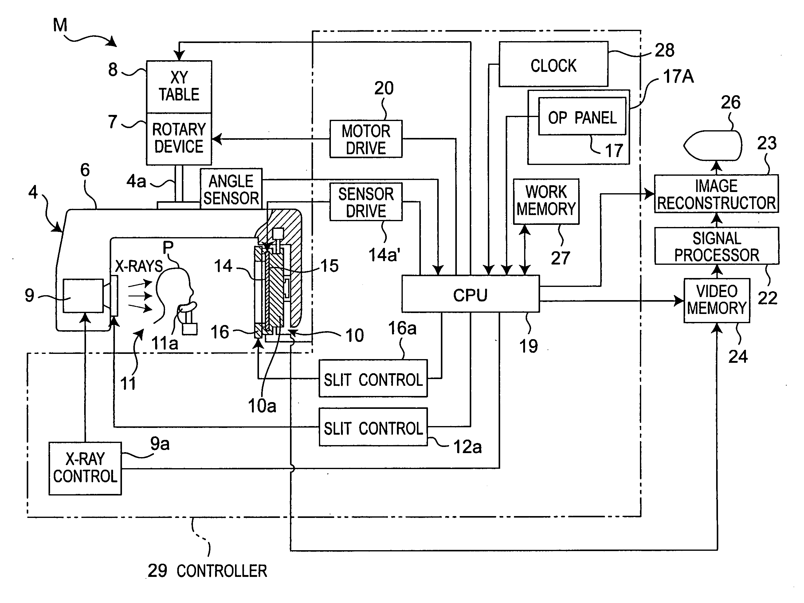
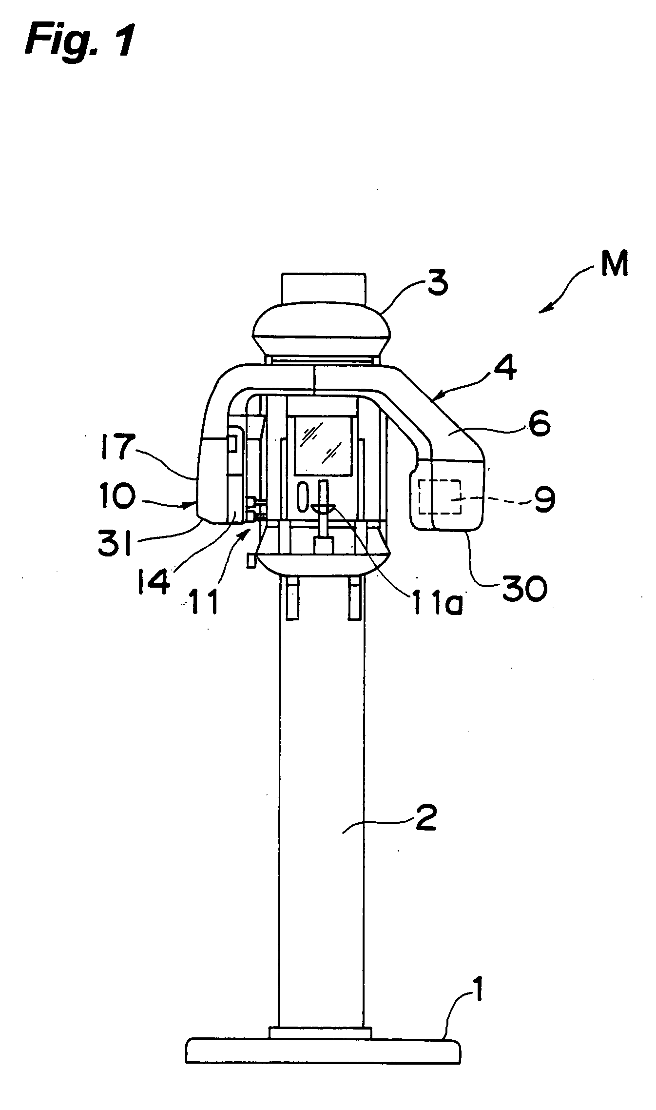
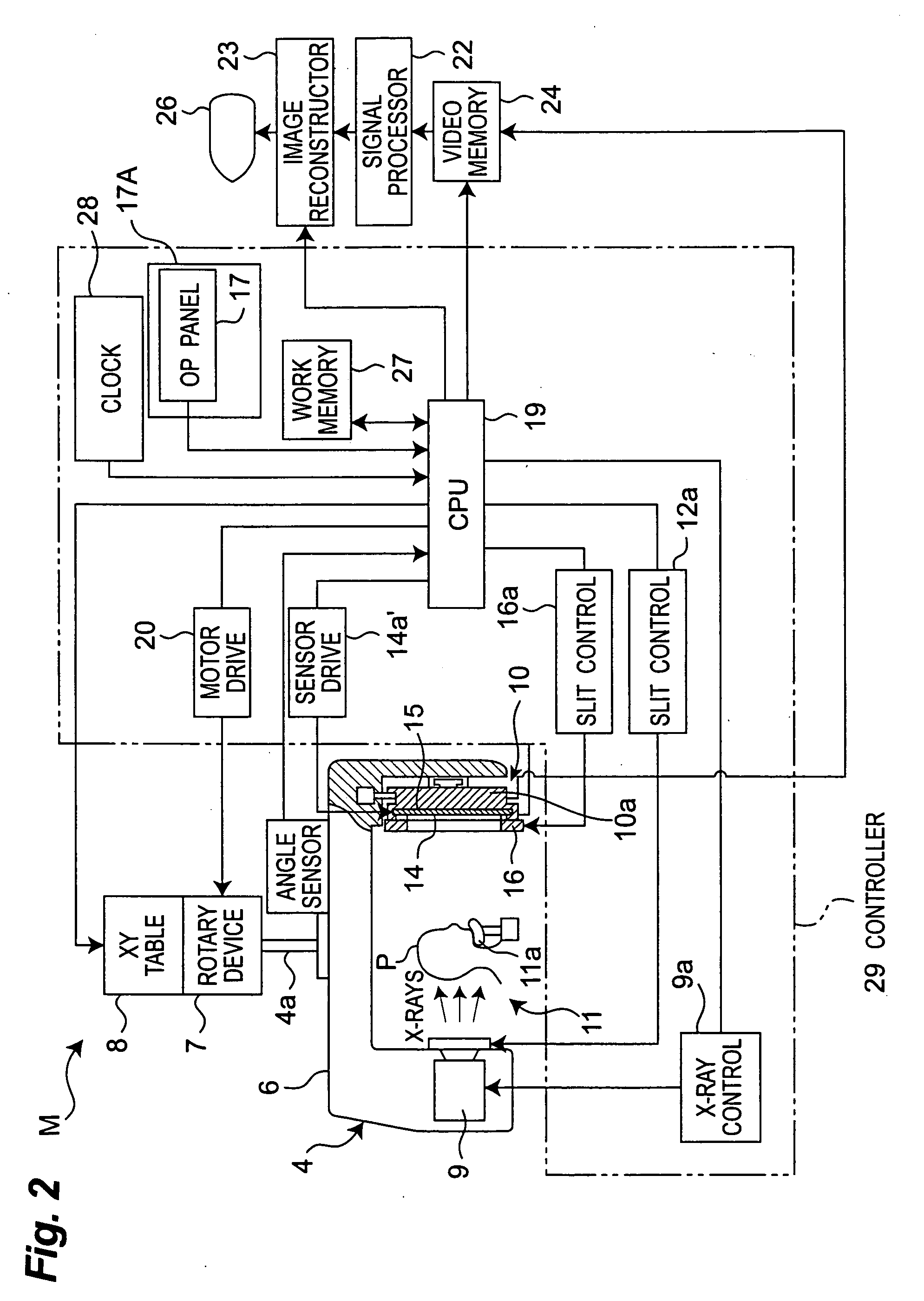
[0053]FIG. 1 shows a digital X-ray radiography apparatus such as a dental X-ray radiography apparatus according to an embodiment of the invention. Though the embodiments are explained below with reference to examples of the dental X-ray radiography apparatus, the invention is not limited to dentistry, but can be used generally for medical uses.

[0054]In FIG. 1, X-rays generated by an X-ray source transmits an object and are detected by a digital X-ray sensor having a detection plane. By limiting an imaging area in the detection plane to a necessary area, a dose of radiations exposed to a patient can be reduced to the lowest limit. Further, reconstruction of image data is performed on a selected range in the imaging area. For example, an irradiation field of X-rays generated by the X-ray source can be changed by adjusting a slit for the X-ray source, and data necessary for image rec...


PUM


Abstract

In a medical digital X-ray imaging apparatus having a plurality of imaging modes including computed tomography mode, a supporter supports an X-ray source and a digital X-ray sensor having a two-dimensional detection plane for detecting X-rays, while interposing an object between them. An image reconstructor acquires data from the digital X-ray sensor and reconstructs an image based on the acquired data. An operator selects one of a first imaging mode and a second imaging mode. The second imaging mode has an irradiation field different from the first imaging mode and has an area to be read in the digital X-ray sensor smaller than that in the first imaging mode.

Description

TECHNICAL FIELD[0001]The invention relates to a medical X-ray imaging apparatus and a medical digital X-ray sensor for digital X-ray imaging.BACKGROUND ART[0002]A digital X-ray sensor having a two-dimensional detection plane is used for a medical X-ray imaging apparatus. There are various imaging modes in X-ray imaging such as computed tomography (CT) imaging, panoramic imaging (imaging of entire jaws), cephalometric imaging (head normalization photographing) and dental imaging (imaging of one or a few teeth in a small irradiation field). A digital sensor is developed to have an appropriate size and performance for each of the imaging modes. It is desirable that one X-ray imaging apparatus can be used in a plurality of modes. For example, in a digital X-ray imaging apparatus disclosed in JP-A H8-257,026 / 1996, an X-ray MOS sensor is used for panoramic tomography or linear tomography. Slit plates are provided for an X-ray source and for an X-ray MOS sensor, and the sizes of the openin...

Claims


Application Information

Patent Timeline
Patent Type & Authority Applications(United States)
IPC IPC(8): H05G1/58
CPCA61B6/032A61B6/06A61B6/501A61B6/4233A61B6/14
Inventor SUZUKIYOSHIKAWA, HIDEKI
Owner MORITA MFG CO LTD
Why Eureka
Try Eureka