
Intraoral endotracheal tube holder and method for intubation

Inactive Publication Date: 2006-06-20
CHRISTOPHER MARCUS
View PDF9 Cites 45 Cited by
  • Summary
  • Abstract
  • Description
  • Claims
  • Application Information

AI Technical Summary

Benefits of technology

[0010]Aspects of the invention are found in a tube holder assembly and method for securing an endotracheal tube to a biteline of a patient's mouth. The tube holder assembly is compact, requires no adhesive support, and facilitates easy adjustment and readjustment of the endotracheal tube by a single medical practitioner.
[0012]A bite tensioner mount is coupled to the tube body and receives a tensile force from a bite tensioner assembly. The bite tensioner assembly features two opposing ends having a tensioner lock at one end and anchor webbing at another end. Operatively, the tensioner lock is secured to the bite tensioner mount of the tube body whereas anchor webbing is fixed to the patient so that a tensile force is exerted along the bite tensioner assembly from the tensioner lock to the anchor webbing. Anchor webbing is advantageous for securing the bite tensioner assembly to the patient's body, especially in instances where hair, oil, skin, and dirt provide slippage at particular locations as compared with the overall surface area contact provided by the anchor webbing.

Problems solved by technology

Unfortunately, today's tube holders create many difficulties for medical practitioners and patients alike.
Many typical tube holders feature a complex array of mounting plate configurations for application to the face of the patient whereby operational setup is time consuming and laborious.
In particular, set up often needlessly involves several physicians and nurses who take turns maintaining the position of the endotracheal tube while the complicated array of mounting plates and adhesives of many typical tube holder are properly situated on to the patient by the team of medical practitioners.
The application of adhesives in combination with a complex mounting plate arrangement allows very little opportunity for readjustment of the tube holder during a medical procedure as relocating mounting plates becomes cumbersome and adhesives lose their initial bonding strength.
Furthermore, the complex arrangements of many typical tube holders today often obstruct clear entry into the mouth, which is critically important for suction, inspection, maintaining the cleanliness of the endotracheal tube as well as maintaining the health of the patient's mouth during long term care.
Dirt and bodily secretions such as saliva, sweat, oils from the skin, and blood delivered by the patient onto the mounting plates and associated adhesives diminish the overall operative effectiveness of a tube holder as well as the overall sterile integrity thereof.
It should be added that prolonged application of pressure by the tube holders against the skin of the face could lead to trauma or ulceration from the mounting plates.
Unfortunately there is no known compact device or method for operatively holding an endotracheal tube during intubation that is simple and easy to use for both the medical practitioner and patient alike.

Method used


Image

  • Intraoral endotracheal tube holder and method for intubation
  • Intraoral endotracheal tube holder and method for intubation
  • Intraoral endotracheal tube holder and method for intubation

Examples


Embodiment Construction

[0034]For a more complete understanding of the present invention, preferred embodiments of the present invention are illustrated in the Figures. Like numerals being used to refer to like and corresponding parts of the various accompanying drawings. It is to be understood that the disclosed embodiments are merely exemplary of the invention, which may be embodied in various forms.

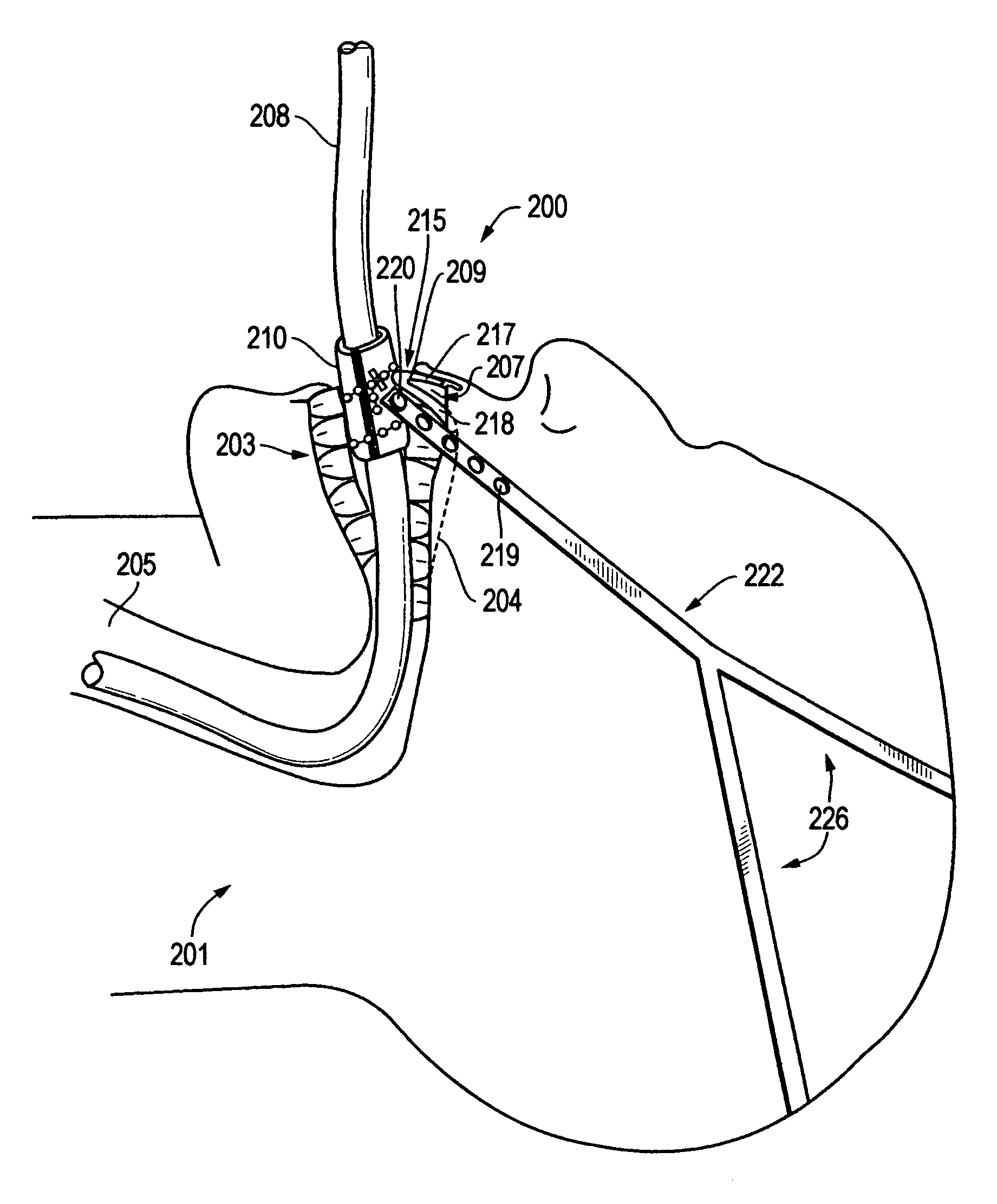
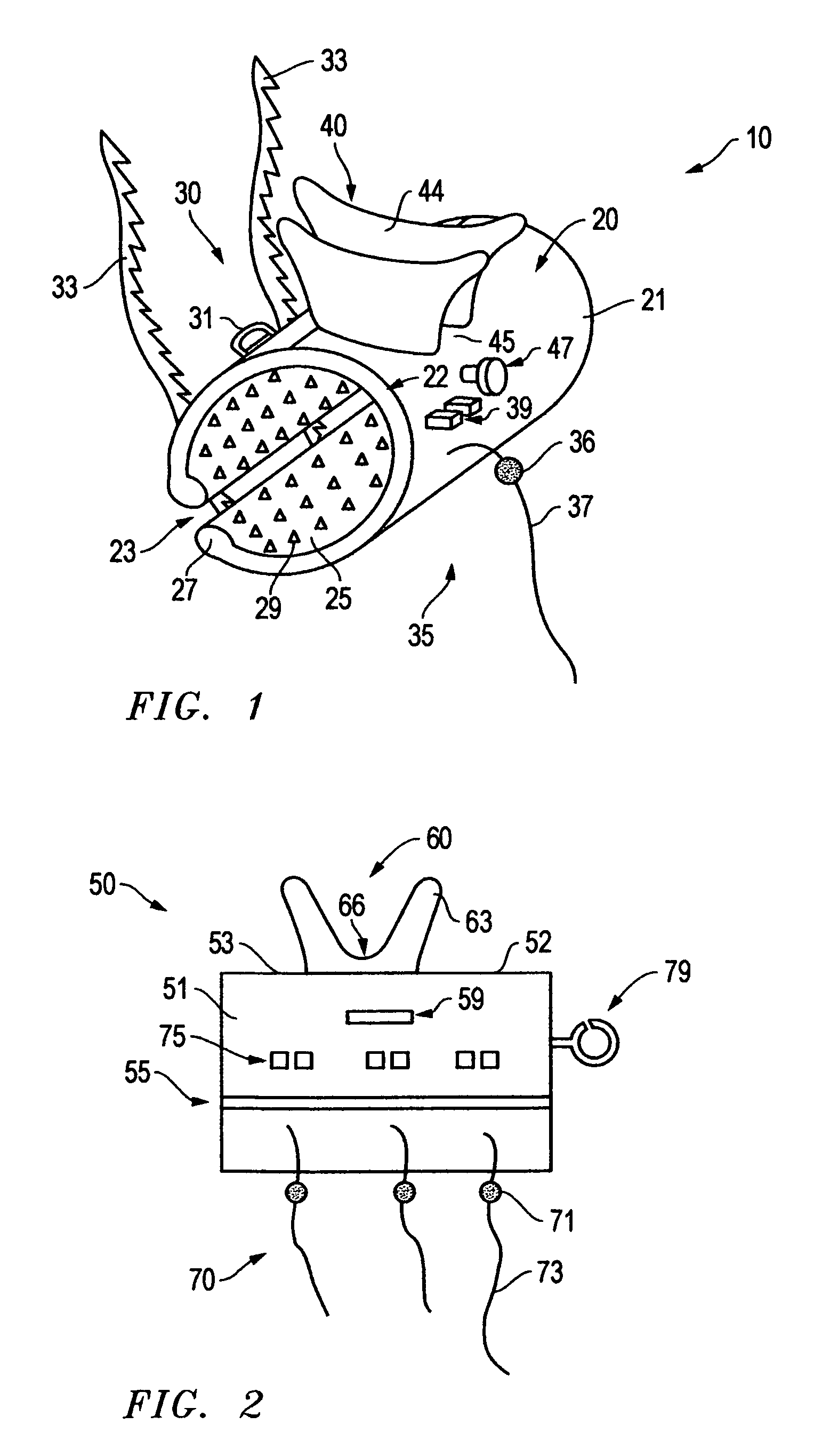
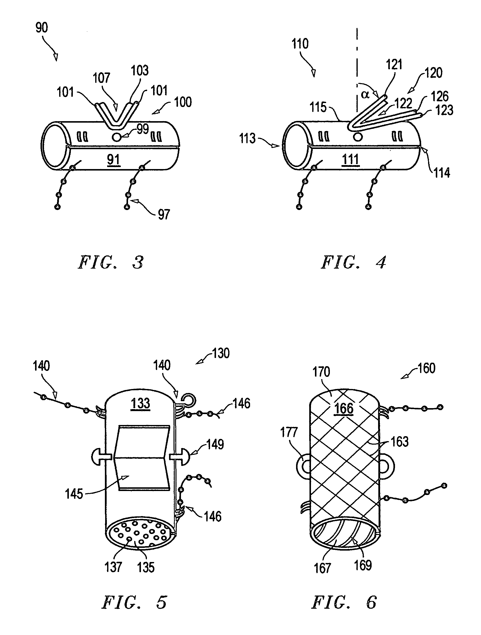
[0035]FIG. 1 illustrates one aspect, among others, of a tube holder assembly 10 for securing an endotracheal tube to a biteline of a patient's mouth. Generally, during a medical procedure, a tube holder assembly maintains a desired position of an endotracheal tube as it is firmly secured onto the biteline of a patient. Illustratively, FIGS. 7 and 8 show an intubation system for attachment to a biteline whereby a bite tensioner assembly applies a tensile force to a tube holder assembly for fixating the tube holder assembly against the biteline of a patient. In this disclosure and appended claims the term “bite...


PUM


Abstract

A tube holder assembly and method for securing an endotracheal tube to a biteline of a patient's mouth. The tube holder assembly features a tube body. A bite support assembly, coupled to the tube body, receives a tensile force and supplies a compressive force to the mouth of the patient. The bite support assembly includes a pair of substantially parallel bite walls extending outwardly from the exterior surface of the tube body. The bite walls define a bite rest therebetween for receiving a biteline of the patient's mouth and thus securing the biteline between a pair of bite walls as the bite rest applies a compressive force to the biteline.

Description

BACKGROUND OF THE INVENTION[0001]1. Field of the Invention[0002]The present invention generally relates to an intraoral tube holder assembly for supporting an endotracheal tube. More particularly, but not by way of limitation, the present invention relates to a system and method for securing an endotracheal tube to a biteline of a patient's mouth wherein the system incorporates the features of both a tube holder and bite block in a single tube holder assembly.[0003]2. Description of the Related Art[0004]Intubation is a well known medical procedure referring to the placement of a tube in to a body orifice. Endotracheal intubation, a very common intubation procedure, directs a tube from either the mouth or the nose through the larynx to the trachea for fluid engagement therein. Most often, tracheal intubation is applied in emergency medicine especially during cardiopulmonary resuscitation, CPR; for intensive care patients under respiratory support; for protecting the airways of comato...

Claims


Application Information

Patent Timeline
IPC IPC(8): A61M16/00
CPCA61M16/0488A61M16/0493A61M16/0497Y10S128/26
Inventor CHRISTOPHER, MARCUS
Owner CHRISTOPHER MARCUS
Learn More