System and method for 3-D digital reconstruction of an oral cavity from a sequence of 2-D images

a digital reconstruction and oral cavity technology, applied in the field of digital dental imaging, can solve the problems of untoward effects, unsatisfactory details, and expensive and time-consuming processes, and achieve the effect of reducing discomfort for patients and relatively short acquisition tim

Inactive Publication Date: 2006-08-01
UNIV OF LOUISVILLE RES FOUND INC
View PDF2 Cites 131 Cited by
  • Summary
  • Abstract
  • Description
  • Claims
  • Application Information

AI Technical Summary

Problems solved by technology

In order to evaluate tooth movement progress, the orthodontist monitors this movement by means of visual inspection, intra-oral measurements, fabrication of casts, photographs, and radiographs; this process is both costly and time consuming.
Moreover, repeated acquisition of radiographs may result in untoward effects.
Obtaining a cast of the jaw is a complex operation for the dentist, an unpleasant experience for the patient, and also may not provide all the necessary details of the jaw.
While oral and maxillofacial radiology is now widely accepted as a routine technique for dental examinations, the equipment is rather expensive and the resolution is frequently too low for 3-D modeling of dental structures.
Furthermore, the radiation dose required to enhance both contrast and spatial resolution can be unacceptably high.

Method used


Image

  • System and method for 3-D digital reconstruction of an oral cavity from a sequence of 2-D images
  • System and method for 3-D digital reconstruction of an oral cavity from a sequence of 2-D images
  • System and method for 3-D digital reconstruction of an oral cavity from a sequence of 2-D images

Examples


Embodiment Construction

[0021]In the following detailed description of embodiments of the invention, reference is made to the accompanying drawings that form a part hereof, and in which is shown by way of illustration specific embodiments in which the invention may be practiced. These embodiments are described in sufficient detail to enable those skilled in the art to practice the invention, and it is to be understood that other embodiments may be utilized and that logical, mechanical, electrical and other changes may be made without departing from the scope of the present invention. The following detailed description is, therefore, not to be taken in a limiting sense, and the scope of the present invention is defined only by the appended claims.

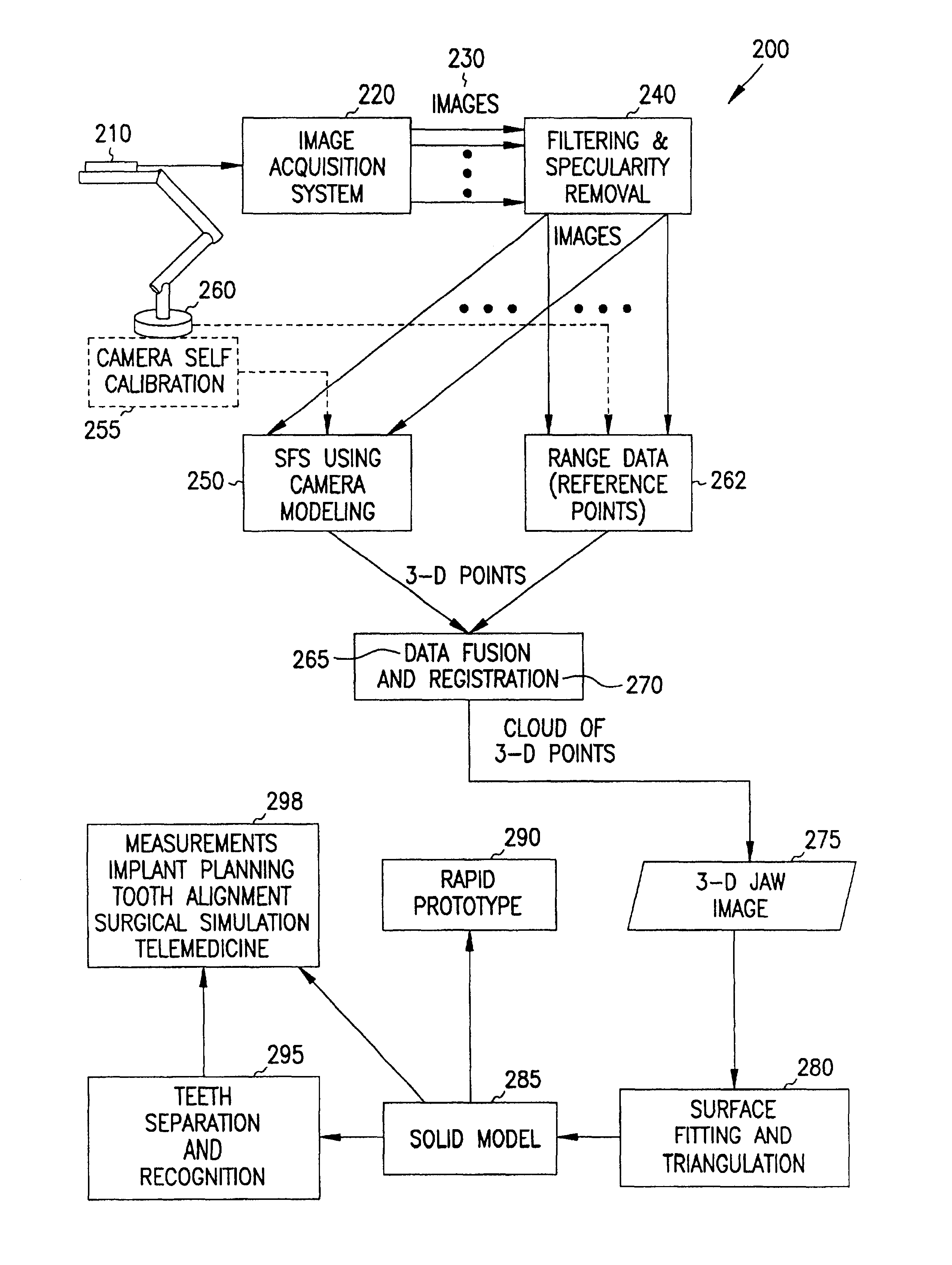
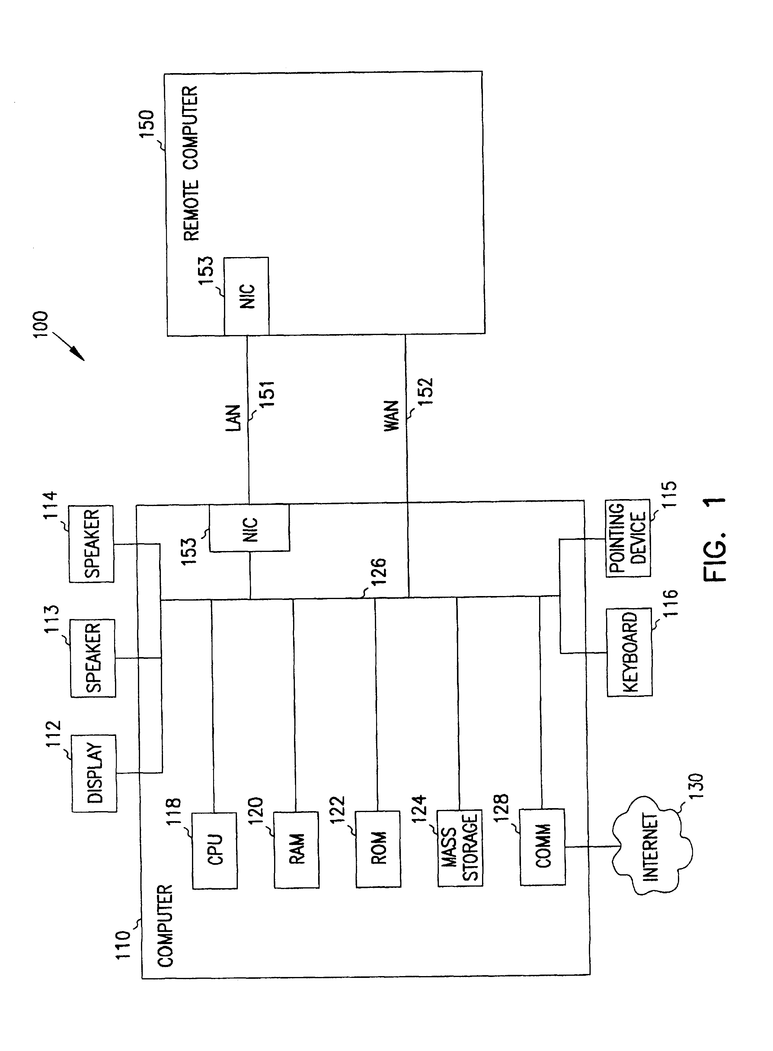
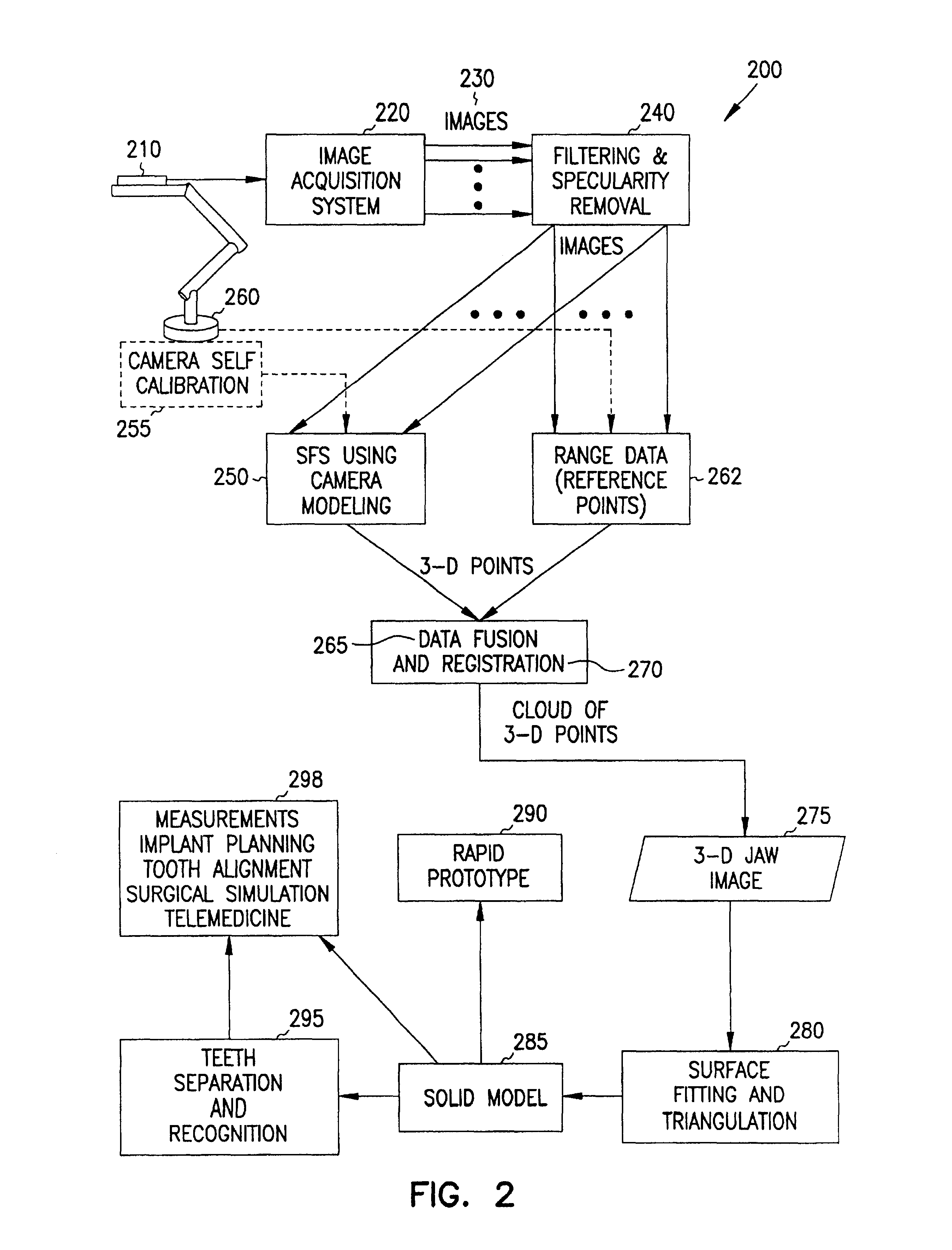
[0022]The detailed description is divided into five sections. In the first section, the hardware and the operating environment in conjunction with which embodiments of the invention may be practiced are described. In the second section, a system level overview of t...


PUM


Abstract

Systems and methods are provided through which a model-based vision system for dentistry which assists in diagnosis, treatment planning and surgical simulation. The present invention includes an integrated computer vision system that constructs a three-dimensional (3-D) model of the patient's dental occlusion using an intra-oral video camera. A modified shape from shading technique, using perspective projection and camera calibration, extracts the 3-D information from a sequence of two-dimensional images of the jaw. Data fusion of range data and 3-D registration techniques develop a complete 3-D digital jaw model. Triangulation of the 3-D digital model is then performed, and optionally, a solid 3-D model is reconstructed.

Description

RELATED APPLICATION[0001]This application claims the benefit of U.S. Provisional Application Ser. No. 60 / 199,913, filed Apr. 26, 2000 under 35 U.S.C. 119(e).FIELD OF THE INVENTION[0002]This invention relates generally to digital dental imaging, and more particularly to three-dimensional dental imaging.BACKGROUND OF THE INVENTION[0003]Dentistry requires accurate 3-D representation of the teeth and jaw for diagnostic and treatment purposes. For example, orthodontic treatment involves the application, over time, of force systems to teeth to correct malocclusion. In order to evaluate tooth movement progress, the orthodontist monitors this movement by means of visual inspection, intra-oral measurements, fabrication of casts, photographs, and radiographs; this process is both costly and time consuming. Moreover, repeated acquisition of radiographs may result in untoward effects. Obtaining a cast of the jaw is a complex operation for the dentist, an unpleasant experience for the patient, a...

Claims


Application Information

Patent Timeline
Patent Type & Authority Patents(United States)
IPC IPC(8): G06T17/00A61C9/00
CPCG06T17/00A61C9/004A61C9/0053
Inventor FARAG, ALY A.TASMAN, DAVIDYAMANY, SAMEH M.
Owner UNIV OF LOUISVILLE RES FOUND INC
Why Eureka
Try Eureka