Template for the localization of lesions in a breast and method of use thereof

a breast and template technology, applied in the field of breast tissue localization template, can solve the problems of difficult for physicians and technologists to precisely localize a lesion, ambiguity of localization lesion on the base of mammogram can be a significant problem, and difficulty in finding the lesion, so as to achieve the effect of allowing the determination of the location of the lesion in a much shorter time and a greater degree of confidence in correlating the lesion

Active Publication Date: 2006-10-24
VARIAN MEDICAL SYSTEMS
View PDF11 Cites 15 Cited by
  • Summary
  • Abstract
  • Description
  • Claims
  • Application Information

AI Technical Summary

Benefits of technology

[0011]Heretofore, there has been a fair degree of imprecision in localizing lesions based upon mammograms—even by the expert mammographers. Most physicians and technologists simply just perform an estimate of the location of the lesion and then scan over the area. A major advantage of the template of the present invention is that it is provides much more precision and allows the determination of the location of the lesion in a much shorter time. It also gives a greater degree of confidence in correlating lesion between ultrasound studies and mammograms.

Problems solved by technology

Because of the non-orthogonal projections of the mammogram views, it could be difficult for the physicians and technologists to precisely localize a lesion (calcification, nodule, or mass) that is seen on the mammogram views.
However, the same problem in translating the location of the lesion on the mammogram into radial coordinate exists as the Cartesian coordinate system because of the obliquity of the MLO view.
The ambiguity of the localizing lesion on the bases of mammogram can be a significant problem when the physician or technician needs to find the lesion on ultrasound.
In general, the ultrasound technologists need to scan the entire half of the breast and still have problem finding the lesion.
This is extremely time consuming and expensive.
Often times it is difficult to be sure that a lesion seen on ultrasound is the same lesion seen on mammogram.
It cannot be used in conjunction with mammograms in localizing lesions within the breast because it does not take into account of how the mammograms are being performed or the direction of the x-ray beam.
The device, therefore, cannot be used to localize a lesion within the women's breast.

Method used


Image

  • Template for the localization of lesions in a breast and method of use thereof
  • Template for the localization of lesions in a breast and method of use thereof
  • Template for the localization of lesions in a breast and method of use thereof

Examples


Embodiment Construction

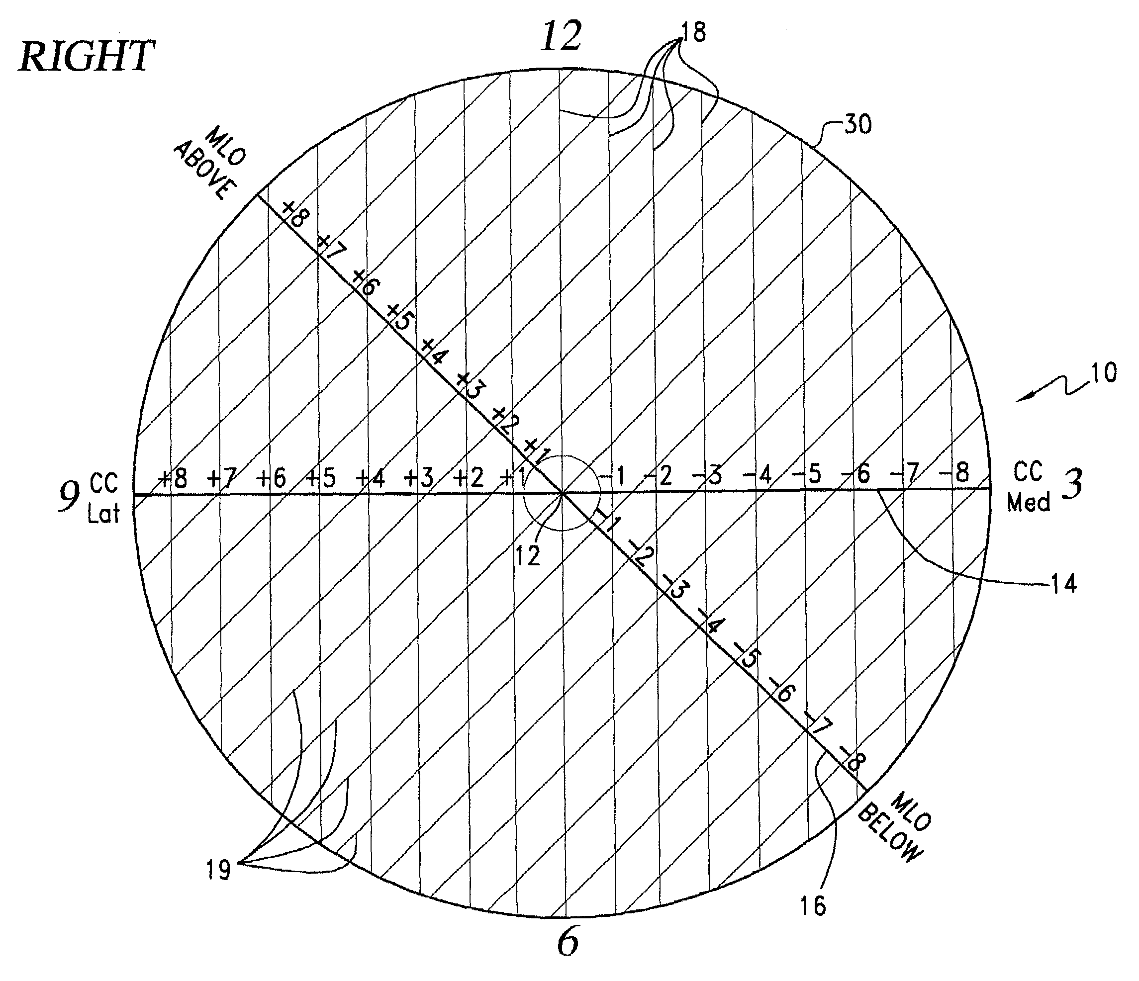
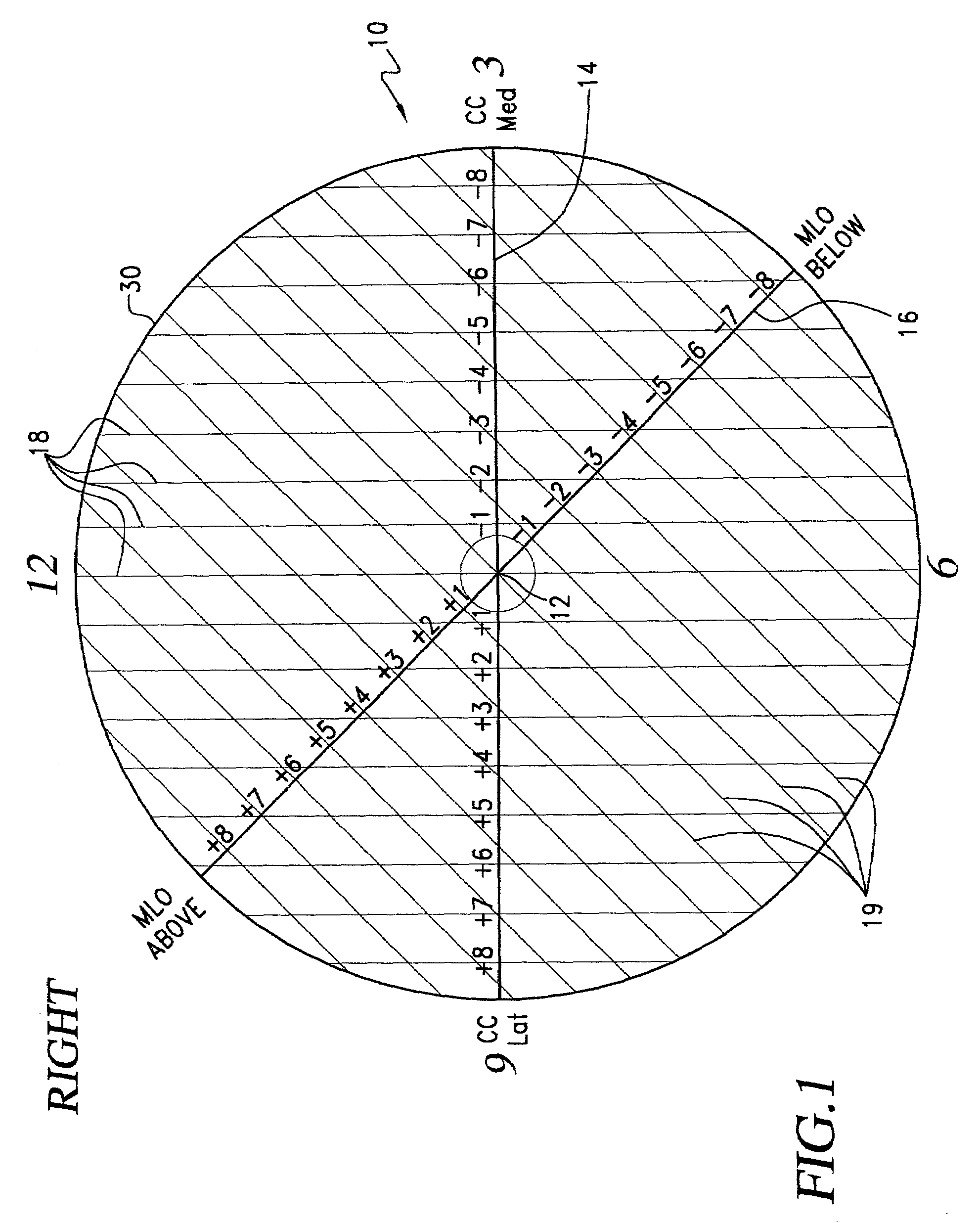
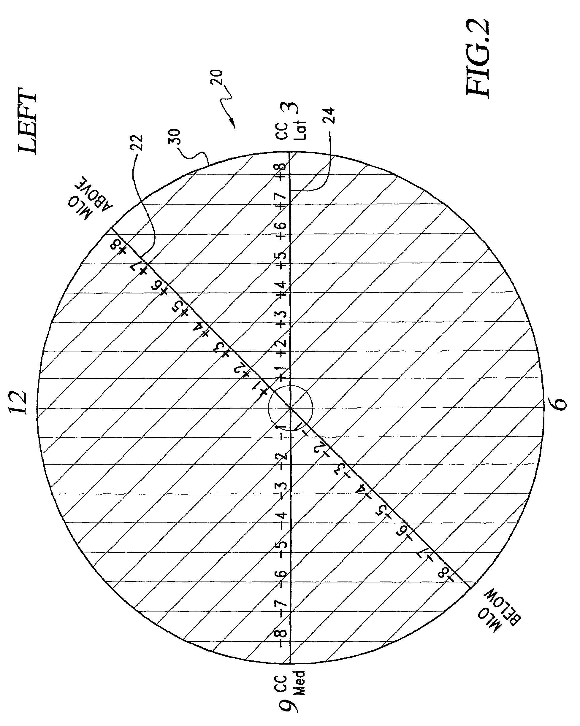
[0018]Referring to the drawings and the characters of reference marked thereon FIGS. 1 and 2 illustrate preferred embodiments of the right and left templates of the present invention, designated generally as 10 and 20, respectively. Each of the templates 10, 20 comprises a substrate of transparent material, preferably flexible plastic. The substrate is preferably planar. It may be formed of various other materials such as glass. The right template 10 includes an origin or central marking 12, a horizontal line 14 extending through the central marking 12 and an oblique line 16 extending approximately a negative 45 degrees from the horizontal line 14 and through the central marking 12. These markings and the additional markings discussed hereinafter may be scratched, grooved, etched, silk screened, printed, painted, molded, or similarly fixed upon the substrate in the manner and relationships indicated to facilitate the measurements to be made.

[0019]The right template 10 includes a fir...


PUM


Abstract

A template for the localization of lesions in a breast. The template includes a substrate of transparent material that includes a central marking; a horizontal line extending through the central marking; a first series of spaced lines, each line of the first series being perpendicular to the horizontal line and each line including marking indicia for indicating the distance of that line from the central marking; an oblique line extending approximately 45 degrees from the horizontal line and through the central marking; and, a second series of spaced lines, each line of the second series being perpendicular to the oblique line and each line including marking indicia for indicating the distance of that line from the central marking. There is a right breast template and a left breast template. The appropriate template is utilized and the CC and MLO views of a mammogram are used to define a CC line and an MLO line that are plotted on that template. The intersection of these two lines is indicative of the location of the lesion. The template can then be placed over the breast of the patient and a lesion-indicative marking can be placed on the patient's skin.

Description

BACKGROUND OF THE INVENTION[0001]1. Field of the Invention[0002]This present invention relates to medical and scientific measuring devices, and more particularly to a template for precisely and efficiently localizing a lesion of the breast from mammogram results.[0003]2. Description of the Related Art[0004]Mammography has been shown to be the most sensitive and most reliable method to detect breast cancer. Mammograms are generally taken in two non-orthogonal projections for each breast. The “cranial caudal” (CC) projection is taken with the breast being compressed in superior to inferior orientation (vertically) with the x ray tube located in the superior aspect of the breast. The second projection is the “medial lateral oblique” (MLO) projection taken at 45 degree from the horizontal projection with the x ray tube shooting from medial towards lateral but in a 45 degree oblique orientation. This is done because this view maximizes the amount of breast tissue that can be included on ...

Claims


Application Information

Patent Timeline
Patent Type & Authority Patents(United States)
IPC IPC(8): A61B19/00A61B5/107
CPCA61B5/107A61B5/4312A61B19/46A61B2019/545A61B90/06A61B2090/395
Inventor WONG, WILSON S.
Owner VARIAN MEDICAL SYSTEMS
Why Eureka
Try Eureka