Automated scanning method for pathology samples

a scanning method and pathology technology, applied in the field of automatic analysis of samples, can solve the problems of missing one patient, normally not being practicable,

Active Publication Date: 2006-11-07
LEICA BIOSYST IMAGING
View PDF19 Cites 54 Cited by
  • Summary
  • Abstract
  • Description
  • Claims
  • Application Information

AI Technical Summary

Benefits of technology

[0011]The present invention provides techniques for high-throughput automated analysis of samples such as biological samples having microscopic features.

Problems solved by technology

All the slides were first manually examined by conventional microscopy (as noted in the introduction, this would normally not be practical).
Further, one patient was missed in the manual examination but found positive upon visual verification of the candidate cells after the automated analysis.

Method used


Image

  • Automated scanning method for pathology samples
  • Automated scanning method for pathology samples
  • Automated scanning method for pathology samples

Examples


Embodiment Construction

Overview

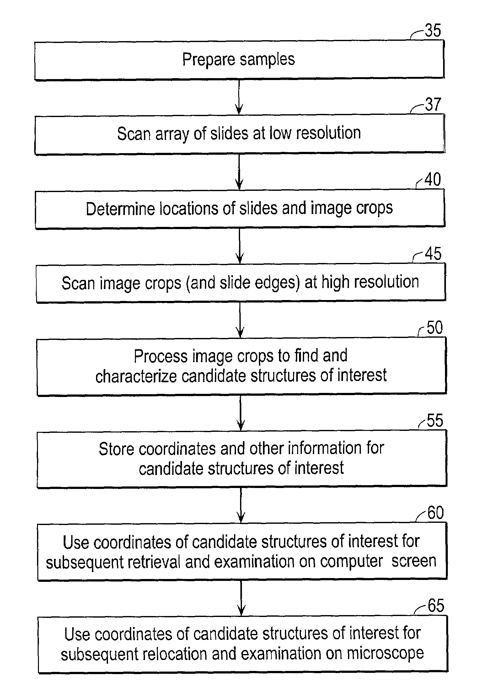
[0025]The present invention provides techniques for the scanning and analysis of large numbers of cytology and histology samples, using a flatbed scanner to capture images of the structures of interest (e.g., cells, groups of cells, and the like). The scanner provides sufficient image resolution to allow for the analysis of samples provided with one or more relevant markers to produce enough brightfield contrast to recognize structures of interest, including samples subjected to such common pathology staining techniques as ICC (immunocytochemistry), IHC (immunohistochemistry), or in situ hybridization.

[0026]A particular application is the detection of micrometastatic (tumor) cells in lymph nodes, but the invention can be used for many applications, especially when large numbers of samples need to be processed. Micrometastatic cells are often referred to as micrometastases or micromets.

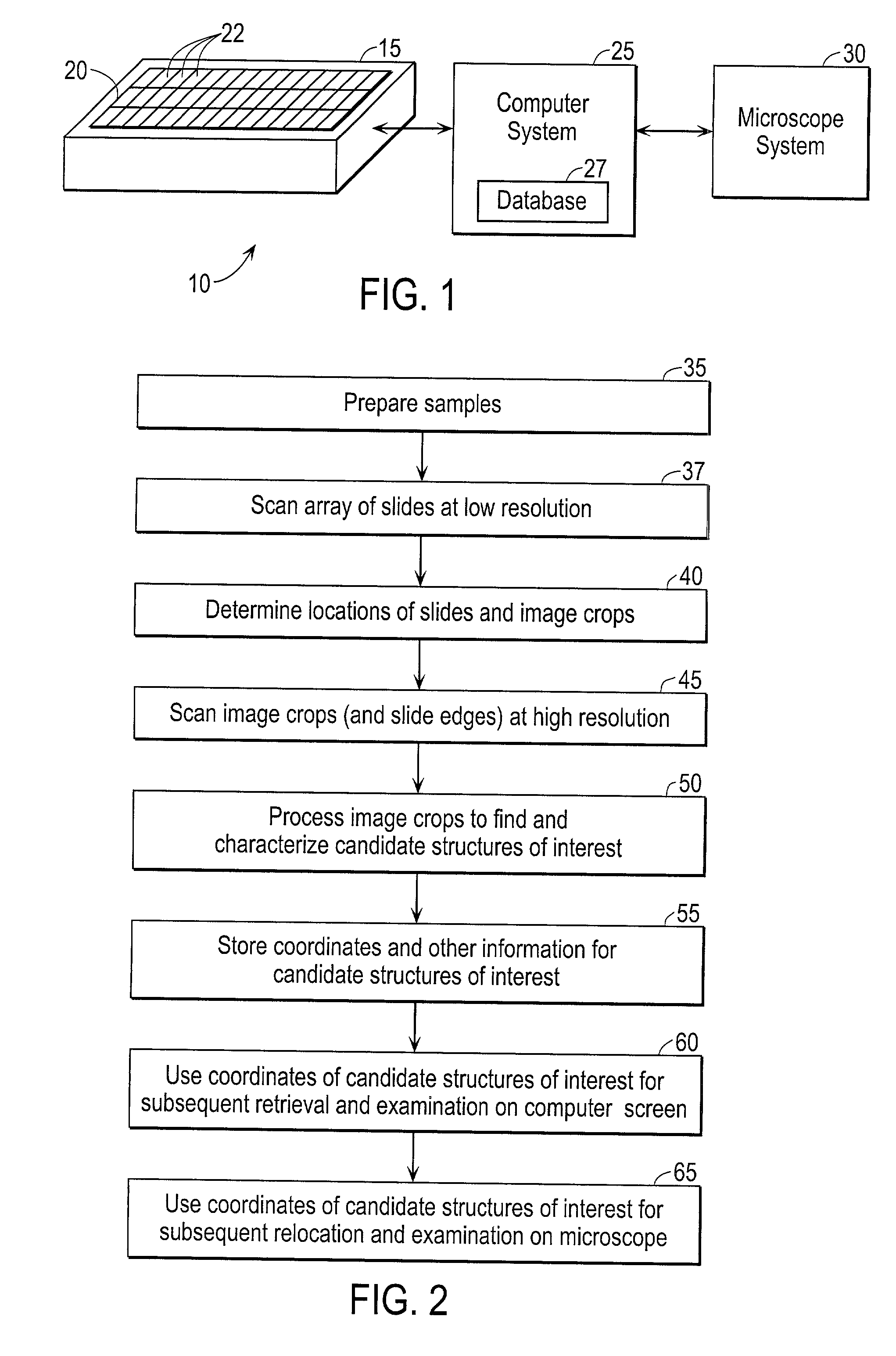
[0027]FIG. 1 is a schematic view of a system 10 implementing an embodiment of the present i...


PUM

PropertyMeasurementUnit
depthaaaaaaaaaa
distanceaaaaaaaaaa
field of viewaaaaaaaaaa

Abstract

Scanning and analysis of cytology and histology samples uses a flatbed scanner to capture images of the structures of interest such as tumor cells in a manner that results in sufficient image resolution to allow for the analysis of such common pathology staining techniques as ICC (immunocytochemistry), IHC (immunohistochemistry) or in situ hybridization. Very large volumes of such material are scanned in order to identify cells or clusters of cells which are positive or warrant more detailed examination, and if analysis at higher resolution is necessary, information regarding these positive events is transferred to a secondary microscope, such as a conventional scanning microscope, to allow further analysis and review of the selected regions of the slide containing the sample.

Description

CROSS-REFERENCES TO RELATED APPLICATIONS[0001]This application claims priority from the following two provisional applications, the entire disclosures of which, including any attached documents, are incorporated by reference in their entirety for all purposes:[0002]Provisional Application No. 60 / 376,775, filed Apr. 29, 2002 of Padraig S. O'Kelly et al. titled “Automated Scanning Method for Pathology Samples;” and[0003]Provisional Application No. 60 / 298,227 filed Jun. 12, 2001 of Padraig S. O'Kelly et al. titled “Automated Scanning Method for Pathology Samples.”BACKGROUND OF THE INVENTION[0004]This invention relates generally to the automated analysis of samples (specimens) such as biological samples having microscopic features, and more specifically to the use of a flatbed scanner in such analysis.[0005]The desirability of analyzing lymph nodes of cancer patients for micrometastatic (tumor) cells is well established, both as a indicator of patient prognosis and as a possible guide a...

Claims


Application Information

Patent Timeline
Patent Type & Authority Patents(United States)
IPC IPC(8): G06K9/00G01N15/14G06T7/00
CPCG06T7/0012G06T7/0042G01N15/1475G06T2207/30024G06T7/73
Inventor VERWOERD, NICO PETERVROLIJK, JOHANNESMESKER, WILHELMINA E.SLOOS, WILLEM C. R.BONNET, JANO'KELLY, PADRAIG S.GREGSON, MARKSHIELDS, KEVINTANKE, HENDRIKUS J.
Owner LEICA BIOSYST IMAGING
Why Eureka
Try Eureka