Use of human plasma hyaluronidase in cancer treatment

a technology of hyaluronidase and human plasma, which is applied in the field of hyaluronidase, can solve the problems of limited success in immunopurification attempts, inability to isolate this acid active hyaluronidase activity from human serum, and inability so as to improve the level of activity and less likely to induce some side effects. , the effect of better control of the activity level

Inactive Publication Date: 2006-12-12
RGT UNIV OF CALIFORNIA
View PDF6 Cites 75 Cited by
  • Summary
  • Abstract
  • Description
  • Claims
  • Application Information

AI Technical Summary

Benefits of technology

[0027]An advantage of the present invention is that purified hpHAse is more appropriate for therapeutic uses than the presently available commercial formulations of hyaluronidase which are from a non-human source, which contain two hyaluronidases (rather than one), and which, as determined by SDS-PAGE analysis, is a very crude mixture that contains various proteins, including several unidentified proteins and proteins having various biological activities including anticoagulant activities (Doctor et al., Thrombosis Res. 30:565–571). Purified hpHAse provides a “clean” source of hyaluronidase, is less likely to induce some of the side effects associated with the presently available commercial formulation, and allows better control of the level of activity associated with specific dosages.
[0028]Another advantage of the invention is that hpHAse can be purified from a lipid fraction of plasma that, according to present commercial practices, is otherwise discarded.
[0029]An advantage of the HAse assay of the invention is that it provides for an assay for HAse activity that is at least 1,000-fold more sensitive than conventional ELISA-like assays. In addition, the invention provides a biotinylated hyaluronic acid substrate that can be easily and efficiently prepared.
[0030]An advantage of the anti-native aaHAse assay of the invention is that the assay provides a rapid screen for antibodies that bind native aaHAse.
[0031]Another advantage of the invention is that specific detection of hpHAse allows a specific means of measuring LuCa-1 expression and correlation of levels of hpHAse in serum or urine of a patient with susceptibility to or presence of a disease, associated with a LuCa-1 defect (e.g., cancer, e.g., small cell lung cell carcinoma).

Problems solved by technology

The bovine extract is not optimum not only because of its non-human source, but also because the extract contains multiple types of hyaluronidases and other as yet undefined components.
1967 supra), attempts to isolate this acid active hyaluronidase activity from human serum have met with limited success due in part to the stability of the purified activity and the inability to obtain an adequately high specific activity.
Immunopurification attempts have been hindered by the inability to identify and isolate antibodies that specifically bind the activity in its native form in plasma.
These conventional methods for detecting hyaluronidase activity are hampered by both the difficulties in producing a detectably-labeled hyaluronic acid substrate and the technical difficulties in achieving rapid, sensitive, and reproducible detection of hyaluronidase activity.
8:571–584), which requires tedious, time-consuming steps.
Furthermore, conventional hyaluronidase assays use assay plates having HA substrate non-covalently bound to the plate surface, which can lead to both false positive and false negative results.
Because the HA substrate is non-covalently bound to the plate surface, the washing step following exposure of the plates to the HAse-containing sample often results in non-specific removal of non-degraded HA substrate on the plate.
Thus, the sensitivity of the conventional HAse assay is compromised.
HAse activities using gel zymography avoid the problem associated with ELISA-like assays, but are time-consuming (e.g., the test sample and the HA-containing gel are normally incubated for about 18 hr to 24 hr) and can result in artifacts if the gel is improperly loaded with excess protein sample.
Moreover, analyses of crude preparations is impossible by gel zymography.

Method used


Image

  • Use of human plasma hyaluronidase in cancer treatment
  • Use of human plasma hyaluronidase in cancer treatment
  • Use of human plasma hyaluronidase in cancer treatment

Examples


example 1

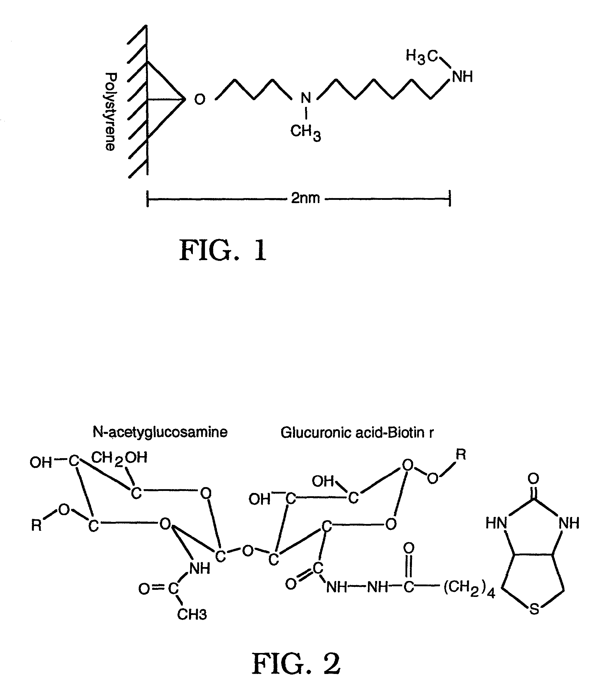
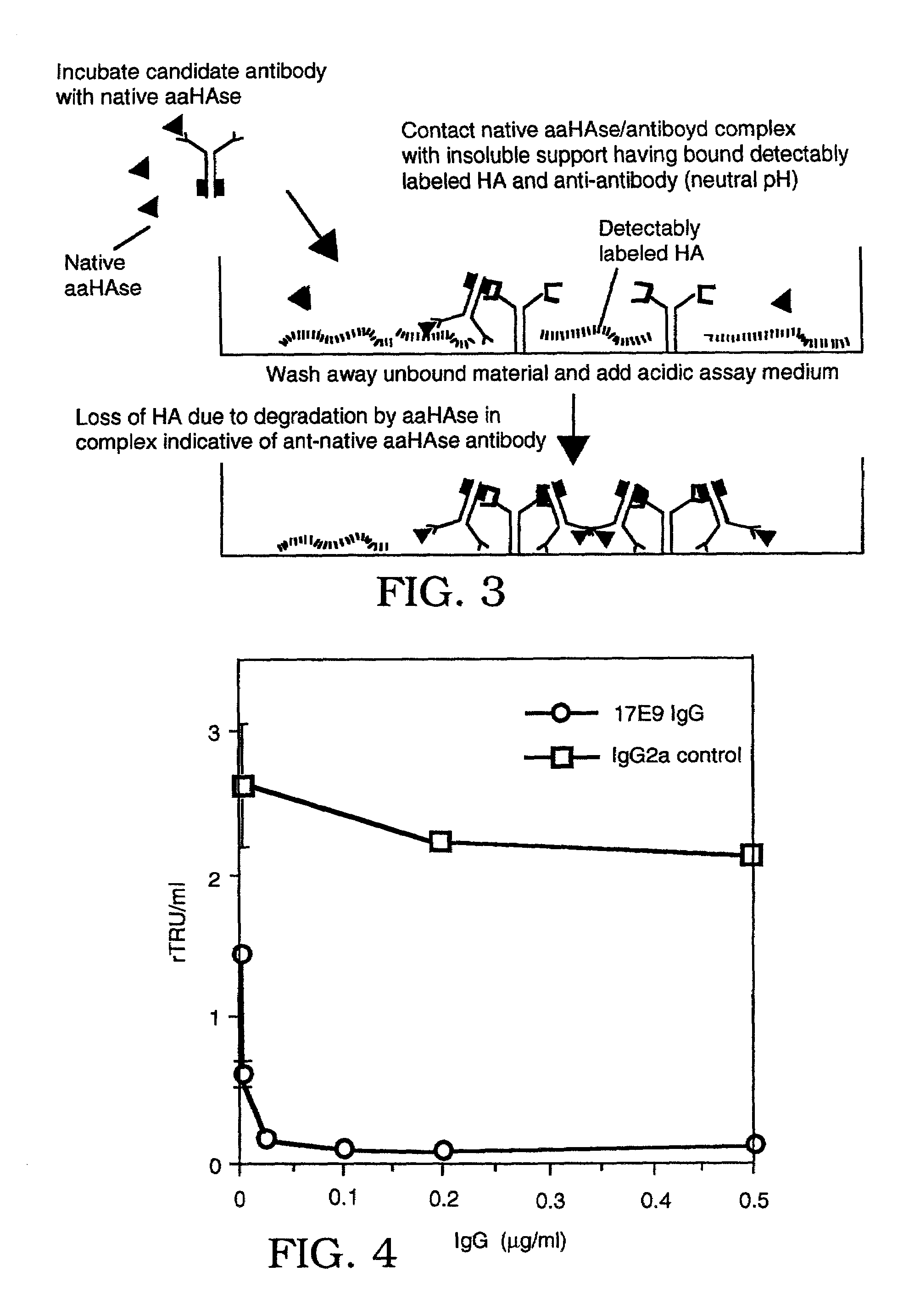
Hyaluronidase (HAse) Activity Assay Using Covalently-Bound Biotinylated Hyaluronic Acid

[0148]Human umbilical cord hyaluronic acid (HA; ICN) was biotinylated through free carboxyl groups with biotin hydrazide (Pierce), coupled using 1-ethyl-dimethylaminopropyl carbodiamide (EDC, Sigma). 100 mg of hyaluronic acid (HA) was dissolved in 0.1 M Mes pH 5.0, and biotin hydrazide (Pierce), dissolved in DMSO, was added to a final concentration of 1 mM. EDC was dissolved into the HA / biotin solution to a final concentration of 0.03 mM and left stirring overnight at 4° C. Uncoupled biotin was then removed through exhaustive dialysis against distilled water (dH20) and was stored at −20° C. at a final concentration of 1 mg / ml.

[0149]Up to about 10 days before performing the hyaluronidase assay, the biotinylated hyaluronic acid (bHA) was then covalently coupled to Covalink-NH microtiter plates (NUNC, Placerville, N.J.) at a final concentration of 5 μg of bHA / well with 9.2 μg / well Sulfo-NHS (Pierce) ...

example 2

Purification of Human Plasma Hyaluronidase Using the Biochemical Purification Method of the Invention

[0152]hpHAse was purified in a three-step biochemical procedure: 1) temperature-induced detergent hpHAse extraction of human plasma; 2) Fast Flow-S cation exchange chromatography; and 3) hydroxyl-apatite resin. Hyaluronidase activity and protein concentration were determined at each stage of purification, and the specific hyaluronidase activity calculated. Hyaluronidase activity was determined using the assay in Example 1. Protein concentration was determined by both absorbance at 280 nm and with the Biorad (Burlingame, Calif.) protein microassay kit (Tengblad 1979 Biochim. Biophys. Acta 578:281–289) in 96 well plates, using crystallized bovine serum albumin as a standard and read at 595 nM.

[0153]1) Temperature-Induced Detergent Phase Extraction

[0154]Two liters of outdated human plasma, obtained from either UCSF or Irwin Memorial blood donor centers, were routinely used for enzyme pu...

example 3

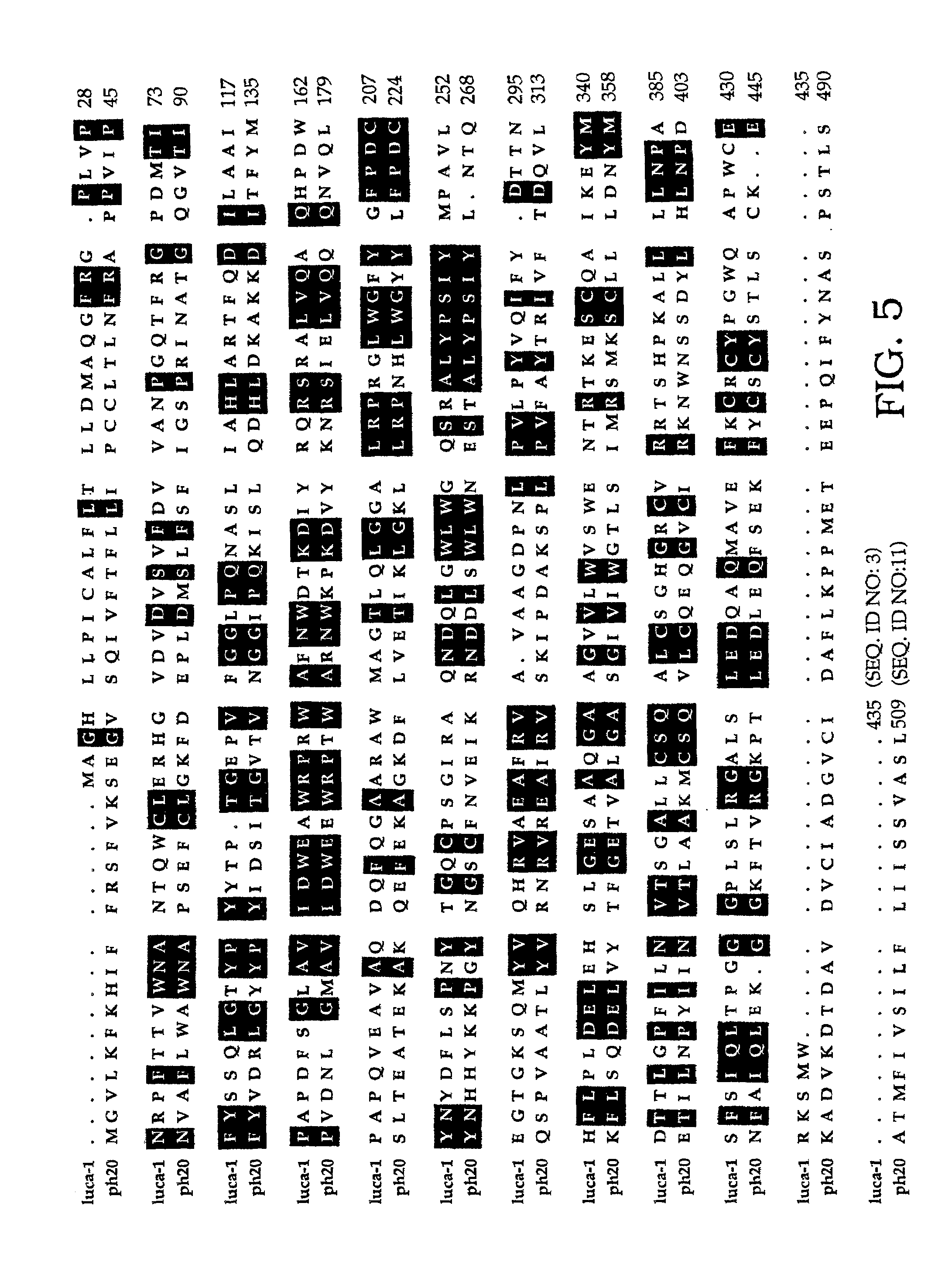
Screening Assay for Anti-Native Acid Active HAse (aaHAse) Antibodies

[0168]Biotinylated HA (bHA) was prepared as described in Example 1. Approximately 5 μg of bHA / well and 1.25 μg / well of goat anti-mouse IgG (Jackson Immunolabs) were covalently coupled to Covalink-NH microtiter plates (NUNC, Placerville, N.J.) with 9.2 μg / well Sulfo-NHS (Pierce) and 6 μg / well fresh EDC overnight at 4° C. Unbound HA and goat antibody were removed through washing plates with wash buffer (PBS with 2M NaCl, 50 mM MgSO4, 0.05% Tween-20).

[0169]Anti-native acid active HAse (anti-native aaHAse) antibodies were screened by incubating candidate antibodies with a sample containing an acid active HAse in a neutral pH buffer composed of 1% Triton X-100 in phosphate-buffered saline (PBS). If the sample was a diluted sample (e.g., a diluted plasma sample), the neutral pH buffer additionally contained 5 mg / ml bovine serum albumin (BSA). The aaHAse-containing sample was incubated with the candidate antibodies to allo...


PUM

PropertyMeasurementUnit
pHaaaaaaaaaa
molecular weightaaaaaaaaaa
pHaaaaaaaaaa

Abstract

The invention is based on the discovery of methods for purification of an acid active hyaluronidase found in human plasma (hpHAse), including both biochemical and immunoaffinity purification methods. The method of immunoaffinity purification of the invention is based on the discovery of a method for identifying antibodies that specifically bind native hpHAse (anti-native hpHAse antibodies), and anti-native hpHAse antibodies identified by this screening method. The invention also features an assay for sensitive detection of HAse activity using biotinylated hyaluronic acid (bHA). Purification and characterization of hpHAse lead to the inventors' additional discovery that hpHAse is encoded by the LuCa-1 gene, which gene is present in the human chromosome at 3p21.3, a region associated with tumor suppression. The invention additionally features methods of treating tumor-bearing patients by administration of hpHAse and / or transformation of cells with hpHAse-encoding DNA.

Description

CROSS REFERENCE TO RELATED APPLICATIONS[0001]This application is a continuation of U.S. patent application Ser. No. 08 / 916,935, filed Aug. 21, 1997, now U.S. Pat. No. 6,193,963, which is a continuation-in-part of U.S. application Ser. No. 08 / 733,360, filed Oct. 17, 1996, now U.S. Pat. No. 6,103,525, which applications are hereby incorporated by reference.STATEMENT REGARDING FEDERALLY SPONSORED RESEARCH OR DEVELOPMENT[0002]This invention was made with government support under grant nos. CA44768, CA58207, and GM46765, awarded by the National Institutes of Health. The government may have certain rights in this invention.FIELD OF THE INVENTION[0003]This invention relates generally to the field of β-1,4-endoglycosidases, particularly hyaluronidases.BACKGROUND OF THE INVENTION[0004]Hyaluronidases (HAses; E.C. 3.1.25) are a group of neutral- and acid-active enzymes found throughout the animal kingdom in organisms as diverse as microbes (e.g, Streptococcus pyogenes, Treponema palladium, and...

Claims


Application Information

Patent Timeline
Patent Type & Authority Patents(United States)
IPC IPC(8): A61K31/70A01N63/00C12N15/63A61K38/43G01N33/53G01N33/50A61K38/00A61K38/46A61K39/395A61P35/00C07K16/40C12N5/10C12N9/16C12N9/24C12N9/26C12N15/09C12P21/08C12R1/91G01N33/15G01N33/573
CPCC07K16/40C12N9/16C12Y302/01035C12Y302/01036G01N33/573C12N9/2474A61K38/00A61P35/00
Inventor STERN, ROBERTFROST, GREGORY I.CSÓKA, ANTHONYWONG, TIM M.
Owner RGT UNIV OF CALIFORNIA
Why Eureka
Try Eureka