Method of and apparatus for detecting diseased tissue by sensing two bands of infrared radiation

a technology of infrared radiation and infrared radiation, which is applied in the field of infrared radiation sensing methods and apparatuses for detecting diseased tissue, can solve the problems of insufficient quantity of ir imagers, breast cancer is very susceptible to detection, and changes in temporal or periodic perfusion, so as to achieve enhanced contrast in the two series of ir images

Inactive Publication Date: 2007-05-08
LOCKHEED MARTIN CORP
View PDF22 Cites 18 Cited by
  • Summary
  • Abstract
  • Description
  • Claims
  • Application Information

AI Technical Summary

Benefits of technology

[0011]In further embodiments, alternative data analysis is possible. This alternative data analysis may include finding contributing frequencies for each subarea and determining that tissue corresponding to a cluster having a spatial distribution of less than a lower threshold frequency or more than an upper threshold frequency is diseased. The data may undergo fast Fourier analysis for this frequency determination. The data in the two series of thermal images can be correlated with diseased tissue having a different correlation intercept than healthy tissue. The contrast in the two series of IR images can be enhanced by subjecting the predetermined area of tissue to thermal stress, such as by directing a cooling flow of air across the area of tissue.

Problems solved by technology

More recently, researchers discovered that dysfunction of the neuronal control of the vasculature due to cancerous lesions leads to temporal or periodic perfusion changes.
In particular, breast cancer appears to be very susceptible to detection through IR imaging.
The small temperature changes associated with neuronal and non-neuronal perfusion require an IR imager sensitivity of less than 0.01° C. While IR imagers having this level of sensitivity have been demonstrated, these IR imagers have not successfully been built in quantity.

Method used


Image

  • Method of and apparatus for detecting diseased tissue by sensing two bands of infrared radiation
  • Method of and apparatus for detecting diseased tissue by sensing two bands of infrared radiation
  • Method of and apparatus for detecting diseased tissue by sensing two bands of infrared radiation

Examples


first embodiment

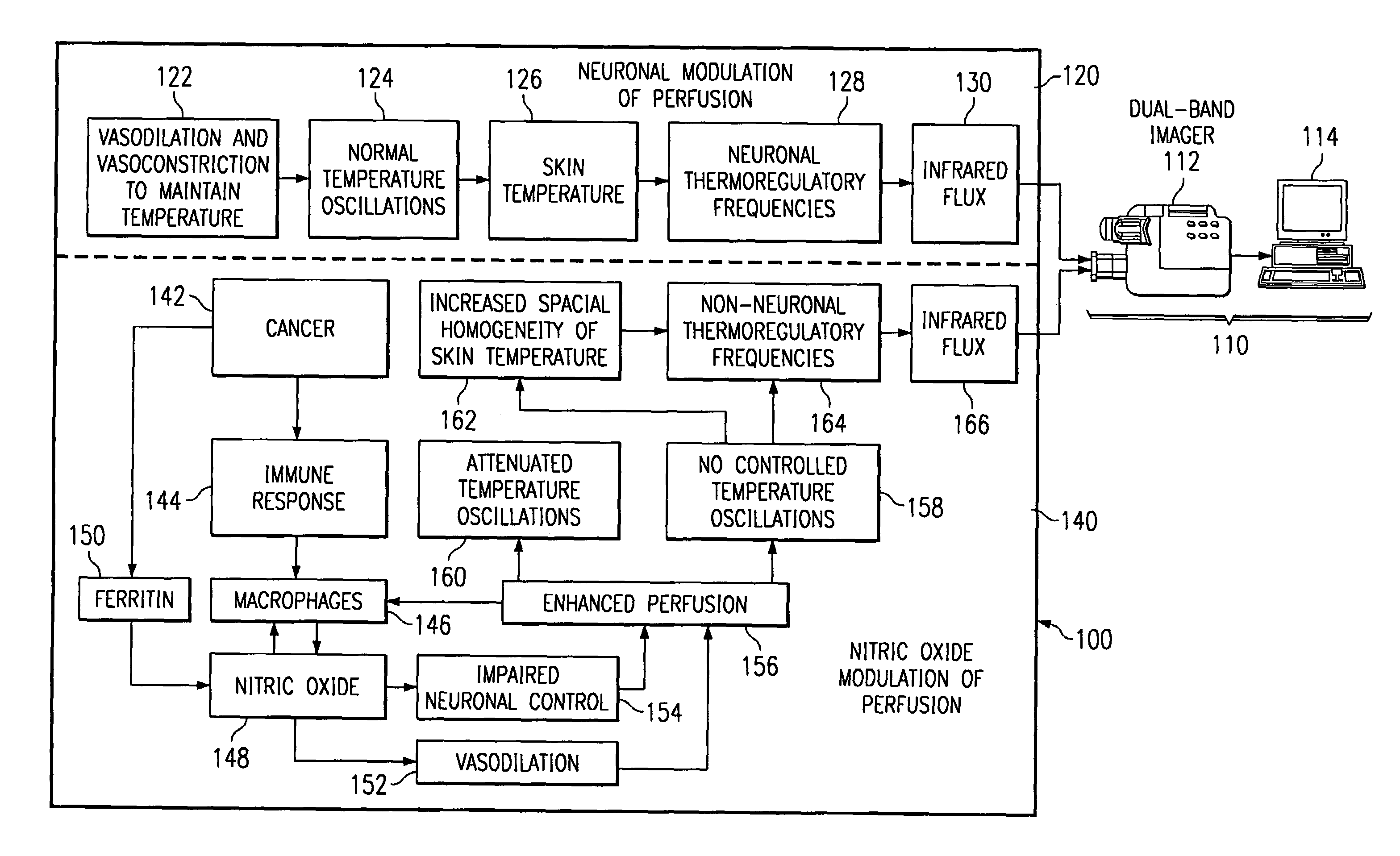
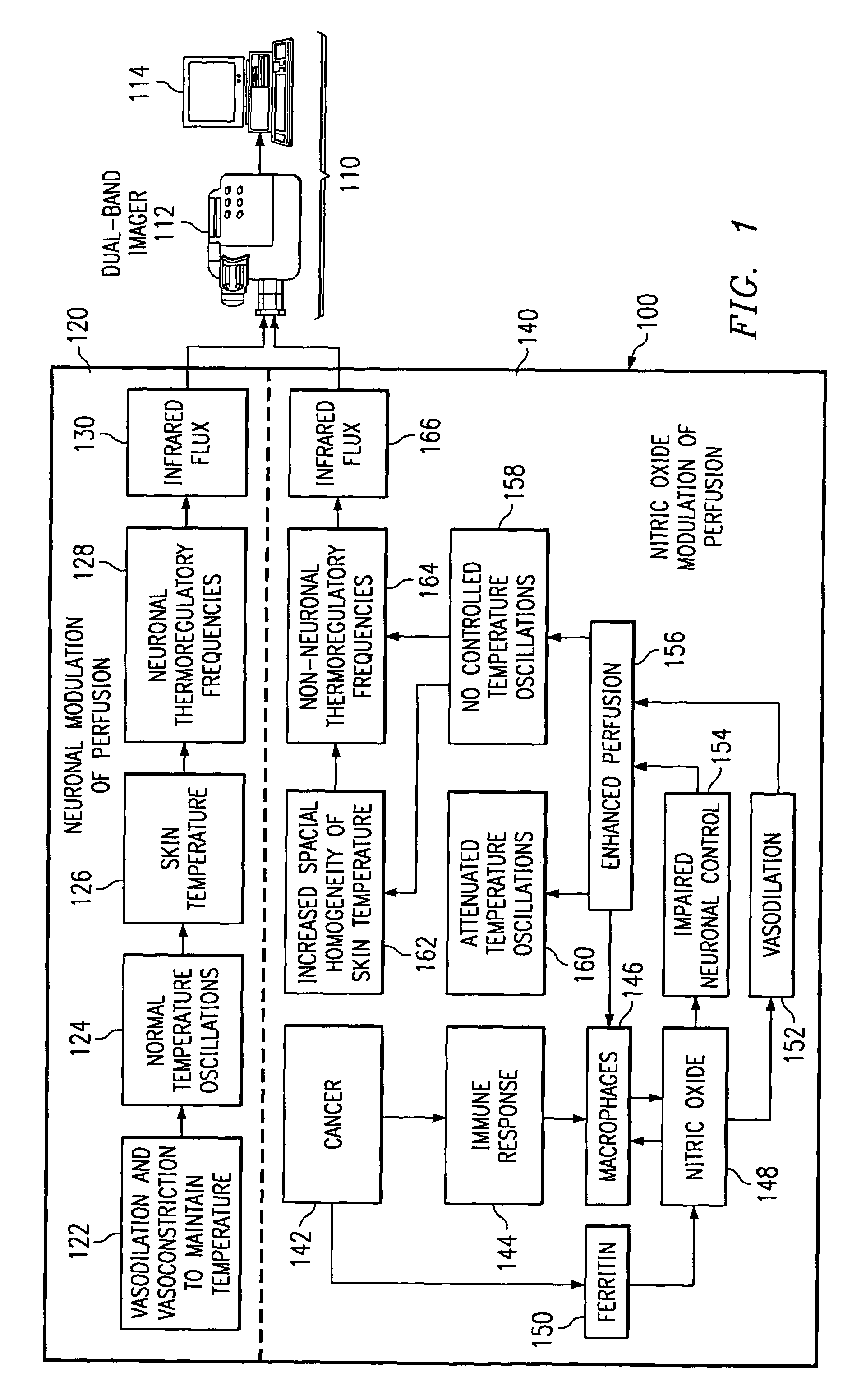
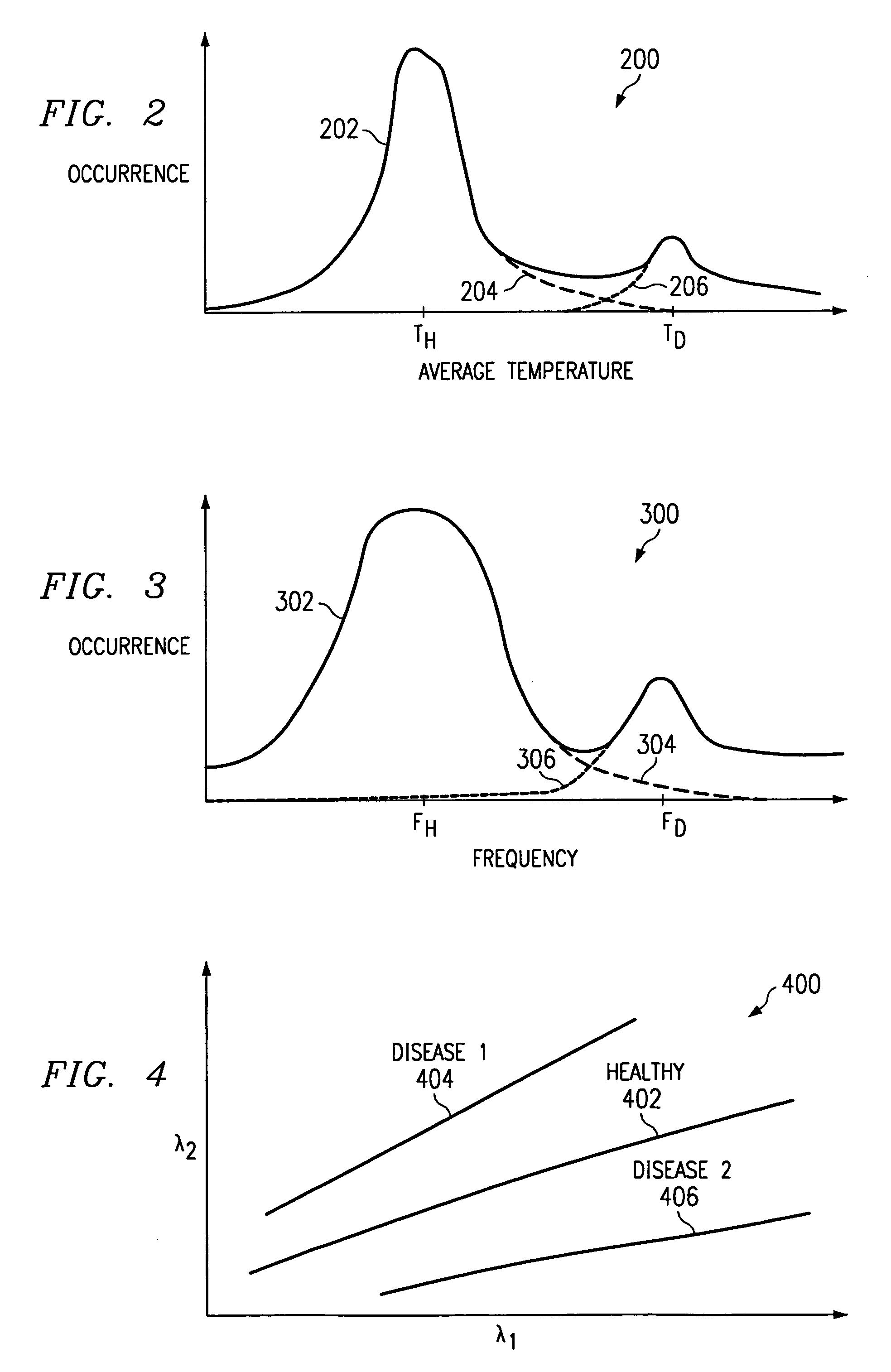
[0031]The first embodiment then finds the average temperature value for each of these subareas. This is done for each individual thermal image in both the first and the second series of thermal images resulting in first and second pluralities of average temperature values. These first and second pluralities of average temperature values are then analyzed in view of FIG. 2. FIG. 2 illustrates a histogram showing all of the average temperature values for the first plurality of average temperature values 200. Curve 202 is the composite curve showing the average temperature values for skin overlying both healthy and diseased tissue. Curve 204 corresponds to the average temperature values for the skin overlying a healthy region of tissue. Curve 204 therefore corresponds to skin whose underlying tissue is thermally regulated by neuronal control of blood perfusion. The peak temperature value for this healthy tissue is denoted TH. In regions of skin overlying diseased or cancerous tissue, t...

second embodiment

[0033]As each of the first and second series of IR images is preferably taken periodically, TRFs can be determined. the present invention makes use of these TRFs. FIG. 3 illustrates a TRF histogram for both healthy and diseased tissue 300. Curve 302 is a composite for both the healthy and diseased tissue while curve 304 corresponds to healthy tissue and curve 306 corresponds to diseased tissue. Curve 304 for healthy tissue reflects neuronal control blood perfusion and generally has a frequency of between 10 and 700 milliHertz. In contrast, curve 306 for diseased tissue reflects NO-based control of blood perfusion and has a higher frequency, generally in the range of 0.8 to 2.0 Hz.

[0034]The second embodiment makes use of the differences in TRFs by finding the contributing frequency for each subarea in the first series of thermal images. This contributing frequency may be determined by analyzing the average temperature value for a subarea based on the known periodic nature of the firs...

fourth embodiment

[0037]The radiance measurements of healthy skin taken for the fourth embodiment may be made as a function of integration time for the dual-band IR imager 112, the temperature of the skin and tissue being imaged, or a combination thereof. The temperature of the skin and tissue can be varied by directing either a warming or a cooling stream of air on the skin and tissue resulting in thermal stress to the skin and tissue. Alternatively, this thermal stress may be induced by directing a flow of water vapor to the skin and tissue. While this thermal stress finds particular application with the fourth (and fifth) embodiments, it can readily be used in conjunction with the other embodiments as well.

[0038]Due to the oscillatory nature of thermal regulation, the sensitivity of the fourth (and fifth) embodiments can be increased. By finding the contributing frequency for each of the subareas, the correlation between the two series of thermal images can be made at neuronal frequencies or at NO...


PUM


Abstract

A method of and apparatus for detecting diseased tissue based upon infrared imaging in two different bands of infrared wavelengths is described. The use of two series of infrared images taken in two different bands of infrared wavelengths increases sensitivity to the subtle temperature changes caused by diseased skin and tissue, especially in the case of cancerous tissue. By sensing skin temperature, the homogeneity thereof, the time variations thereof and the correlation between the two series of infrared images, the present invention decreases the rate of false positives and false negatives. The increased discrimination due to two series of infrared images allows for reliable detection of diseased or cancerous tissue even in the presence of skin tone variations such as birthmarks, tattoos and freckles. The present invention finds special application in the field of breast cancer detection where subtle skin temperature variations may readily be sensed using two series of infrared images.

Description

FIELD OF THE INVENTION[0001]The present invention relates to a method of and apparatus for detecting diseased tissue. In paticular, the present invention provides a noninvasive method of detecting diseased tissue by sensing skin temperature, the homogeneity thereof, the temporal variation thereof and the correlation thereof using a sensor detecting two different bands of infrared wavelengths.BACKGROUND OF THE INVENTION[0002]In the field of diseased or cancerous tissue detection, many methods require subjecting the patient to doses of X-ray radiation or to painful biopsies, especially for breast cancer detection. More recently, researchers discovered that dysfunction of the neuronal control of the vasculature due to cancerous lesions leads to temporal or periodic perfusion changes. By measuring, recording and analyzing these periodic perfusion changes, typically through infrared (IR) imaging, diseased or cancerous tissue can be detected. While these periodic perfusion changes appear ...

Claims


Application Information

Patent Timeline
Patent Type & Authority Patents(United States)
IPC IPC(8): A61B5/00A61B5/026
CPCA61B5/015A61B5/415A61B5/418A61B5/026
Inventor MITRA, PRADIP
Owner LOCKHEED MARTIN CORP
Why Eureka
Try Eureka