Endoscopic surgical access port and method

a technology of endoscopic surgery and access port, which is applied in the field of endoscopic surgical apparatus and methods of tissue dissection, can solve the problems of the length of the hollow body, and achieve the effect of facilitating the insufflation of an anatomical spa

Inactive Publication Date: 2007-10-02
MAQUET CARDIOVASCULAR LLC
View PDF51 Cites 243 Cited by
  • Summary
  • Abstract
  • Description
  • Claims
  • Application Information

AI Technical Summary

Benefits of technology

[0005]In accordance with embodiments of the apparatus and method of the present invention, an access port is provided which obviates the need for flapper valves and selectable sliding seals that snap toggle into position over the proximal end of the hollow body. This facilitates insufflation of an anatomical space during the interval while an endoscopic instrument is positioned through the hollow body. In recognition of the typically small spatial volume of a channel or anatomical space formed along the saphenous vein, it has been discovered that insufflation need only be established during insertion and manipulation of an endoscopic instrument through the access port, and that re-pressurization of the small volume can be satisfactorily restored within a very brief interval following insertion of an endoscopic instrument through the sliding seal of the access port.

Problems solved by technology

However, the versatility of selectable seals and flapper valves to accommodate endoscopic instruments of various diameters significantly extends the length of the hollow body and requires additional manual re-configuration to position a selected seal over the proximal end of the hollow body to provide a sliding seal of appropriate dimensions to accommodate the diameter of a selected endoscopic instrument.

Method used


Image

  • Endoscopic surgical access port and method
  • Endoscopic surgical access port and method
  • Endoscopic surgical access port and method

Examples


Embodiment Construction

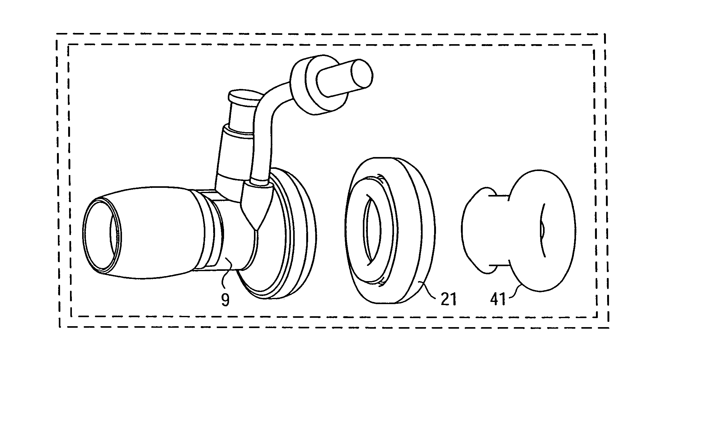
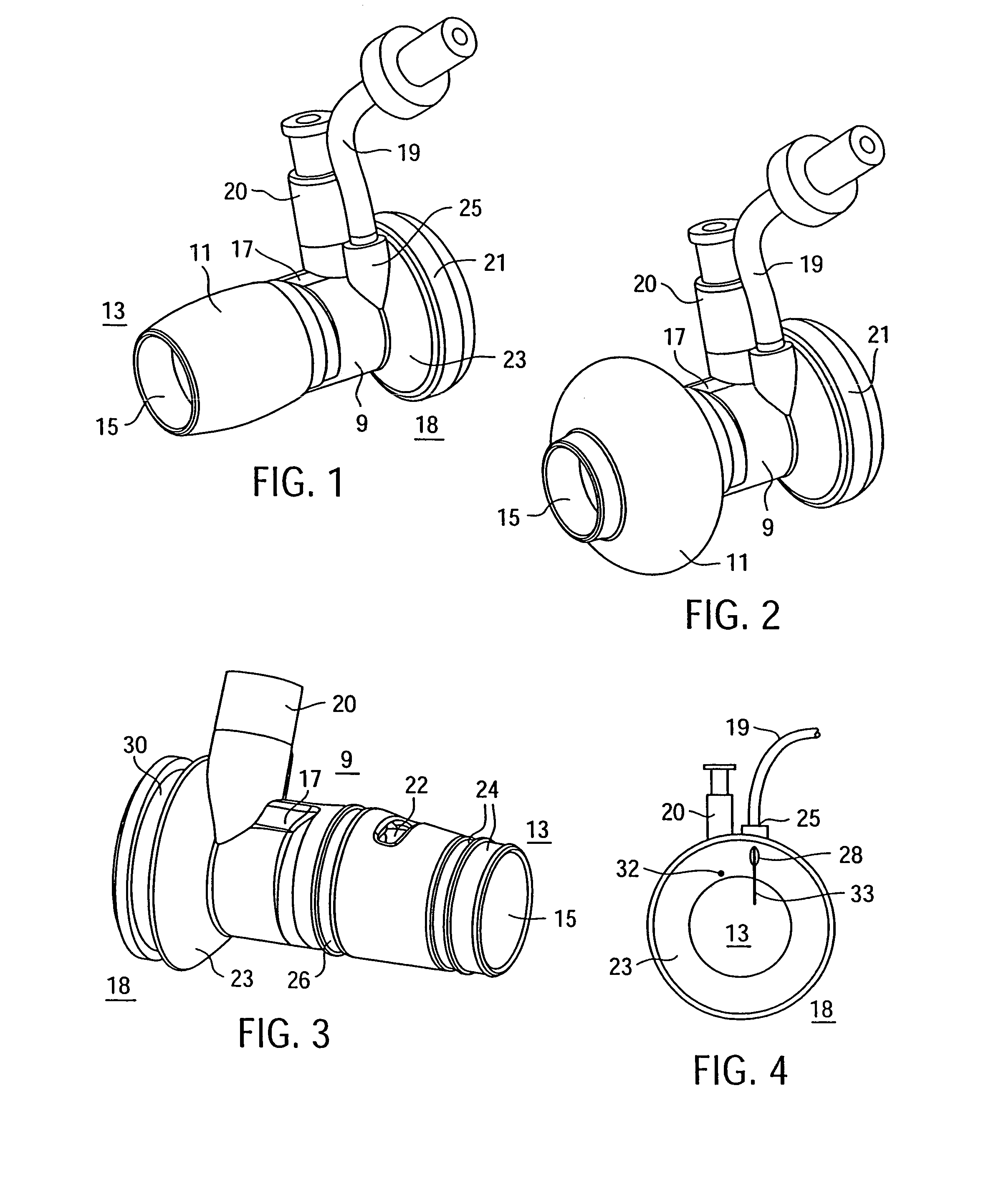
[0013]Referring now to FIG. 1, there is shown a perspective view of the access port according to one embodiment of the present invention in which the hollow body 9 of fluid-impervious material includes a central bore 15 and a generally toroidally-shaped balloon 11 disposed about the outer periphery of the body 9 near the distal end 13 thereof. The interior diameter of the central bore 15 through the hollow body 9 is sized to accommodate the largest diameter of endoscopic instrument therein and may be about 0.6″ at the distal end 13, and may flair out to a wider diameter of about 0.9″ at the proximal end 18. A fluid or air passage 17 along an outer wall of the body 9 connects to an external fluid-tight fitting 20 for coupling to a source of gas under pressure, such as a syringe, in order to selectively inflate the balloon 11 within the confines of an initial cutaneous incision near a saphenous vein that is to be harvested. Inflating the balloon 11 with fluid under pressure, as shown ...


PUM


Abstract

A sliding gas-tight seal on an access port promotes insufflation of an anatomical space formed in tissue at a surgical site only during insertion of an endoscopic instrument through the access port into the anatomical space, and promotes deflation of the inflated space upon removal of the endoscopic instrument from within the access port. An inflatable balloon disposed about the port near the distal end may be selectively expanded to seal and anchor the access port within an incision through which a surgical procedure with insufflation is to be performed. Multiple resilient seals may be attached to the body of the port, and an auxiliary resilient seal may be inserted within the aperture of a seal attached to the body to accommodate a wide range of endoscopic instruments of various exterior dimensions inserted through the seals.

Description

RELATED APPLICATION[0001]This application is a continuation of application Ser. No. 09 / 648,660, now U.S. Pat. No. 6,811,546, entitled “Endoscopic Surginal Access Port and Method”, filed on Aug. 25, 2000 by Peter Callas, et al.FIELD OF THE INVENTION[0002]This invention relates to endoscopic surgical apparatus and methods of tissue dissection, and more particularly to a sliding gas seal for controlling insufflation of an endoscopic surgical site on a patient.BACKGROUND OF THE INVENTION[0003]Coronary bypass surgery commonly requires a length of the saphenous vein of the patient to form a shunting vessel around a site of stenosis or other blockage in a coronary artery. The saphenous vein was conventionally ‘harvested’ from the patient's leg through an incision extending the length of the section of saphenous vein to be harvested. Recently, endoscopic surgical procedures have replaced open-incision harvesting procedures and have significantly reduced patient trauma, discomfort, complicat...

Claims


Application Information

Patent Timeline
Patent Type & Authority Patents(United States)
IPC IPC(8): A61M29/00A61B17/00A61B17/34
CPCA61B17/3423A61B17/00008A61B17/3421A61B2017/3486A61B17/3462
Inventor CALLAS, PETERLUNSFORD, JOHN P.CHIN, ALBERT K.WEI, MICHAEL
Owner MAQUET CARDIOVASCULAR LLC
Why Eureka
Try Eureka