Calculation of additional projection data from projection data acquired with a divergent beam

a technology of divergent beams and projection data, applied in the field of computed tomography, can solve the problems of transmission profiles, inability to achieve real-time accuracy of ct numbers, and inability to reconstruct images from stored transmission profiles, so as to increase the resolution of each projection view, reduce the angle between views, and increase the in-plane resolution of images

Active Publication Date: 2007-10-09
WISCONSIN ALUMNI RES FOUND
View PDF1 Cites 5 Cited by
  • Summary
  • Abstract
  • Description
  • Claims
  • Application Information

AI Technical Summary

Benefits of technology

[0017]Another general object of the invention is to increase the in-plane resolution of images produced by a divergent beam medical imaging system. Rather than replace corrupted data using the present invention, new projection data may be calculated using the acquired projection views. Additional projection views may be calculated to reduce the angle between views, or additional data may be calculated for each of the acquired projection views to increase the resolution of each projection view.
[0018]The present invention alters the trade-off made between image resolution on the one hand and scan time on the other hand. It should be apparent that rather than increasing in-plane image resolution the present invention may also be used to reduce the x-ray dose in a CT system. That is, an image of the same resolution can be reconstructed with fewer acquired projection views thus shortening the scan and reducing x-ray dose.

Problems solved by technology

The reconstruction of an image from the stored transmission profiles requires considerable computation and cannot be accomplished in real time.
As a result, when all of the transmission profiles are back projected to determine the CT number of each voxel in the reconstructed field of view, the CT numbers will not be accurate.
A similar problem is presented when transmission profiles are affected by metal objects such as dental filings in the patient being scanned.
In this situation x-rays passing through the metal object are strongly absorbed and the attenuation measurement is very noisy causing strong artifacts in the reconstructed image.
The data truncation problem and the x-ray absorption problem each corrupt the acquired attenuation data set in a unique way.
In the first problem a select few of the acquired views are significantly affected and in the second problem all or nearly all the acquired views are affected in a more limited manner.
However, this strategy results in a longer scan time since it requires a finite amount of time to acquire each view.
However, this results in an increase in the number of required detector elements to span the same fan beam angle and the supporting electronics with a consequent increase in scanner cost.
There are a number of situations that result in the corruption of acquired divergent beam projection views.
There are a number of situations that result in the corruption of acquired divergent beam projection views when different energy x-ray beams are utilized in CT imaging.

Method used


Image

  • Calculation of additional projection data from projection data acquired with a divergent beam
  • Calculation of additional projection data from projection data acquired with a divergent beam
  • Calculation of additional projection data from projection data acquired with a divergent beam

Examples


Embodiment Construction

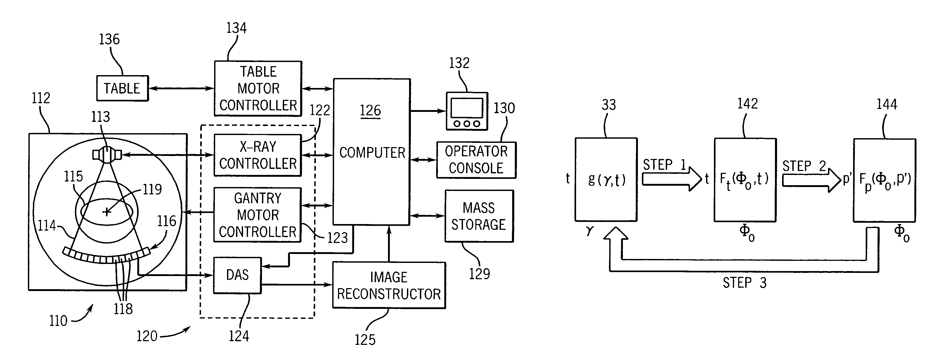
[0062]With initial reference to FIG. 10, a computed tomography (CT) imaging system 110 includes a gantry 112 representative of a “third generation” CT scanner. Gantry 112 has an x-ray source 113 that projects a fan-beam of x-rays 114 toward a detector array 116 on the opposite side of the gantry. The detector array 116 is formed by a number of detector elements 118 which together sense the projected x-rays that pass through a medical patient 115. Each detector element 118 produces an electrical signal that represents the intensity of an impinging x-ray beam and hence the attenuation of the beam as it passes through the patient. During a scan to acquire x-ray projection data, the gantry 112 and the components mounted thereon rotate about a center of rotation 119 located within the patient 115.

[0063]The rotation of the gantry and the operation of the x-ray source 113 are governed by a control mechanism 120 of the CT system. The control mechanism 120 includes an x-ray controller 122 th...


PUM


Abstract

A data consistency condition is derived for an array of attenuation values acquired with a divergent beam. Using this data consistency condition, estimates of selected attenuation values can be calculated from the other attenuation values acquired during the scan. Such estimates may be used to replace corrupted data in the acquired data set, or attenuation values may be added to the acquired data set to increase in-plane resolution of a reconstructed image.

Description

CROSS-REFERENCE TO RELATED APPLICATIONS[0001]This application is a continuation-in-part of U.S. patent application Ser. No. 10 / 955,459 filed on Sep. 30, 2004, now U.S. Pat. No. 7,050,528, and entitled “CORRECTION OF CT IMAGES FOR TRUNCATED OR INCOMPLETE PROJECTIONS”.STATEMENT REGARDING FEDERALLY SPONSORED RESEARCH[0002]This invention was made with government support under Grant No. 1R21 EB001683-01 awarded by the National Institute of Health. The United States Government has certain rights in this invention.BACKGROUND OF THE INVENTION[0003]The field of the present invention is computed tomography and, particularly, computer tomography (CT) scanners used to produce medical images from x-ray attenuation measurements.[0004]As shown in FIG. 1, a CT scanner used to produce images of the human anatomy includes a patient table 10 which can be positioned within the aperture 11 of a gantry 12. A source of highly columinated x-rays 13 is mounted within the gantry 12 to one side of its apertur...

Claims


Application Information

Patent Timeline
Patent Type & Authority Patents(United States)
IPC IPC(8): A61B6/00
CPCG06T11/005Y10S378/901G06T2211/432G01N2223/401G01N2223/419A61B6/032A61B6/482A61B6/5205
Inventor CHEN, GUANG-HONG
Owner WISCONSIN ALUMNI RES FOUND
Why Eureka
Try Eureka