Blood vessel sealing system

a sealing system and blood vessel technology, applied in the field of blood vessel sealing system, can solve the problems of blood clots in no practical way to positively limit the protrusion of collagen into the interior of the blood vessel, and the reclosure of the opening in the blood vessel wall, and achieve the effect of accurate positioning and simple us

Inactive Publication Date: 2008-02-19
ST JUDE MEDICAL
View PDF27 Cites 95 Cited by
  • Summary
  • Abstract
  • Description
  • Claims
  • Application Information

AI Technical Summary

Benefits of technology

[0006]These and other problems and disadvantages associated with the prior art are overcome by the invention disclosed herein by providing a technique for implanting a collagen plug in the tissue access passage to the percutaneous puncture or opening in a blood vessel which insures that the end of the plug does not extrude through the blood vessel wall opening into the blood vessel lumen. A tamponading means located inside the blood vessel lumen during installation of the collagen plug serves to positively prevent the collagen plug from being inadvertently pushed through the blood vessel wall opening as it is installed but which is removed after installation of the collagen plug so that no protrusion or other dislodgable material is left in the interior of the blood vessel after the procedure is completed. The tamponading means is selectively expandable to an expanded size and shape such that it will not pass back out through the blood vessel wall opening and its position in the blood vessel is controlled by a control member extending exteriorly of the patient for easy manipulation. For removal, however, tamponading means is selectively collapsible to a collapsed condition small enough to pass back out through the installed collagen plug without interfering with the coagulum forming capability of the collagen plug. The technique is simple to use and the location of the collagen plug is inherently accurately positioned at the blood vessel wall opening independently of variances in the patient's skin and tissue thickness at the puncture site and without premeasuring or adjustment of the applicator.
[0007]The method of the invention is directed to the sealing a percutaneous puncture through a blood vessel wall comprising the steps of inserting a collapsed expandable tamponading member attached to a control member into the blood vessel lumen through the blood vessel wall puncture so that the control member extends out of the patient through the access passage to the skin surface; expanding the tamponading member to a size larger than the blood vessel wall puncture; pulling the expanded tamponading member back against the blood vessel wall opening using the control member to form a seal at the interior end of the blood vessel wall puncture; inserting a collagen plug into the access passage around the control member while the seal is maintained at the blood vessel wall puncture until the leading end of the collagen plug is located at the exterior end of the blood vessel wall puncture; then collapsing the tamponading member to the collapsed condition; and retracting the collapsed tamponading member back through the collagen plug. The collagen plug has a preformed passage therethrough which slidably receives the control member to allow placement of the collagen plug along the control member. The collagen serves to induce a coagulum that seals the outside end of the puncture in the blood vessel wall. After the collapsed tamponading member is pulled back through the collagen plug, the collagen plug self seals any opening through the plug left by the passage of the expandable member. The collagen plug may be housed in a retractable barrel while the collagen plug is inserted into the access passage and the barrel then retracted from around the collagen plug while maintaining the collagen plug in a substantially fixed position in the access passage. The barrel may be retracted fully or partially from around the collagen plug before the expanded tamponading member is collapsed and withdrawn. The method may also include visually indicating when the projecting end of the collagen plug is located at the exterior end of the blood vessel puncture. The collagen plug may also be backed up while the collapsed tamponading member is withdrawn through the plug to insure that the plug remains in place. The collagen plug may also be urged slightly toward the blood vessel wall puncture to compensate for any loss in volume of the collagen plug as it turns into a gelatinous mass.

Problems solved by technology

One of the difficulties in these procedures, however, is reclosure of the opening in the blood vessel wall after the procedure.
The difficulty with this procedure is that there is no practical way to positively limit the protrusion of the collagen into the interior of the blood vessel.
This sometimes results in the collagen being inserted or subsequently expanding through the blood vessel wall opening into the vessel lumen and causing a blood clot to form in the interior of the blood vessel.
This is undesirable because the clot and / or the protruding collagen can dislodge and move along the blood vessel to cause an embolus.
The problem with this technique is that the sealing plug may break loose before it is absorbed by the body and cause a blockage of the blood flow through the blood vessel.
The sealing plug may be a nidus for a blood clot to form and subsequently dislodge resulting in embolization.
Another potential problem in leaving a device inside the vessel wall is that the device, like any foreign body, may erode the wall of the vessel or promote extensive intravascular scaring both of which potentially will disrupt vessel architecture.

Method used


Image

  • Blood vessel sealing system
  • Blood vessel sealing system
  • Blood vessel sealing system

Examples


second embodiment

of Applicator

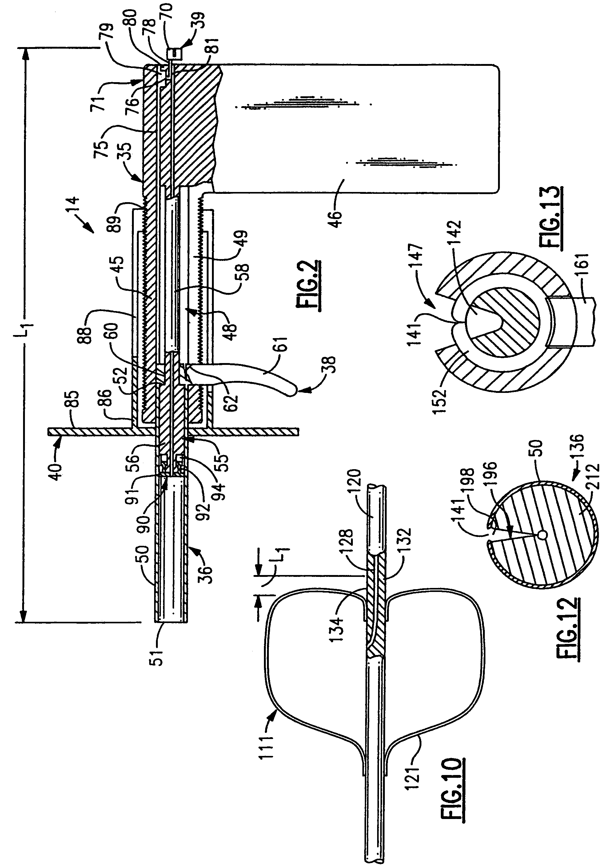
[0047]The second embodiment of the applicator is designated 114 and is illustrated in FIGS. 11-15. The basic difference between the applicator 114 and the applicator 14 is that the applicator 114 has an open section through which the control member on the temporary sealing assembly can be installed without feeding the control member axially through the collagen plug and applicator. The applicator 114 like the applicator 14, includes a housing assembly 135, and introducer assembly 136, a retraction mechanism 138, an interconnect assembly 139, and a locator mechanism 140. These assemblies and mechanisms operate similarly to the corresponding assemblies and mechanisms of the first embodiment of the invention.

[0048]The cylindrical body 145 defines a V-shaped cutout 147 therein down to the passage through which the control member on the temporary sealing assembly passes so that control member can be laid into the passage as it is being operated to install the collagen plug. ...

third embodiment

of the Invention

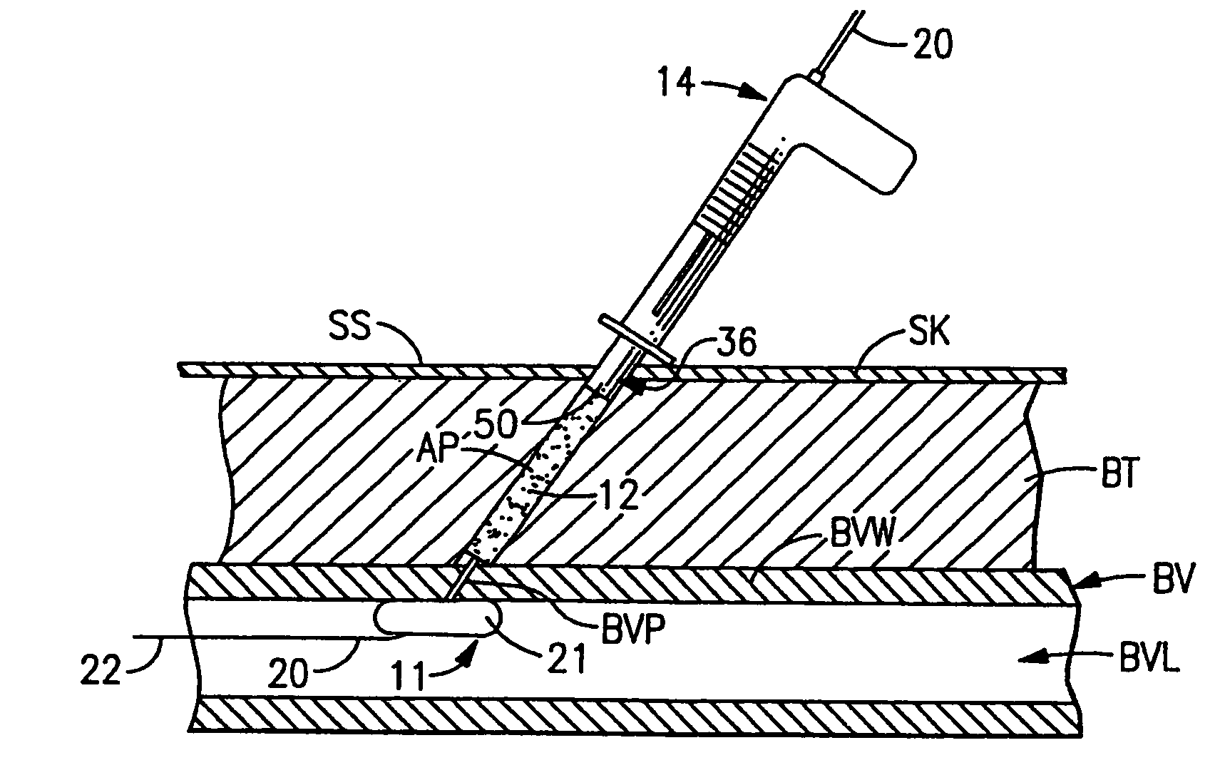
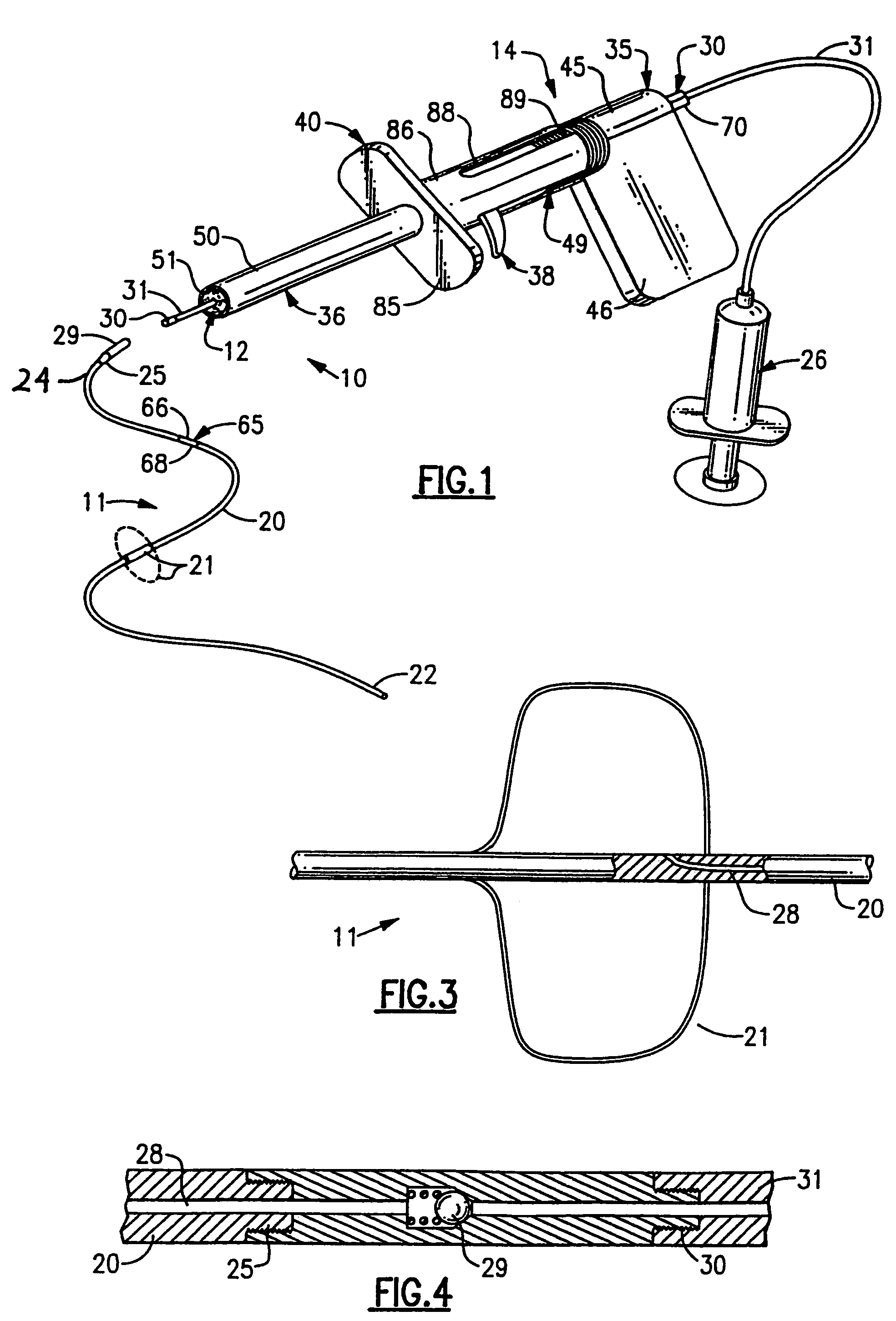
[0050]FIGS. 16 and 17 illustrate a third embodiment 210 of the invention which includes a collagen plug 212 and an applicator tube 214. The collagen plug 212 is installed on the control member and the extension 31 to the syringe 26 as described with the first embodiment of the invention. The applicator tube 214 is a cylindrical tube defining a central passage therethrough to just slidably receive the control member of the temporary sealing arrangement. The physician simply slides the collagen plug 212 and the tube 214 onto the control member of the temporary sealing arrangement so that the leading end of the collagen plug 212 faces the access passage AP in the patient. The physician then uses the applicator tube 214 to push the collagen plug 212 down to the outside end of the puncture BVP. The combined overall length of the collagen plug 212 and the applicator tube 214 corresponds to that of the first embodiment of the invention so that the physician can use the indi...

fourth embodiment

of the Applicator

[0052]FIG. 18 illustrates an alternate embodiment of the applicator which has been designated by the reference of 314. The applicator 314 include an insertion barrel 150 with the projecting end 351 thereon adapted to be inserted into the access passage AP in the patient. The barrel 350 defines an internal passage therethrough adapted to slidably receive the collagen plug 12 therein. A one-way check member 353 is mounted in the barrel 350 behind the plug 12 and has a face thereon abutting the plug 12 to maintain it lengthwise of the barrel 350 as will become more apparent. The check member 353 defines a tamponading member receiving chamber 352 which opens onto the trailing end of the plug 12 and trailing end of the check member 353 is provided with a one-way gripping assembly 354 which grips the control member of the temporary sealing arrangement to allow the check member 353 to be moved along the control member toward the expanded tamponading member but prevents mov...


PUM


Abstract

A method of and apparatus for sealing access openings into blood vessels using an expandable member in the blood vessel on a control member that extends out through the access opening and an applicator which inserts a preformed sealing material into the access opening over the control member to keep the control member and expandable member centered in the access opening and withdrawing the collapsed expandable member back through the sealing material after the access opening is sealed.

Description

CROSS-REFERENCE TO RELATED APPLICATIONS[0001]This is a continuation of prior U.S. patent application Ser. No. 09 / 656,643 filed Sep. 7, 2000 (now U.S. Pat. No. 6,699,261), which is a continuation of U.S. patent application Ser. No. 08 / 383,256 filed Feb. 3, 1995 (now U.S. Pat. No. 6,162,240) which is a continuation of U.S. patent application Ser. No. 07 / 817,587 filed Jan. 7, 1992 (now U.S. Pat. No. 6,056,768), the disclosures of which are incorporated herein by reference in their entirety.BACKGROUND OF THE INVENTION[0002]This invention relates generally to the stopping of bleeding from blood vessels through openings in the vessel wall and more particularly through surgically produced punctures for different medical procedures.[0003]Certain medical procedures require the percutaneous puncturing of a blood vessel to gain access to the interior of the vascular system of the patient for the procedure to be conducted. Procedures that commonly require such an percutaneous punctures are ball...

Claims


Application Information

Patent Timeline
Patent Type & Authority Patents(United States)
IPC IPC(8): A61B19/00A61B17/00
CPCA61B17/0057A61B2017/00637A61B2017/00654A61B2017/00659A61B2017/00672
Inventor CATES, CHRISTOPHER U.KNOPF, WILLIAM D.WHITNEY, DOUGLASS G.
Owner ST JUDE MEDICAL
Why Eureka
Try Eureka