Method and apparatus for computer controlled cell based diagnosis

a cell-based diagnosis and computer-controlled technology, applied in the field of computer-controlled cell-based diagnosis, can solve the problems of high risk to the fetus, and high risk of highly specialized procedures, so as to minimize the time and minimize the time.

Inactive Publication Date: 2008-03-18
IKONISYS INC
View PDF20 Cites 34 Cited by
  • Summary
  • Abstract
  • Description
  • Claims
  • Application Information

AI Technical Summary

Benefits of technology

[0031]The method further comprises contacting a body fluid or tissue sample at a location corresponding to each candidate blob represented in the subset of the first image data set, with a reagent to generate a medically significant signal. This method provides the advantage of being able to remove from further processing a body fluid or tissue sample for which no subset of the first data set representing a candidate blob is created. The signal can be measured to determine whether it is a significant signal level. The first and / or the second image data subsets can be transformed into a representation that is more suitable for control and processing by a computer as described herein. In a preferred embodiment, the image data is transformed from an RGB (Red Green Blue) signal into an HLS (Hue Luminescence Saturation) signal. Filters and / or masks are utilized to distinguish those cells that meet pre-selected criteria and eliminate those that do not, and thus identify rare cells.
[0034]In general, a subset of a first image data set can be created as described above. The method can further produce an image file of red, green and blue pixels representative of red, green and blue intensities at respective pixel locations within the color image received According to some aspects of the invention, the processing further includes manually selecting a plurality of pixels within the background; determining color intensity value ranges corresponding to the portion of the background; and identifying as the background those areas of the image having color intensity values within the ranges determined. The method may include before the step of measuring, processing in the computer to filter the color image to make color intensity values of dark pixels in the color image lighter and to make color intensity values of light pixels in the color image darker. Moreover, the filtering may include passing the color image through a hole filling filter; passing the filled color image through an erosion filter; performing a separate operation on the eroded filled color image, to define outlines around areas; selecting pixels within the outlines by performing a logical NOT operation, and performing a logical AND operation between the selected pixels and the filled color image. The steps further encompass contacting a body fluid or tissue sample at a location corresponding to each candidate blob represented in the subset of the first image data set, with a reagent to generate a medically significant signal. This provides the advantage of being able to remove from further processing a body fluid or tissue sample for which no subset of the first data representing a candidate blob is created. There is an optional step by which the signal can be measured to determine whether it is of a significant level. Another optional step encompasses transformation of one or both of the first and the second image data subsets into a representation that is more suitable for control and processing by a computer as described herein. In a preferred embodiment, the image data is transformed from an RGB (Red Green Blue) signal into an HLS (Hue Luminescence Saturation) signal. Filters and / or masks are utilized to distinguish those cells that meet pre-selected criteria and eliminate those that do not.
[0052]According to yet another aspect of the invention, there is provided a device for screening fetal cells contained within a smear of an unenriched sample of maternal blood containing a naturally present concentration of fetal cells, comprising: a flexible film having thereon a smear of at least 250 μl of maternal blood. In one embodiment, the flexible film has thereon a smear of at least 500 μl of maternal blood. In one important embodiment, the flexible film is an elongated film, the length being at least 10 times the width. It is particularly preferred that the flexible film include marking coordinates, whereby the computerized microscopic vision system described herein can locate a cell relative to a point on the film, permitting that cell to be returned to at a later time, if desired.
[0053]According to yet another aspect of the invention, there is provided a device for screening rare cells contained within a sample of cells at a concentration of no greater than one rare cell for every 10,000 cells in the sample of cells. The device is a flexible film having fixed thereon the sample of cells, wherein the flexible film is at least five inches long. In one preferred embodiment the flexible film has a length at least 10 times its width. In another important embodiment, the flexible film includes marking coordinates, whereby the computerized microscopic vision system described herein can locate a cell relative to a point on the film, permitting the cell to be returned to at a later time, if desired.
[0054]According to another aspect of the invention, there is provided a device for dispensing materials to a specific location on a slide. The device includes a microscopic vision system for detecting a signal indicative of the presence o a rare cell in a sample of cells. The device also includes means for identifying the coordinates of the rare cell in an optical field. The device further has attached to it a dispenser for dispensing a volume of material and means for moving the dispenser to the coordinates whereby the volume of material may be dispensed upon the rare cell. The material dispensed can be reagents such as a label, PCR, primers, and the like. According to another important embodiment of the invention, the need for scanning large areas of microscopic preparations in the minimum possible amount of time is met by the use of an apparatus or system that provides a “composed” image. It is based on the simultaneous use of an array of computer controlled objective lenses, arranged on a support system and having the capacity to focus on a microscopic preparation. Each of the objective lenses is connected to a charge coupled device camera, herein referred to as a CCD camera, being connected to image acquisition hardware installed in a host computer. The images are stored in the computer memory and they are combined in an appropriate side to side fashion, so that a “composed” image is formed in the computer memory. The “composedp” image can be further processed as a unity, using any kind of imaging procedures to detect specific features that are in question. The significant advantage of the described system consists in its capacity to acquire images simultaneously from a number of objective lenses, thus minimizing the time needed to process large areas of the sample in a manner that is inversely proportional to the number of objectives used.

Problems solved by technology

The disadvantages of these modern methods are at least two-fold.
These highly specialized procedures carry a small, but significant, risk to the fetus.
Early in pregnancy, the level of risk to the fetus is high and the number of cells obtained is low.
Therefore, results of these procedures often are not obtained until 18-20 weeks of pregnancy.
Second, accurately assessing, quantifying, and assigning a significance to images of cells is difficult, time-consuming and unacceptably subjective.
However, nucleated erythrocytes have generated the greatest degree of interest as sources of genetic material for prenatal diagnosis due to their rarity in the adult blood circulation, their abundance in fetal blood and their limited life span.
Although they are consistently present in the maternal blood circulation, fetal cells are very rare, severely limiting their diagnostic utility.
These methods are extremely difficult to perform, often fail to isolate a sufficient number of fetal cells to be diagnostically significant and sometimes fail to provide a sample of a sufficient number of undamaged fetal cells of adequate purity for reliable subsequent diagnosis.
In contrast, an abnormal number of autosomes typically results in early death.
Although Down's syndrome is easily diagnosed after birth, prenatal diagnosis is problematic.
Despite the above-described advances in the development of fluorescent in situ hybridization methods for the diagnosis of genetic abnormalities, the analysis of the fluorophor-labeled sample remains labor-intensive and involves a significant level of subjectivity.
As will be readily apparent, the above-described procedure is quite time-consuming.
Moreover, because the results are visually-perceived, the frequency of erroneous results can vary from one experiment to the next, as well as from one observer to the next.

Method used


Image

  • Method and apparatus for computer controlled cell based diagnosis
  • Method and apparatus for computer controlled cell based diagnosis
  • Method and apparatus for computer controlled cell based diagnosis

Examples


Embodiment Construction

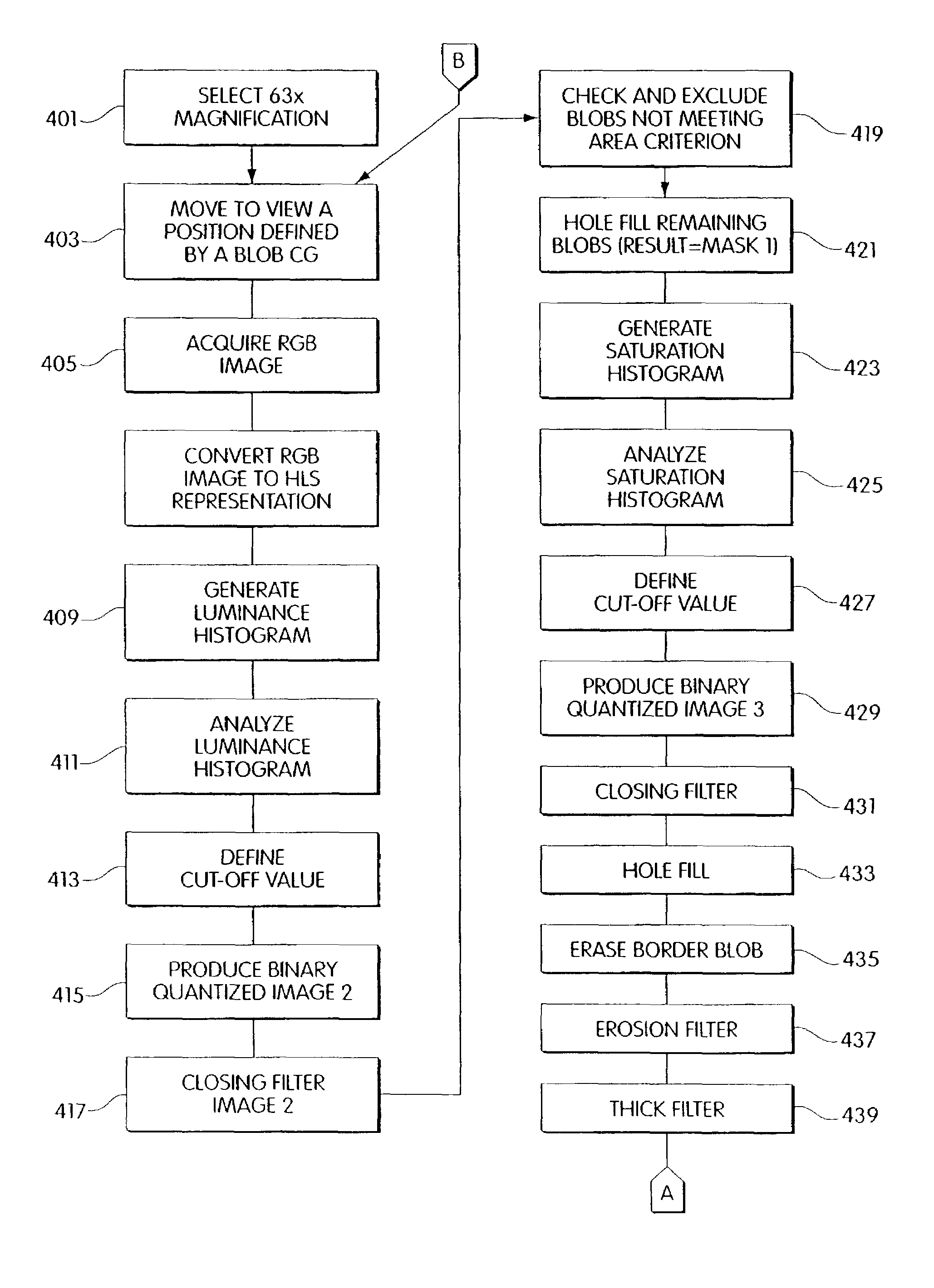
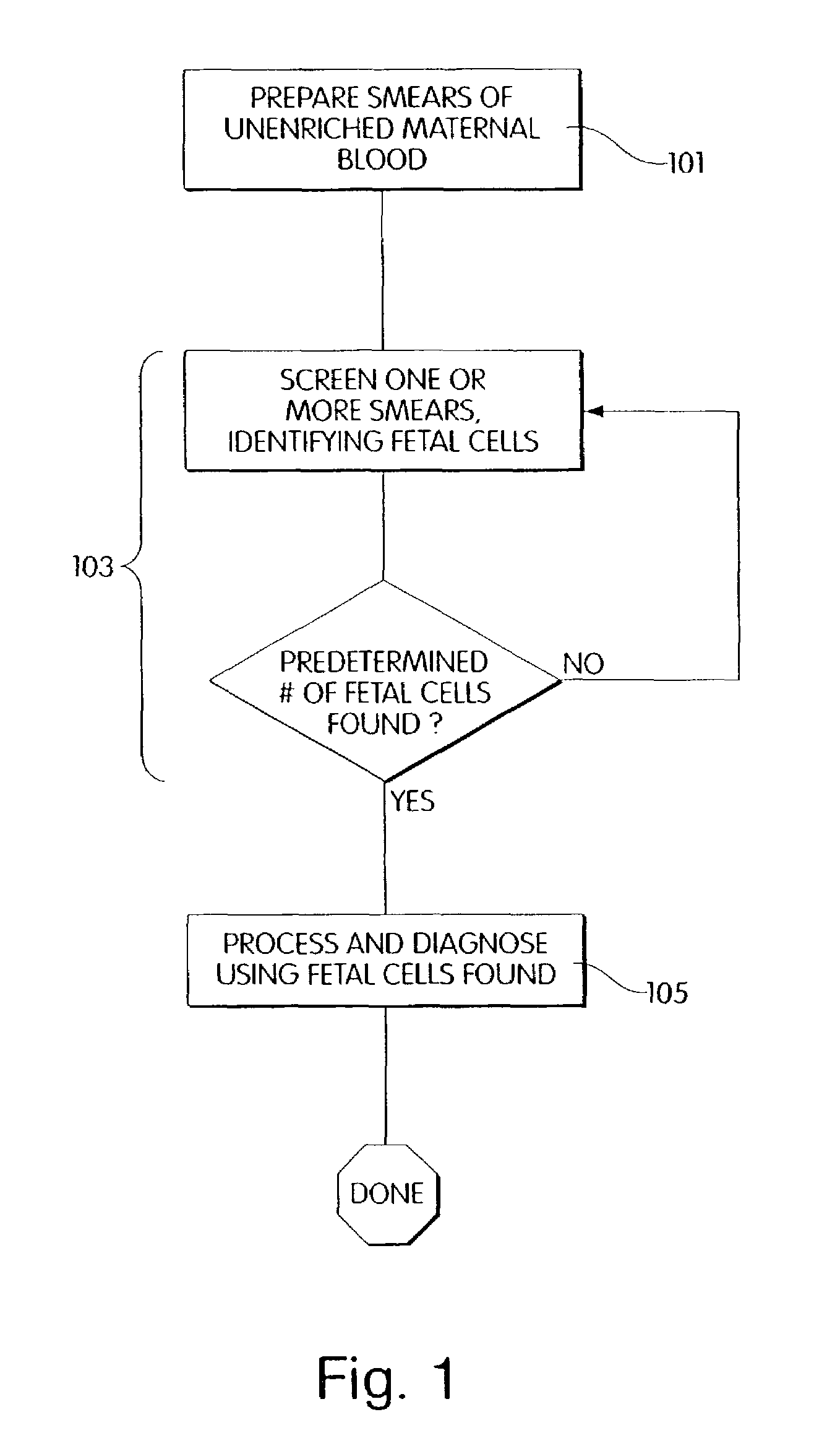
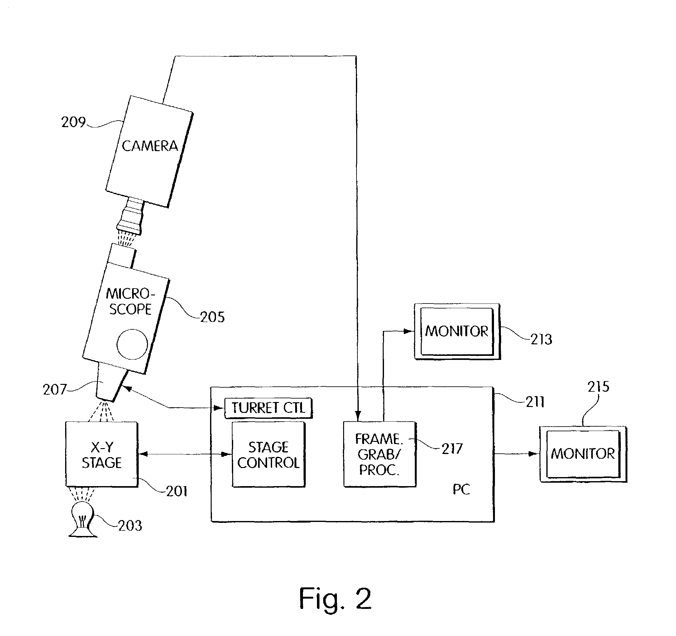
[0069]The invention will be better understood upon reading the following detailed description of the invention and of various exemplary embodiments of the invention, in connection with the accompanying drawings. While the detailed description explains the invention with respect to fetal cells, a rare cell type, and blood as the body fluid or tissue sample, it will be clear to those skilled in the art that the invention can be applied to and, in fact, encompasses diagnosis based on any cell type and any body fluid or tissue sample for which it is possible to create a monolayer of cells on a substrate.

[0070]Body fluids and tissue samples that fall within the scope of the invention include but are not limited to blood, tissue biopsies, spinal fluid, meningeal fluid, urine, alveolar fluid, etc.. For those tissue samples in which the cells do not naturally exist in a monolayer, the cells can be dissociated by standard techniques known to those skilled in the art. These techniques include...


PUM

PropertyMeasurementUnit
pHaaaaaaaaaa
pHaaaaaaaaaa
sizeaaaaaaaaaa

Abstract

A computer controlled method for detecting and diagnosing a rare cell type in a tissue sample is provided, said method comprising treating the tissue sample such that it generates a first signal indicative of the presence at a location of a rare cell, detecting the first signal, treating the location at which the first signal is detected to generate a second signal indicative of a diagnostically useful cellular characteristic and detecting the second signal. The first signal can be morphological or a color present in a sought cell either before or after staining. The second signal can be generated by in situ PCR or PCR in situ hybridization. In one preferred embodiment, the rare cell type is fetal cell in a maternal blood tissue sample, said sample consisting of a smear of unenriched maternal blood. In another embodiment, the method is used to diagnose or genotype cancer cells in a blood or tissue biopsy sample.

Description

[0001]This Application Claims Priority to PCT / US99 / 27608 Filed Nov. 18, 1999.1.FIELD OF THE INVENTION[0002]The present invention relates to computer controlled methods and apparatus for obtaining and preparing cell samples and for identifying a rare cell of interest from a field of cells and making a diagnosis based on a characteristic of a rare cell selected in the field. In one important embodiment, the invention relates to obtaining and preparing a maternal blood sample for fetal cell based prenatal diagnosis. Diagnosis performed by embodiments of the invention includes employing a compute to identify, characterize, and count objects in the optical field which are tagged using fluorescence in situ hybridization to visualize selected objects in the optical field.2. BACKGROUND OF THE INVENTION[0003]The advent of DNA based prenatal diagnosis for human genetic disorders has led to the development of a number of new diagnostic methods. These diagnostic methods permit early detection a...

Claims


Application Information

Patent Timeline
Patent Type & Authority Patents(United States)
IPC IPC(8): G06K9/00G01N1/28G01N1/31G01N15/10G01N15/14
CPCG01N1/312G01N15/1475G06K9/00127G01N2001/282G01N2001/317G01N2015/1006G01N2015/1472G01N2015/1477G01N2015/1488G01N2800/36G06V20/69
Inventor TSIPOURAS, PETROSTAFAS, TRIANTAFYLLOS P.
Owner IKONISYS INC
Why Eureka
Try Eureka