High speed spectral domain functional optical coherence tomography and optical doppler tomography for in vivo blood flow dynamics and tissue structure

a functional optical coherence and tissue structure technology, applied in the field of biomedical imaging, can solve the problems of limiting the spatial resolution to approximately 200 m, limiting the spatial resolution of flow measurements by ldf, and high spatial resolution noninvasive techniques for imaging in vivo blood flow dynamics and tissue structure are currently not available as diagnostic tools, so as to improve the system sensitivity, eliminate low frequency noise, and double the imaging range

Active Publication Date: 2008-04-15
RGT UNIV OF CALIFORNIA
View PDF8 Cites 265 Cited by
  • Summary
  • Abstract
  • Description
  • Claims
  • Application Information

AI Technical Summary

Benefits of technology

[0012]A method is described to increase the speed and sensitivity of optical Doppler tomography (or Doppler Optical coherence tomography) by measuring the OCT signal in the spectral (Fourier) domain. The invention can be used for imaging blood flow for ocular diseases, cancer diagnosis, burn depth determination and many other clinic applications involving blood flow.
[0013]It is accomplished by two methods. First, instead of scanning the reference mirror and measuring the time dependent interference fringe, the reference mirror is fixed and the full or at least a portion of the spectrum of the fringe signal is acquired. The more of the full spectrum which is obtained, the better will be the resolution of the structure and the velocity images. Once the spectral signal is obtained, the time (or location) dependent fringe signal can be recovered by Fourier transformation of the spectral signal. Once the time dependent signal is determined, the Doppler shift and variance is determined by a phase resolved method using cross correlation algorithm or wavelet analysis. Because the spectral signal represents the signal over the full depth, the sensitivity is increased. Furthermore, spectral signal is acquired in parallel, which can significantly increase the imaging speed.
[0014]Second, a spectral signal can also be acquired by scanning the laser wavelength and using a single detector. In the past, the time domain signal was acquired when the reference arm is scanned. The disadvantage is that only the coherence gated area of the signal is acquired. Therefore, both signal sensitivity and imaging speed is limited. Because the spectral signal represents the signal over the full depth, sensitivity is increased. Furthermore, the spectral signal is acquired in parallel, which can significantly increase the imaging speed.
[0016]A swept source based polarization-sensitive Fourier domain optical coherence tomography (FDOCT) system is disclosed that can acquire the Stokes vectors, polarization diversity intensity and birefringence images in biological tissue by reconstruction of both the amplitude and phase terms of the interference signal. The Stokes vectors of the reflected and backscattered light from the sample are determined by processing the analytical complex fringe signals from two perpendicular polarization-detection channels. Conventional time domain OCT (TDOCT) and spectrometer based FDOCT systems are limited by the fact that the input polarization states are wavelength dependent. The swept source based FDOCT system overcomes this limitation and allows accurate setting of the input polarization states. From the Stokes vectors for two different input polarization states, the polarization diversity intensity and birefringence images are obtained.
[0017]The disclosed swept laser based Fourier domain optical coherence tomography (FDOCT) system has an imaging range which was doubled by cancellation of the mirror image. The elimination of low frequency noises due to DC and autocorrelation terms increased the system sensitivity by 20 dB.

Problems solved by technology

High spatial resolution noninvasive techniques for imaging in vivo blood flow dynamics and tissue structure are currently not available as a diagnostic tool in clinical medicine.
However, the relatively long acoustic wavelengths required for deep tissue penetration limits the spatial resolution to approximately 200 μm.
Although LDF has been used to measure mean blood perfusion in the peripheral microcirculation, strong optical scattering in biological tissue limits spatially resolved flow measurements by LDF.
Early ODT systems were unable to achieve high imaging speed, high velocity sensitivity and high spatial resolution simultaneously.
In the time domain ODT, mechanical devices are required for axial scanning (A-line scanning) and limit the imaging speed and velocity dynamic range.
However, these non-instantaneous algorithms require high stability of the systems and limit the imaging speed due to mechanical translation.
The phase shift of an N by N (N>2) fiber coupler has been proposed to access the complex image but it has the drawback of phase drift due to temperature sensitivity of the coupler splitting ratio.

Method used


Image

  • High speed spectral domain functional optical coherence tomography and optical doppler tomography for in vivo blood flow dynamics and tissue structure
  • High speed spectral domain functional optical coherence tomography and optical doppler tomography for in vivo blood flow dynamics and tissue structure
  • High speed spectral domain functional optical coherence tomography and optical doppler tomography for in vivo blood flow dynamics and tissue structure

Examples


Embodiment Construction

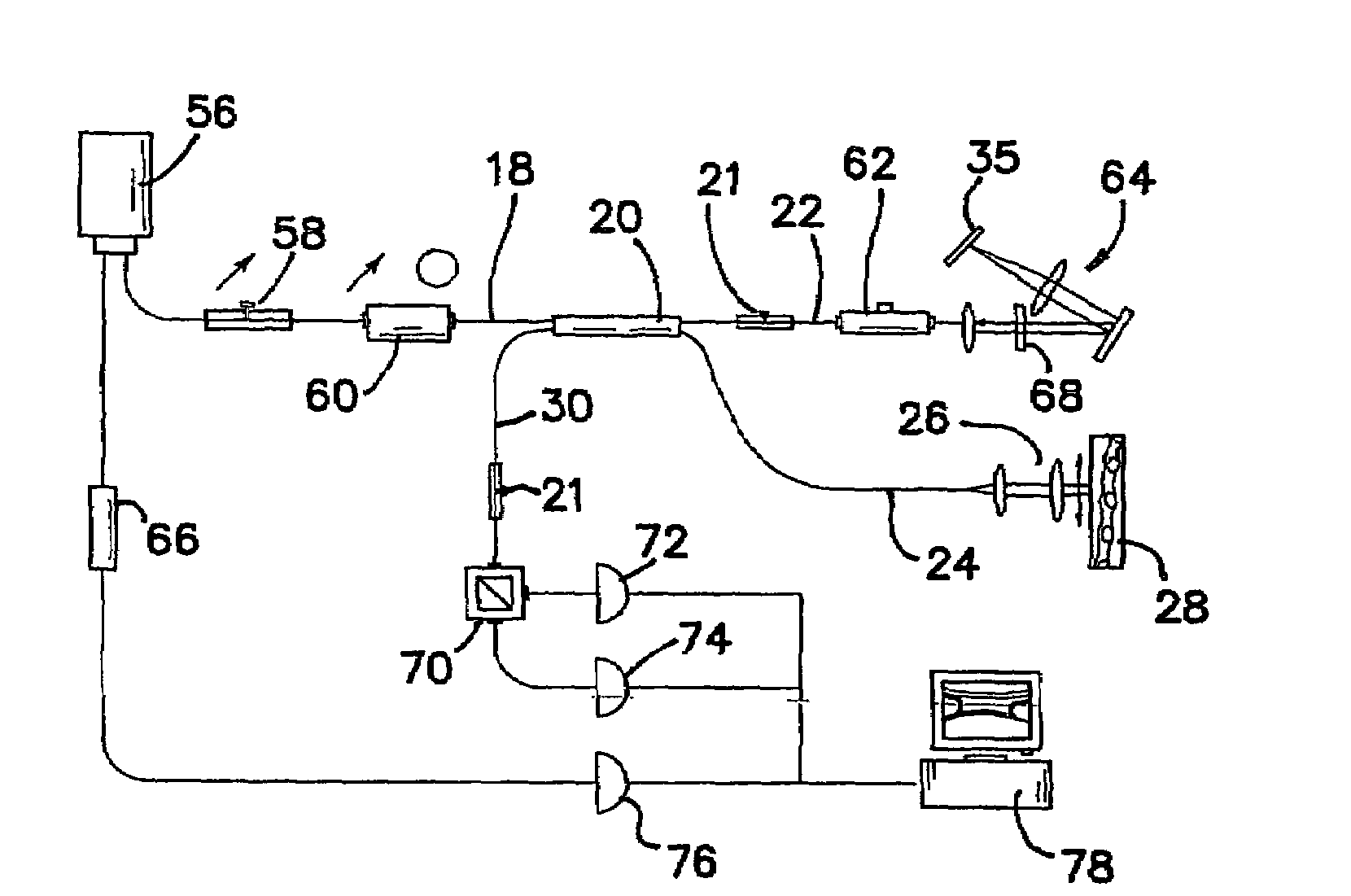
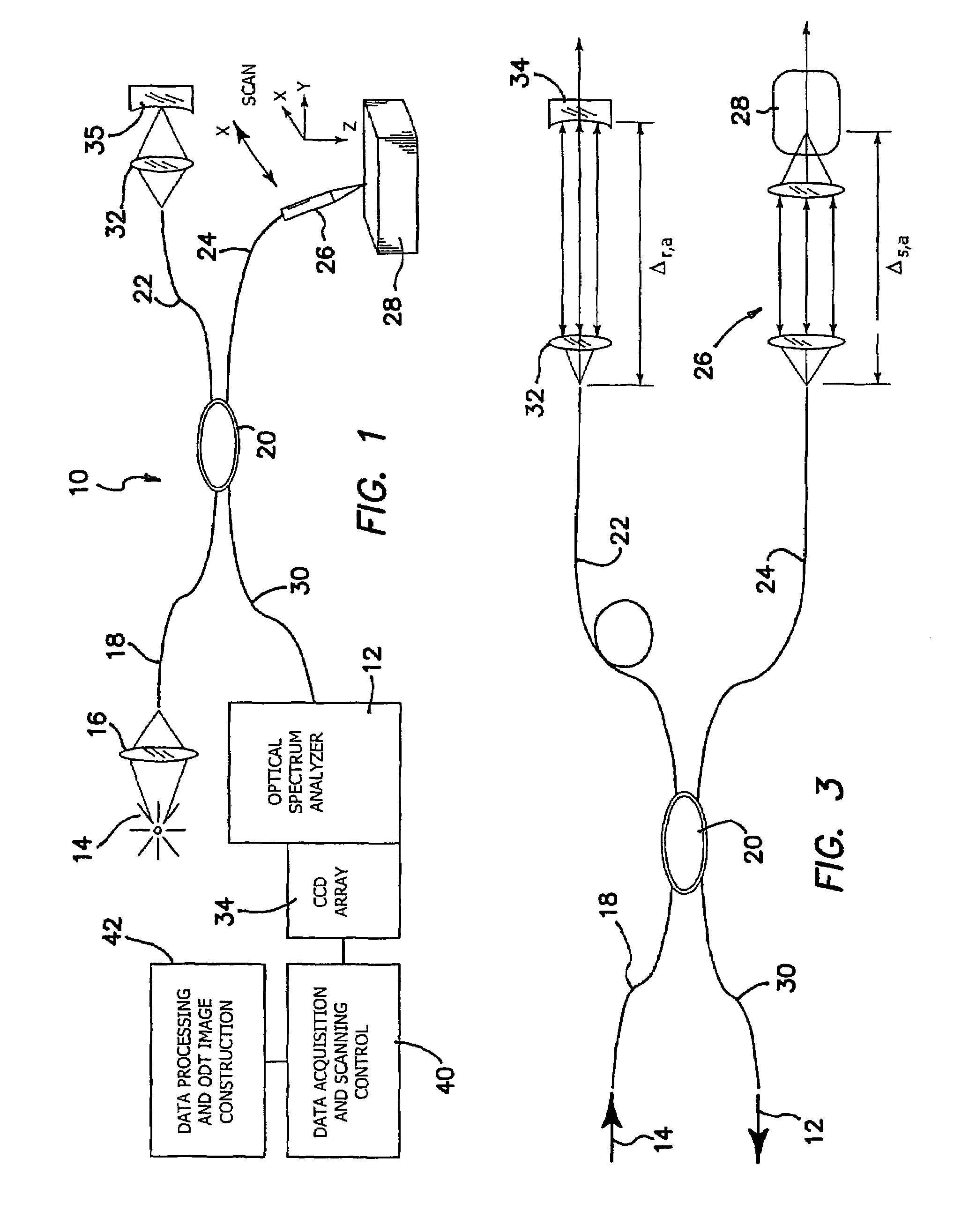
[0034]Although high resolution ODT structural and velocity images of static and moving constituents in turbid samples have been obtained using a time domain instrument, clinical application of the technique requires higher imaging speed. The invention is directed to a high speed FDOCT system for imaging in vivo blood flow. This is achieved and demonstrated according to the approach of the invention by using an FDOCT system based on spectral interferometry, an imaging construction algorithm, Monte Carlo simulation of multiple scattering and coherence gating to gain insight into and provide a model for the FDOCT imaging process.

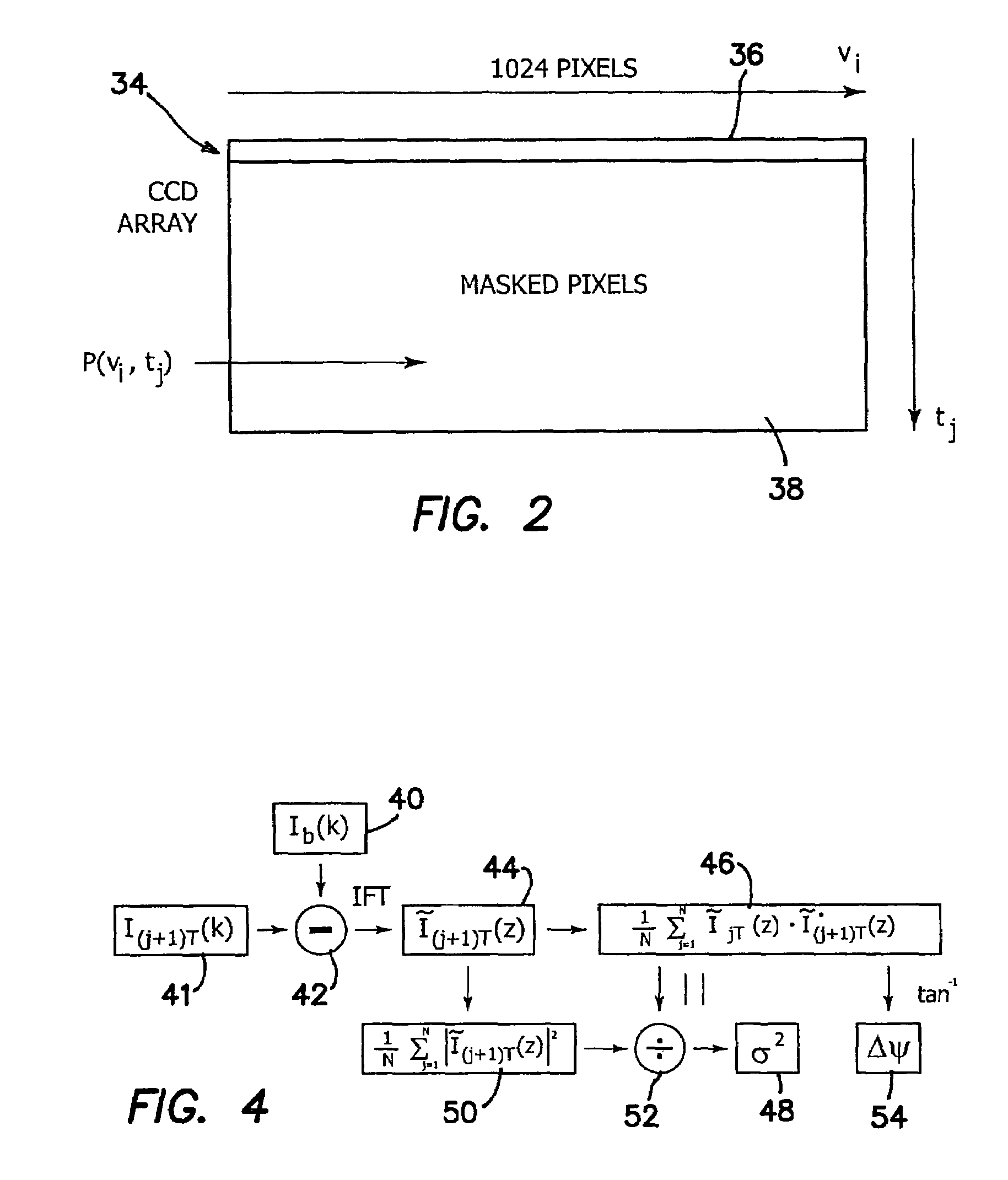
[0035]The prior temporal domain ODT instrument acquires data for each pixel serially by a sequential two-dimensional scan, which limits imaging speed. Image acquisition time for our prototype device is given by T=NxNzΔtp, where Nx and Nz are the number of pixels in the lateral and depth dimensions, and Δtp is the pixel acquisition time. Inasmuch as detection of...


PUM


Abstract

A system for tomographic imaging includes a source of at least partially coherent radiation, a frequency-swept laser source and an interferometer. The radiation in the interferometer is phase modulated at a modulation frequency for elimination of DC and autocorrelation noises as well as the mirror image. The interference fringes of the radiation backscattered from the sample into the interferometer are detected to obtain a spectral signal. The spectral signal of the detected backscattered interference fringes is transformed to obtain a location dependent signal at each pixel location in a data window. A tomographic image of the fluid flow in the data window is generated for display and of the structure of the scanned fluid flow sample in the data window from the location dependent signal is generated.

Description

RELATED APPLICATIONS[0001]The present application is related to U.S. Provisional Patent Application Ser. No. 60 / 528,024, filed on Dec. 9, 2003, which is incorporated herein by reference and to which priority is claimed pursuant to 35 USC 119.[0002]This invention was made with Government support under Grant Nos. WF23281, GM58785, and Rr01192 awarded by the National Institutes of Health. The Government has certain rights in this invention.BACKGROUND OF THE INVENTION[0003]1. Field of the Invention[0004]The invention relates to the field of biomedical imaging, and in particular to functional optical coherence tomography and functional optical Doppler tomography.[0005]2. Description of the Prior Art[0006]Direct visualization of physiological anatomy provides important information to the diagnostician and therapist for the evaluation and management of disease. High spatial resolution noninvasive techniques for imaging in vivo blood flow dynamics and tissue structure are currently not avai...

Claims


Application Information

Patent Timeline
Patent Type & Authority Patents(United States)
IPC IPC(8): G01B9/02A61B5/026
CPCA61B5/0066A61B5/0261A61B5/7203G01B9/02004G01B9/0201G01B9/02091G01B9/02078A61B5/7257A61B5/726
Inventor CHEN, ZHONGPINGZHANG, JUNNELSON, J. STUART
Owner RGT UNIV OF CALIFORNIA
Why Eureka
Try Eureka