Systems and methods for proactive detection of imaging chain problems during normal system operation

a technology of imaging chain and system operation, applied in the field of medical imaging, can solve problems such as system downtime, imaging system cannot be used for normal operation, and internal defects of objects may be detected

Active Publication Date: 2008-08-19
GENERAL ELECTRIC CO
View PDF1 Cites 1 Cited by
  • Summary
  • Abstract
  • Description
  • Claims
  • Application Information

AI Technical Summary

Problems solved by technology

For instance, internal defects in an object may be detected.
Running these diagnostics results in downtime for the users of the system.
In other words, while the diagnostic system is being utilized, the imaging system cannot be used for its normal operations.
However, current systems are unable to actively monitor the status of the image chain.
That is, failures, errors, and / or artifacts in the imaging chain that may be indicative of larger or developing system failures are not easily monitored in current systems.

Method used


Image

  • Systems and methods for proactive detection of imaging chain problems during normal system operation
  • Systems and methods for proactive detection of imaging chain problems during normal system operation
  • Systems and methods for proactive detection of imaging chain problems during normal system operation

Examples


Embodiment Construction

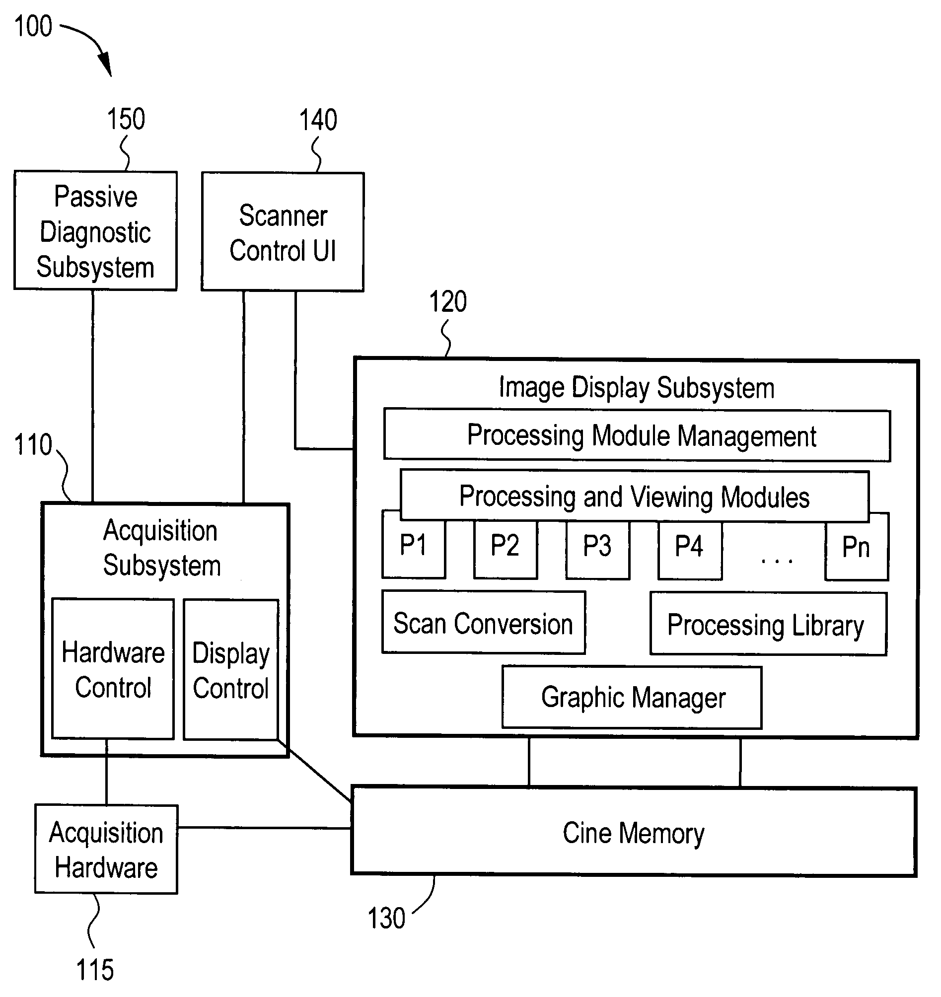
[0020]FIG. 1 illustrates a medical imaging system 100 according to an embodiment of the present invention. The medical imaging system 100 includes an acquisition subsystem 110, acquisition hardware 115, an image display subsystem 120, a memory component 130, an interface component 140, and a passive diagnostic subsystem 150. The acquisition subsystem 110 is in communication with the acquisition hardware 115 and the memory component 130. The acquisition hardware 115 is in communication with the acquisition subsystem 110 and the memory component 130. The image display subsystem 120 is in communication with the memory component 130 and the interface component 140. The interface component 140 is in communication with the acquisition subsystem 110 and the image display subsystem 120. The passive diagnostic subsystem 150 is in communication with the acquisition subsystem 110.

[0021]The acquisition subsystem 110 may include hardware control and / or display control components. The hardware co...


PUM


Abstract

Certain embodiments of the present invention provide a method for performing diagnostics in a medical imaging system during the normal operating mode of the imaging system including firing a diagnostic vector with a probe, collecting signal data based at least in part on the diagnostic vector, and processing the signal data to determine a diagnostic status. The diagnostic status indicates an operating condition of at least one component of the medical imaging system.

Description

BACKGROUND OF THE INVENTION[0001]The present invention generally relates to medical imaging. In particular, the present invention relates to systems and methods for proactive detection of imaging chain problems during normal system operation.[0002]Imaging systems encompass a variety of imaging modalities, such as x-ray systems, computerized tomography (CT) systems, ultrasound (US) systems, electron beam tomography (EBT) systems, magnetic resonance (MR) systems, and the like. Imaging systems generate images of an object, such as a patient. The images may be generated, for example, through exposure to an energy source or wave. For example, ultrasound beams may travel into a patient and produce echo signals reflected from bone and tissue inside the patient. As another example, an x-ray source may be used to generate x-rays that pass through an object. The x-rays may be partially absorbed by the object and an image may be generated based on the energy that passes through the object.[000...

Claims


Application Information

Patent Timeline
Patent Type & Authority Patents(United States)
IPC IPC(8): G06F19/00
CPCG01S7/5205A61B8/00A61B2560/0276
Inventor SABOURIN, THOMAS JAMESSTRATTON, GREGORY CHARLESMORTIZ, MICHAEL RICHARD
Owner GENERAL ELECTRIC CO
Why Eureka
Try Eureka