Slide photograph data creation system, and slide photograph data

a technology of which is applied in the field of slide picture, i. e ., photograph, data creation system and slide picture data, can solve the problems of large amount of memory, large amount of preparation time, and large time and labor expenditure, and achieve high-speed processing and display, efficient performance of examination and photography, and high speed

Inactive Publication Date: 2008-09-16
CLARO
View PDF16 Cites 20 Cited by
  • Summary
  • Abstract
  • Description
  • Claims
  • Application Information

AI Technical Summary

Benefits of technology

[0014]Another aspect of the present invention is that the preparate storage magazine is capable of being freely attached to and detached from the third movable body. The preparate storage magazine includes an attachment / detachment lever, which is capable of being manually switched for displacement between a locked position, where it is fixed to the third movable body, and an unlocked position, where it becomes separable from the third movable body; and a movable stopper, which is operationally linked to the movement of the attachment / detachment lever so as to be in a position preventing slipping-out of the preparates from the preparate storage magazine when the attachment / detachment lever is in the unlocked position, and to be in a position enabling insertion and removal of preparates when the attachment / detachment lever is in the locked position.
[0016]As a result of the present invention, it is possible to photograph at high speed only the areas necessary for photography by microscope. Furthermore, by storing the picture data in one file, there is no need to open and process large numbers of picture files when browsing, a large amount of memory is not necessary, and high-speed processing and display are possible.
[0018]Also, the work of removing sample preparates stored in the preparate storage magazine, moving them between the object lens and the condenser lens of the microscope, and again returning the preparates to the storage magazine upon having finished examination and photography, can be performed automatically. It becomes possible, therefore, to efficiently perform the work of examination and photography of large numbers of samples.
[0019]Also, because the preparate storage magazine can be separated, the operations of insertion and removal of sample preparates into and from the preparate storage magazine become easier. In addition, by preparing plural preparate storage magazines and using these by successive replacement, it becomes possible to perform examination and photography of large numbers of samples more efficiently.
[0020]Furthermore, when separating and carrying the preparate storage magazine, any accidents such as having the front face of the magazine be accidentally turned downward, and having the precious samples fall from the magazine, can be avoided.

Problems solved by technology

When creating large numbers of picture data, much time and labor was expended for replacement of the preparates.
Also, in order to open and process large numbers of picture files when creating and browsing the pictures, a large amount of memory was necessary, and substantial processing time was required.
Also, because basically only two-dimensional picture information could be photographed, the sample structure on the Z axis parallel to the optical axis of the microscope could not be discriminated from the photographed picture.

Method used


Image

  • Slide photograph data creation system, and slide photograph data
  • Slide photograph data creation system, and slide photograph data
  • Slide photograph data creation system, and slide photograph data

Examples


first embodiment

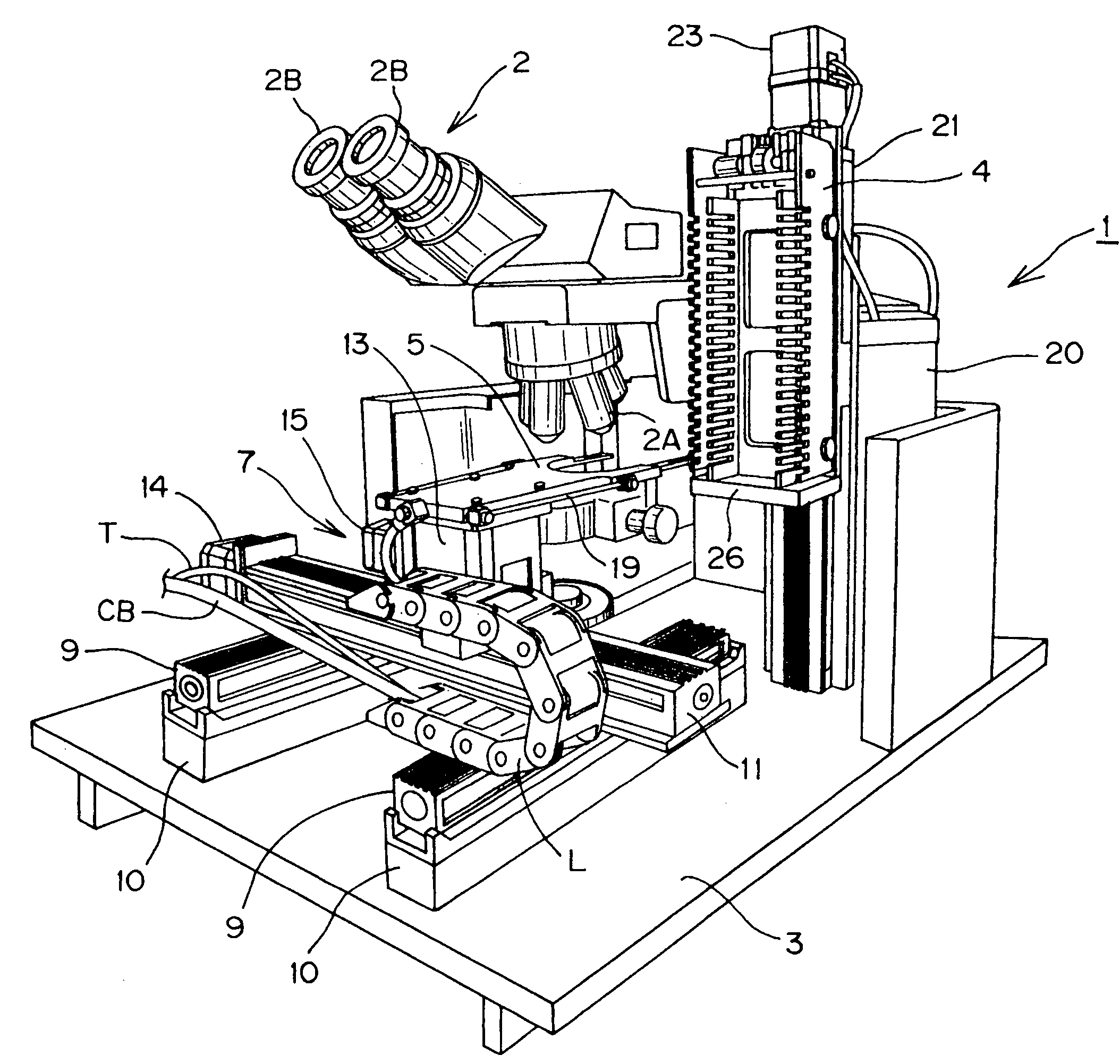
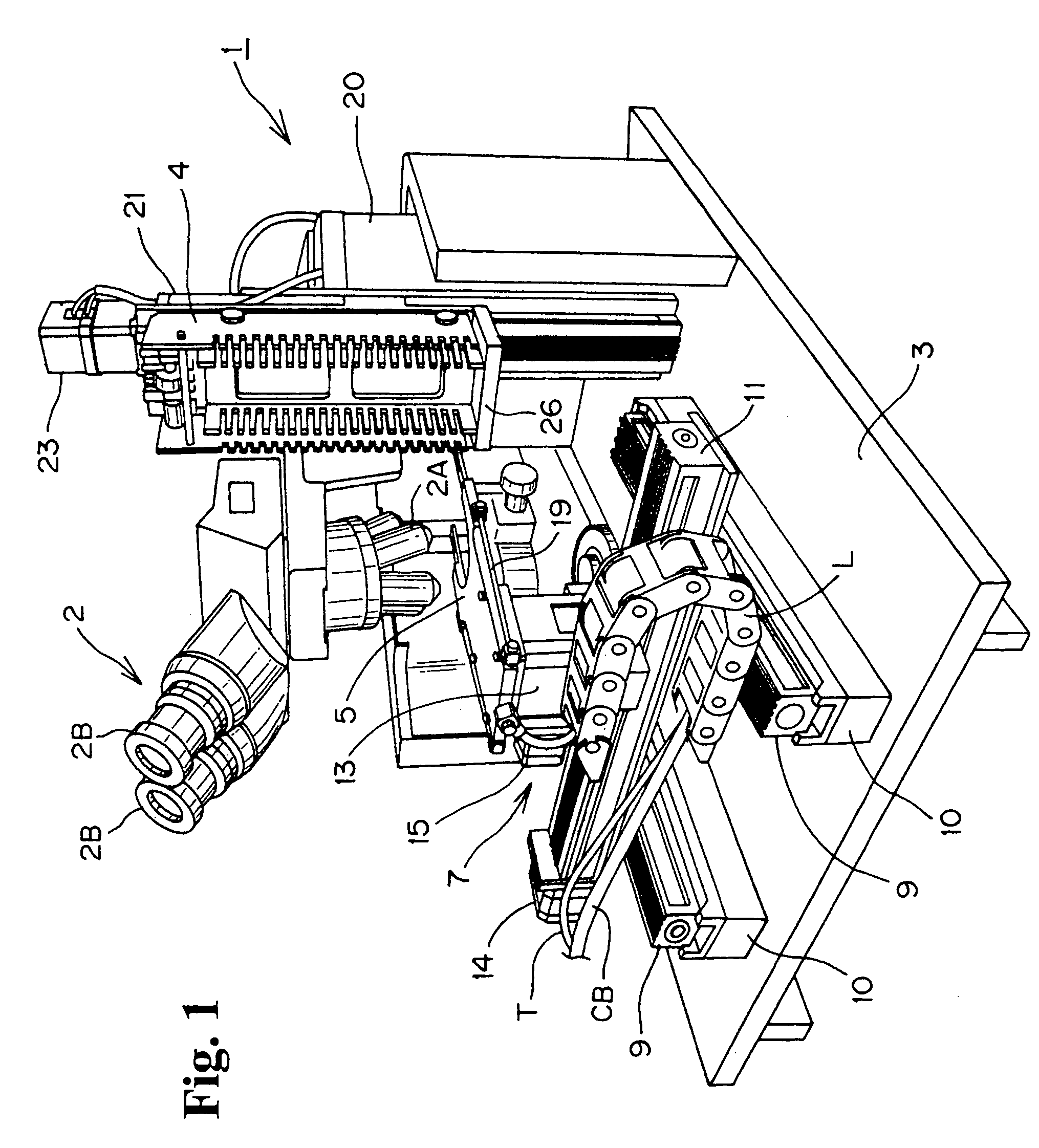
[0052]In the invention, as shown in FIG. 1, a freely bendable cable guide L enables the suction hose T communicating with the ventilation tubes 19, and a power supply / control cable CB connected to the step motor 15 provided on the movable frame 13, to smoothly follow the movement of the movable frame 13 following the guide frame 11. The cable guide L is depicted only in FIG. 1.

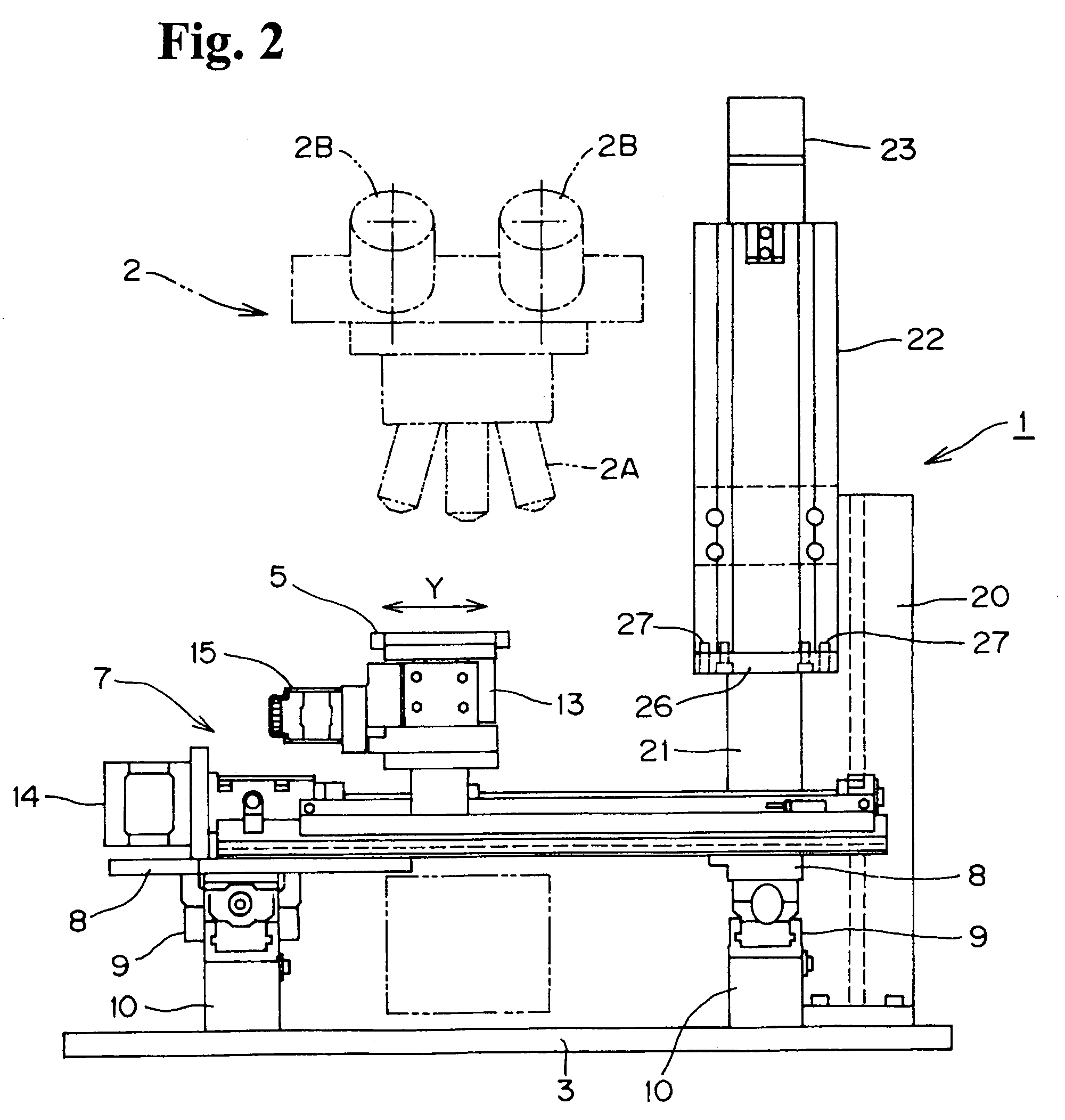
[0053]Meanwhile, a support frame 20 is fixed on the upper surface of the stand 3 in a position to the side of the microscope 2, and a guide frame 21 (third guide member) is attached to this support frame 20. A linear bearing is built into the guide frame 21, and it supports and guides a movable frame 22 (third movable body) to be displaceable to ascend and descend following the longitudinal direction of the guide frame 21.

[0054]Also, a step motor 23 (fourth drive source) is provided on the upper end of the guide frame 21. The step motor 23 is drive-coupled to the movable frame 22 guided by the guide frame 21, ...

second embodiment

[0120]Therefore, according to the invention, in S100, the number-of-preparates information input by the user is read in. In S101, the three-dimensional movement mechanism 7 is controlled and one unprocessed preparate 90 is removed and is placed beneath the digital camera 81. In S102, the two-dimensional bar code is read using the digital camera 81.

[0121]In S103, a loupe picture is photographed using the digital camera 81, and the preparate is stored in the original storage position. In S104, the photographic area is automatically recognized. In S105, it is determined as to whether or not there is an unprocessed preparate, and if the result of the determination is positive, the process moves to S101, and if the result is negative, the process moves to S106.

[0122]In S106, the loupe pictures and automatically recognized areas of all of the preparates are displayed. The user here marks only the parts requiring photography of high-magnification pictures for the loupe pictures where one w...


PUM


Abstract

A slide photograph data creation system includes a digital camera for making high-magnification photographs of samples, a sample transporter for holding and transporting samples, a controller for controlling the camera and the sample transporter so as to provide overlapping photographs, a pasting information generator for recognizing the margin by which the photographs overlap and for generating pasting-together information, and a photograph file generator for storing in a single file a plurality of high-magnification photographs and the pasting-together information. The system facilitates the examination and photography of large numbers of samples, and enables discrimination of the three-dimensional structure of a sample. Since the photographic data is managed with a database, virtual slide photographs and their attributes information can be browsed over a network or the Internet.

Description

BACKGROUND OF THE INVENTION AND RELATED ART STATEMENT[0001]The present invention relates to a slide picture, i.e., photograph, data creation system and slide picture data. In particular, the present invention relates to a slide picture data creation system for setting preparates (slide glass) containing samples in microscopes, photographing the samples at high magnification, and generating and managing slide picture data, in pathological histological studies, hematological studies, genetic studies, cytological studies, and the like, as well as the generated slide picture data.[0002]Recently, pathological histological, hematological, and genetic microscopic studies, and the like, have been performed by photographing samples of extracted pathological tissues and cells and blood, and the like, with a photographic apparatus equipped on a microscope, taking the pictures into a computer, and creating picture data that can be observed on a display. Such technology is described in, for exam...

Claims


Application Information

Patent Timeline
Patent Type & Authority Patents(United States)
IPC IPC(8): H04N7/18B65G1/00G02B21/26G02B21/36G02B23/00H04N9/47G06K9/00G09G5/00
CPCG02B21/367G01N1/312
Inventor TAKAMATSU, TERUMASASATO, TATSUSUKEMIURA, TOMISATONOZAKA, HIROYUKIYASUIKE, HIDEKATSUMIURA, HIROMIZHONGXI, ZHENG
Owner CLARO
Why Eureka
Try Eureka