Acquisition window compensation for nuclear medical image reconstruction attenuation coefficient maps

a nuclear medical image and acquisition window technology, applied in the field of computed tomography system and method analysis, can solve the problems of difficult measurement of attenuation in emission imaging, difficult to obtain “effective” transmission energy, and no definition of densities greater than

Active Publication Date: 2009-01-06
SIEMENS MEDICAL SOLUTIONS USA INC
View PDF1 Cites 7 Cited by
  • Summary
  • Abstract
  • Description
  • Claims
  • Application Information

AI Technical Summary

Problems solved by technology

However, measurement of attenuation in emission imaging is more difficult, because the accurate amount of emission source radiation being generated in the subject that results in a quantity of radiation being detected outside the subject cannot be measured directly.
Because a CT scanner emits a continuous spectrum of x-rays, an “effective” transmission energy is not easily obtained, and usually involves actual measurement of the scan object penetrating radiation spectrum.
However, there is no definition for densities greater than water.
However, there are numerous problems with this approach.
For example, this method does not take into account existing variations in tissues densities for the same tissue classes, it is limited to the errors of the segmentation routine, it may require an educated user to segment the data accurately, and it is not user-friendly.
This is often difficult because pixels may contain more than one tissue type (“partial volume”), or an educated user may be needed to identify the tissue type of each pixel (see, D. R. White, M. Fitzgerald, “Calculated attenuation and Energy Absorption Coefficients for ICRP Reference Man (1975) Organs and Tissues”, Health Physics, 33, pp.
Therefore, this method suffers from similar problems.
While the above method takes into account the dependencies of linear attenuation coefficients on tissue type and photon energy, the method does not take into account variations in the energy window of SPECT data acquisitions.

Method used


Image

  • Acquisition window compensation for nuclear medical image reconstruction attenuation coefficient maps
  • Acquisition window compensation for nuclear medical image reconstruction attenuation coefficient maps
  • Acquisition window compensation for nuclear medical image reconstruction attenuation coefficient maps

Examples


Embodiment Construction

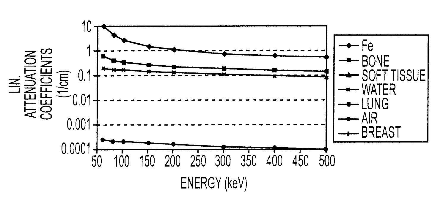
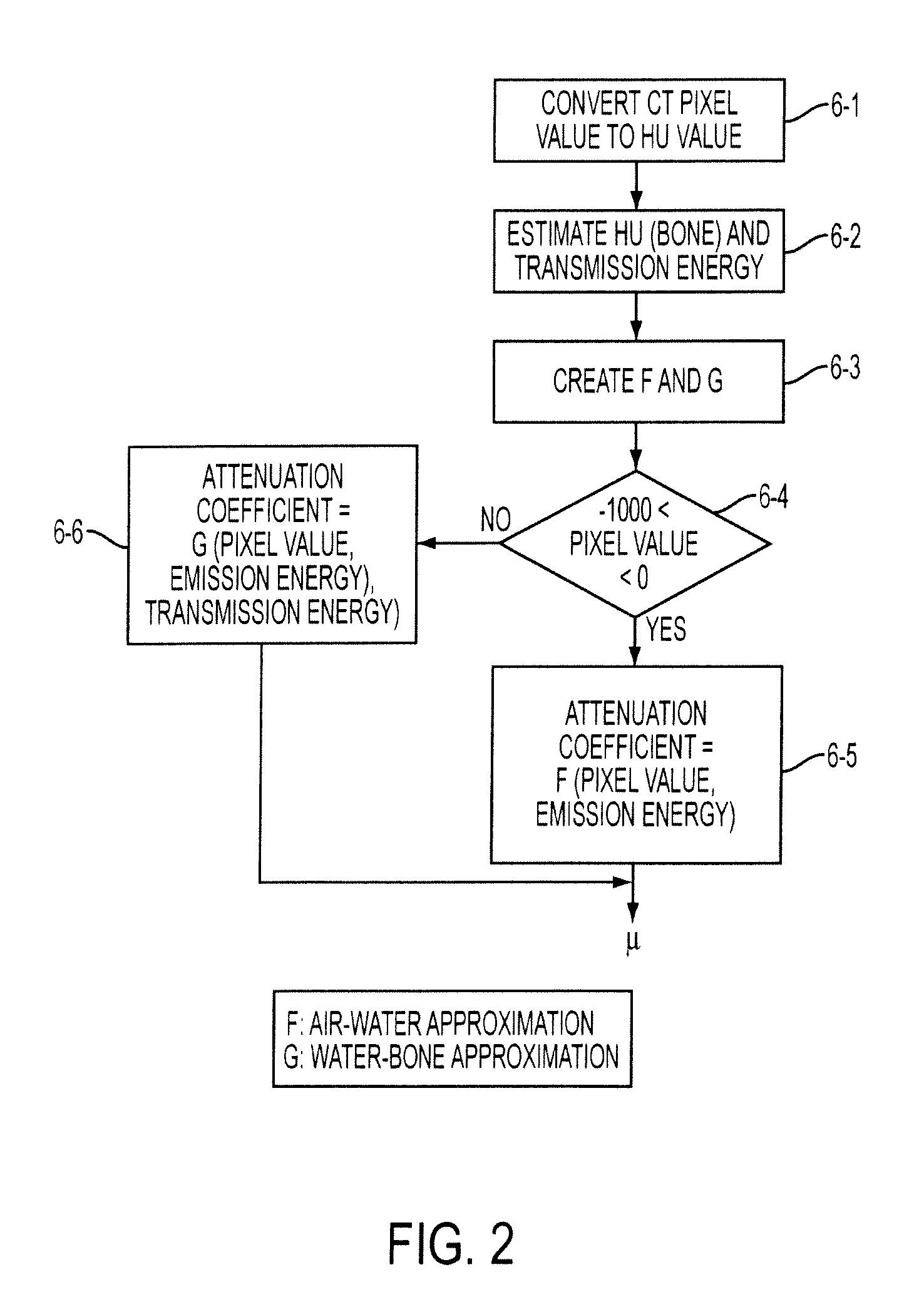
[0033]The basic concepts upon which the present invention builds are disclosed in U.S. Pat. No. 6,950,494 incorporated by reference hereinabove. As explained therein, FIG. 2 is a flow diagram of a method of generating linear attenuation correction factors μ also in accordance with the present invention. It will be understood by those skilled in the art that the following processes may be programmed onto central processor or computer, which may be coupled with a CT scanning device, or which may receive CT data in any fashion. Accordingly, a program written to perform the following may be stored conventionally in memory. Accordingly, the present invention is not limited to any particular system configuration.

[0034]Beginning with step 6-1, CT pixel values are converted to HU values. CT devices may output pixel data a number of ways. Typically, a device outputs data in a proprietary fashion that must be converted to HU. The conversion is usually provided by the vendor. For example, CT d...


PUM


Abstract

Generation of attenuation maps for nuclear medicine image reconstructions based on the use of anatomical image data, such as CT data, take into account variations caused by variations in acquisition energy width and emission energy of the radioisotope used in the clinical imaging procedure, as coefficient correction factors that are stored together with such maps.

Description

CROSS-REFERENCE TO RELATED APPLICATIONS AND CLAIM FOR PRIORITY[0001]This application claims priority under 35 U.S.C. § 119(e) from copending U.S. Provisional Patent Application Ser. No. 60 / 691,335 filed Jun. 16, 2005. This application is also related to U.S. Pat. No. 6,950,494 to Vija et al., issued Sep. 27, 2005 and assigned to the same assignee herein. The '494 patent is incorporated by reference herein in its entirety.BACKGROUND OF THE INVENTION[0002]1. Field of the Invention[0003]This invention generally pertains to systems and methods for analyzing computed tomography data. More particularly, the present invention is directed to a method of converting computed tomography (CT) data to linear attenuation coefficient data for use in nuclear medicine, such as to compensate for attenuation in SPECT and PET imaging. The present invention is specifically directed to methods for automatically adjusting correction factors for linear attenuation coefficient maps based on a finite acquisi...

Claims


Application Information

Patent Timeline
Patent Type & Authority Patents(United States)
IPC IPC(8): G01T1/166
CPCG01T1/1611G01T1/2985G01T1/1615A61B6/037
Inventor VIJA, A HANS
Owner SIEMENS MEDICAL SOLUTIONS USA INC
Why Eureka
Try Eureka