Method of assessing localized shape and temperature of the human body

a technology of localized shape and temperature, applied in the field of medical assessment, can solve the problems of subjective process, unreliability, and no longer used as an outcome measure in most clinical studies, and achieve the effect of improving clinical trial outcome measures

Inactive Publication Date: 2009-04-14
CARNEGIE MELLON UNIV +1
View PDF6 Cites 46 Cited by
  • Summary
  • Abstract
  • Description
  • Claims
  • Application Information

AI Technical Summary

Benefits of technology

[0006]The present invention utilizes various existing imaging sensor technologies, including 3D imaging, thermal imaging, near-infrared imaging and visual range imaging to quantify the major physical examination components of a joint. These include joint swelling 10, range of motion 20, heat distribution 30, and erythema (redness of the skin) 40, as shown in FIG. 1. This provides a highly-reproducible, consistent means of assessing the health of a joint and for tracking the effectiveness of treatment for arthritis, as well as for other applications requiring precise measurement of the body. In addition, because several components of the ACR 20 / 50 / 70 and the ACR Pediatric 30 algorithm rely on these objective measurements, the overall effectiveness and accuracy of these measure will be improved.
[0010]In the present invention, a thermal infrared (TIR) camera is used to obtain a thermogram of the joint, which is capable of measuring subtle differences in temperature of surfaces with high spatial resolution. A TIR camera converts emitted infrared radiation into electrical impulses that are visualized as either a color or a grayscale image called a thermogram. In addition to making detailed measurements in the examination room, thermal imaging allows quantitative comparison, both spatially and thermally, of a patient's current skin temperature profile with earlier profiles.
[0012]Arthritis patients exhibit limited range of motion, which frequently deteriorates over time. Clinicians measure range of motion either visually or using a goniometer placed against the patient's hand while s / he extends and flexes the joint. This process is subjective because the exact placement of the measuring device is physician-dependent. On the other hand, video-based systems can reliably track body motion and accurately estimate the position of a patient's joints, and these methods can be easily adapted to quantifying the range of motion.
[0013]In the case of a rheumatologic examination, tracking of the range of motion of a joint with a video based system can be accomplished by positioning a camera such that its imaging plane is parallel to the joint's plane of motion, thereby imaging the joint in two-dimensions. For example, when viewing the human wrist in profile, it is straightforward to analyze the image to calculate joint angle. For range of motion, measurement of joint angle using cameras has been found to be superior to the clinician's use of a goniometer, since it is non-contact and multiple movements of the patient can be easily captured and averaged in real-time.
[0016]With the four sensing modalities outlined above, the quantitative measures of the joint examination and their corresponding numerical measures and combined global score will exceed the sensitivity and reliability of the qualitative measures currently used to monitor disease activity and response to treatment. Clinicians should find the objective measurements to be an effective surrogate for direct physical examination. The sensor-based measurements are of the present invention are deterministic, repeatable and consistent over time, and, as such will improve clinical trial outcome measures for arthritis.

Problems solved by technology

This process is subjective and so unreliable that it is no longer used as an outcome measure in most clinical studies.

Method used


Image

  • Method of assessing localized shape and temperature of the human body
  • Method of assessing localized shape and temperature of the human body
  • Method of assessing localized shape and temperature of the human body

Examples


Embodiment Construction

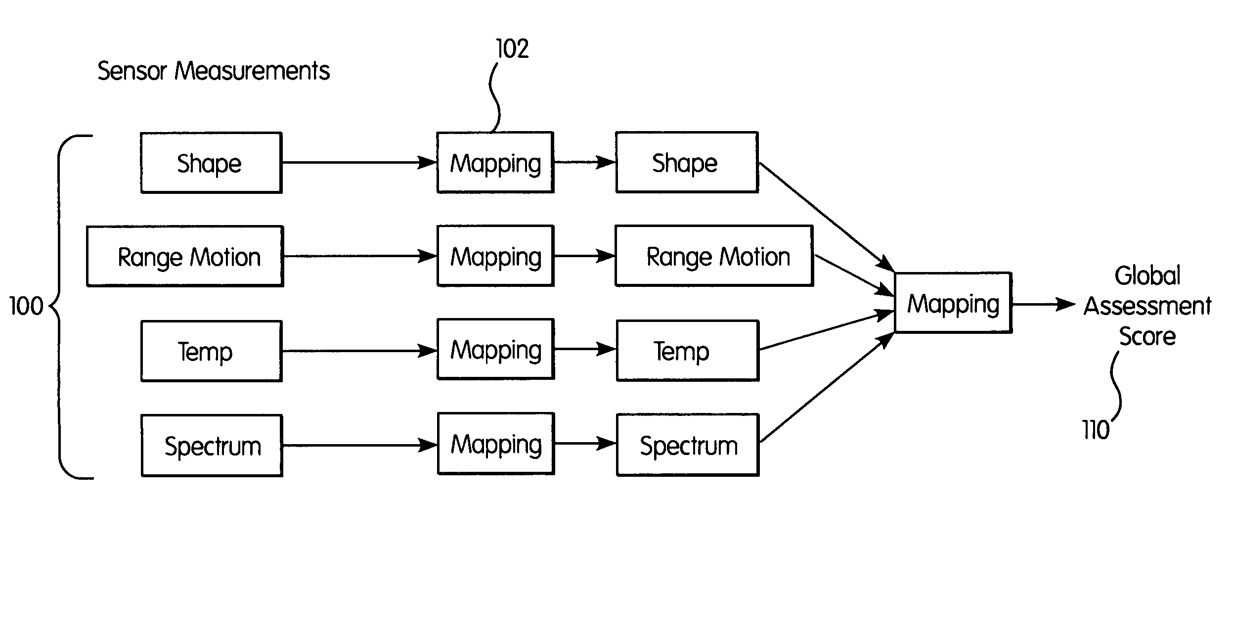
[0034]Existing modalities for measuring outcome in the diagnosis and treatment of arthritis are cumbersome, time-consuming, and often unreliable. In contrast, sensor-based technologies can be quantifiable, reproducible and highly reliable. The present invention establishes ways to apply known sensor technology to the diagnosis of arthritis, as well as the assessment and evaluation of joints. In addition, the same technology can be used to image and measure other areas of the body. The methods are being explained in the context of joint evaluation in arthritis patients merely as exemplars of the technology.

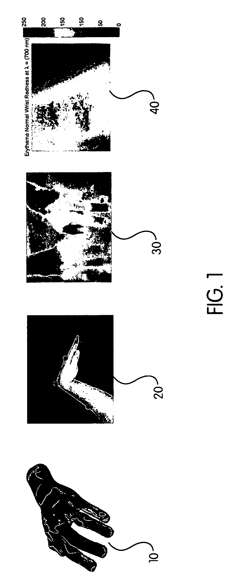
[0035]The first modality, 3D imaging utilizing a 3D scanner, is useful for both shape and volume measurement. Shape measurements are useful in comparing recent scans to baseline scans taken during previous examinations. The difference in the shape of the joint can be useful for tracking the effectiveness of therapy and for assessing the presence of swelling in the area of the joint...


PUM

PropertyMeasurementUnit
wavelengthsaaaaaaaaaa

Abstract

A method of objectively quantifying assessments of joints having arthritis for purposes of assessment, diagnosis and treatment. The objective measurements utilize known imaging modalities including 3D scanning, thermal imaging, visible and near-infrared imaging and two-dimensional imaging to quantify swelling, heat distribution, erythema, and range or motion. The objective measurements can be combined in various ways to assess the extent of the disease and can be used to adjust treatment protocols.

Description

RELATED APPLICATIONS[0001]This application claims the benefit of U.S. Provisional Application 60 / 608,507, filed Sep. 9, 2004.FIELD OF THE INVENTION[0002]This invention relates to the field of medical assessment, and, in particular, relates to the measurement of localized body shape and temperature using sensor technology for purposes of assessing medical conditions and tracking treatment progress.BACKGROUND OF THE INVENTION[0003]Physicians diagnosing and treating disease involving inflammation of the joints, such as arthritis, including osteoarthritis (OA), rheumatoid arthritis (RA) or juvenile rheumatoid arthritis (JRA) have experienced difficulty in quantifying the extent of inflammation or establishing the diagnosis. A typical arthritis patient may present with one or more various symptoms relating to synovitis, including swelling, heat, redness and reduced mobility in one or more joints. However, it has proven remarkably difficult to measure these signs in arthritis patients in ...

Claims


Application Information

Patent Timeline
Patent Type & Authority Patents(United States)
IPC IPC(8): G06K9/00G01N23/223G06V10/70
CPCA61B5/0059A61B5/0062A61B5/015A61B5/103A61B5/4528G06K9/00382G06K9/6217G06T7/0012G06T2207/20212G06T2207/30004A61B5/0064G06V40/11G06V10/70G06F18/21
Inventor HIRSCH, RAPHAELOSBORN, JAMESKWOH, CHIAN KENTDENES, LOUIS J.HUBER, DANIEL
Owner CARNEGIE MELLON UNIV
Why Eureka
Try Eureka