
Method for expression of proteins on spore surface

a protein and spore technology, applied in the field of protein expression on the spore surface, can solve the problems of high method expenditure and long time, low antibody yield, and insufficient hybridoma technology to find antibodies exhibiting new binding properties

Inactive Publication Date: 2009-09-01
GENOFOCUS CO LTD
View PDF3 Cites 2 Cited by
  • Summary
  • Abstract
  • Description
  • Claims
  • Application Information

AI Technical Summary

Benefits of technology

[0023]Under such situation, the present inventors have made intensive studies to be from the shortcoming of conventional display methods, and as a result, we have developed novel display system using a spore as host and a coat protein as motif of surface display. Surprisingly, the developed display system has been found to have excellent stability to a variety of physiochemical stresses in surrounding environment and have much broader applicability.
[0033]The principle of the present invention lies in the employment of microbial spore as host for surface display and spore coat protein as surface display motif. The present inventors have been compelled to select a system for spore surface display since the spore has a following advantages (Driks, 1999): 1) a higher heat stability, 2) a significant stability to radioactivity, 3) a stability to toxins, 4) a higher stability to acid and base, 5) a significant stability to lysozyme, 6) a resistance to dryness, 7) a higher stability to organic solvents, 8) a fusion protein between a surface display motif and a protein of interest is displayed on spore surface immediately after expression without secretion in host cell, 9) no metabolic activity, and 10) shorter time for obtaining spore, e.g. within several hours.
[0034]In particular, the spore coat proteins used in this invention circumvent a necessity for passage across cell membrane, so that they do not need secretion signal and target signal which are prerequisites of surface display motif, thereby ensuring a surface display of protein such as β-galactosidase, in orderly fashion, which is difficult to pass across cell membrane.
[0062]As demonstrated in Examples, the stability of spore displaying the foreign protein of interest an its surface is very high in the present invention, indicating maintenance of the integrity of spore surface structure formed by cooperation of coat proteins while the foreign protein is displayed.
[0071]In addition, the conventional process for preparing protein array needs tedious and repetitive works such as cloning genes of several thousands to tens of thousands of proteins and immobilizing of the proteins expressed. Therefore, there remains a need to improve simplicity and rapidity of the works.
[0077]According to preferred embodiment, the transformant is derived from a variant mutated to enhance spore surface display. For example, the mutation to enhance spore surface display eliminates a production of extracellular secretory protease in the transformant, so that the protein of interest displayed on spore surface is stably maintained. In addition, the mutation to enhance spore surface display eliminates a production of intracellular protease in the transformant. It is also preferred that a gene or genes involved in spore forming is subject to mutation in order to the rate of spore forming (Perego, M., et al., Mol. Microbiol. 19: 1151-1157 (1996)).

Problems solved by technology

However, the conventional method needs high expenditure and long time for performance whereas the yield of antibody is very low.
In addition to this, to screen novel antibody, more than 1010 antibody libraries is generally used, as a result, the hybridoma technology has been thought to be inadequate in finding antibody exhibiting new binding property.
In addition, the lasting use renders the enzyme inactive or gives a problem on transference of substrate and product, thus dropping the productivity of overall process.
Generally, it is difficult to prepare the bioabsorber.
In the developed phage display system, the concentration of the desired clone from phage library has been found to be difficult and the antibody selected from phage library displaying has usually exhibited very low expression rate.
According to a surface display system of Gram negative bacteria, the incorporation of foreign polypeptide into surface structure results in not only its steric limitation which makes it impossible to have stable membrane protein (Charbit et al., 1987; and Agterberg et al., 1990) but also drop of the stability of cell outer membrane and its viability.
In addition, in surface display system of yeast, because the vector used has usually shown a low rate of transformation, which is unfavorable to surface display of library.
However, the phage display technology is encountered to difficulty in concentration of the desired clones from phage library.
However, the over-expression of cell outer membrane protein fused to foreign protein is likely to bring about structural instability of cell outer membrane, consequently, diving the viability of host cell (Georgiou et al., 1996).
Gram positive bacteria as host are relatively rigid and permit stable display of the desired protein; however, transformation efficiency is exhibited low, which results in smaller size of library than E. coli.
The host organisms having been developed are likely to be sensitive to a variety of physiochemical treatments, which makes it impossible to select proteins displayed on surface by virtue of direct physiochemical treatment.
In addition, the host organisms used conventionally have a complicated and weak structure of cell surface, which drops adaptability to extreme environment such as high temperature and high pressure.
However, in the surface display methods having been developed, the host organism does not have resistance to chemicals and physiochemical change such as pH change, and displaying protein on its surface in excess leads to disadvantages in cell surface, finally reducing the viability of host cell largely (Georgiou et al., 1996).

Method used


Image

  • Method for expression of proteins on spore surface
  • Method for expression of proteins on spore surface
  • Method for expression of proteins on spore surface

Examples


example i

Isolation of the Gene Encoding Coat Proteins

I-1: Construction of the Vector for Spore Surface Display

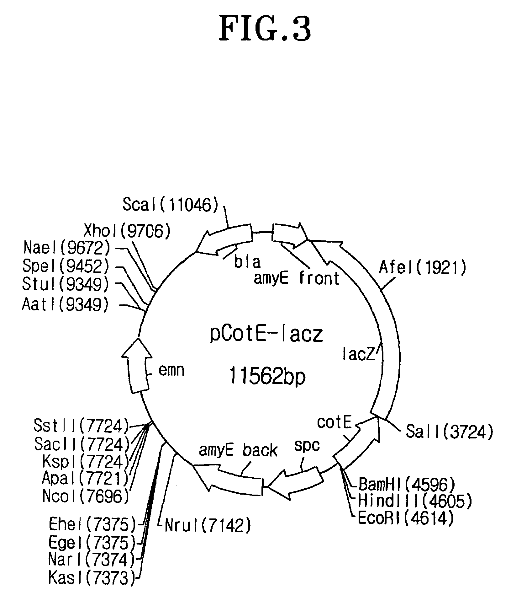
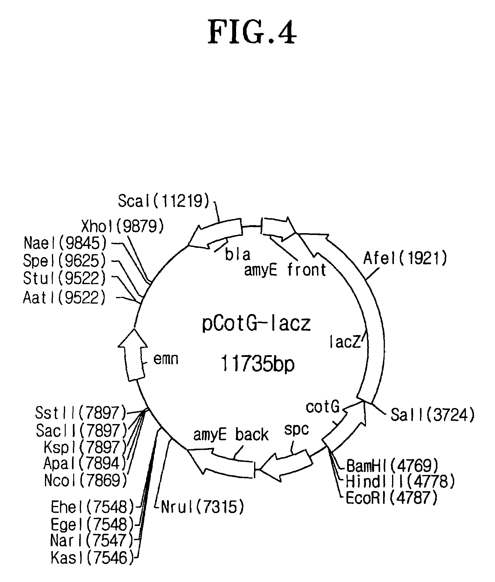
[0102]To isolate the most appropriate coat protein for spore surface display among coat proteins consisting of spore, the recombinant vector having the gene encoding a fusion protein between coat protein and β-galactosidase was constructed as follow:

[0103]To begin with, the DNA was extracted from the Bacillus subtilis 168 strain provided from Dr. F. Kunst (Kunst F., et al., Nature, 390: 249-256(1997)) by Kalman's method (Kalman S., et al., Appl. Environ. Microbiol. 59, 1131-1137(1993)), and the purified DNA was served as template for PCR to spoIVA primers (SEQ ID NOs: 1 and 2), cotB primers (SEQ ID NOs: 3 and 4), cotC primers (SEQ ID NOs: 5 and 6), cotD primers (SEQ ID NOs: 7 and 8), cotE primers (SEQ ID NOs: 9 and 10), cotG primers (SEQ ID NOs: 11 and 12), cotH primers (SEQ ID NOs: 13 and 14), cotM primers (SEQ ID NOs: 15 and 16), cotV primers (SEQ ID NOs: 17 and 18), cotX primers (...

example ii

Spore Production Depending on Culture Time

[0115]As shown in FIG. 7, it is required to stop incubation on a specific time point and isolate spores. In DB104, the enzyme activity of spores after 38 hr of incubation is significantly low comparing to that after 24 hr of incubation. Thus, it is demonstrated that the adjustment of incubation time makes it possible to yield spores displaying enzyme on its surface with the greatest enzyme activity.

example iii

Characterization of Spores Dispalying β-Galactosidase

[0116]Heat resistance was measured as follow in spores displaying β-galactosidase: 100 μl of spores isolated by renografin gradients in Example I-2 were heated for 15 min and then spread on LB plates to evaluate viability of spores (FIG. 8). As shown in FIG. 8, spores displaying CotG-LacZ show similar heat resistance to spores without surface protein. In a result, the display of the foreign protein fused to coat protein on spore surface does not affect on its inherent characteristics such as heat resistance. Moreover, these results provide the promising usage of spore displaying on its surface enzyme in chemical reactions at high temperature. In addition, from these results, it is suggested that the spores transformed according to the present invention remain their inherent resistances to lysozyme, a bacterial cell wall-degrading enzyme and solvent.


PUM

PropertyMeasurementUnit
thicknessaaaaaaaaaa
culture timeaaaaaaaaaa
pHaaaaaaaaaa

Abstract

The present invention relates to a method for display of proteinson spore surface and a method for improving protein with rapidity using the same, which comprises the steps of (i) preparing a vector for spore surface display comprising a gene construct containing a gene encoding spore coat protein and a gene encoding a protein of interest, wherein, when expressed, the gene construct expresses a fusion protein between the spore coat protein and the protein of interest, (ii) transforming a host cell with the vector for spore surface display; (iii) displaying the protein of interest on a surface of a spore of the host cell; and (iv) recovering the spore displaying on its surface the protein of interest.

Description

[0001]This application is the U.S. national phase of international application PCT / KR01 / 02124 filed Dec. 7, 2001 which designated the U.S.FIELD OF THE INVENTION[0002]The present invention relates to a method for display of proteins on spore surface, in particular to a method for surface display using spore coat proteins as surface display motif and a high throughput method for improving protein.DESCRIPTION OF THE RELATED ART[0003]The technology of surface display in which organism displays on its surface the desired proteinaceous substance such as peptide and polypeptide has wider application fields depending on the types of protein displayed or host organism (Georgiou et al., 1993, 1997; Fischetti et al., 1993; and Schreuder et al., 1996). The gene of protein to be displayed is contained in host organism and thus the host can be selectively screened using the characteristics of the protein displayed, thereby obtaining the desired gene from the selected host with easiness. Therefore...

Claims


Application Information

Patent Timeline
Patent Type & Authority Patents(United States)
IPC IPC(8): C12Q1/68C12N1/14C12N9/36C12N15/00C12N15/10C12N15/75C40B40/02
CPCC12N15/1037C40B40/02C12N15/75C12N15/10
Inventor KIM, JUNE-HYUNGKIM, BYUNG-GEECHOI, SOO-KEUNJUNG, HEUNG-CHAEPAN, JAE-GU
Owner GENOFOCUS CO LTD
Learn More