Apparatus for localizing a focal lesion in a biological tissue section

a technology for biological tissue and focal lesion, which is applied in the field of applicative for localizing a focal lesion in a biological tissue section, can solve the problem of not being able to conclude clearly about the malignity of the lesion, and achieve the effect of facilitating a function diagnosti

Inactive Publication Date: 2009-09-01
SIEMENS HEALTHCARE GMBH
View PDF6 Cites 80 Cited by
  • Summary
  • Abstract
  • Description
  • Claims
  • Application Information

AI Technical Summary

Benefits of technology

[0011]In an embodiment of the invention the computer classifies the lesion dependent on the frequency response of the dipole distributions. Conclusions can be made about the tissue type from the frequency response of the dipole moments in the area of the lesion. The admittance measurements are then effected in a frequency range in which the cited conductivity differences and consequently also polarization differences, between malignant and benign tissue, are characteristic. The apparatus thus facilitates a function diagnostic using frequency-dependent admittance data. This ensues, for example, by a classification in which the dipole distribution is associated with a benign or malignant lesion.

Problems solved by technology

The presence of a peak, however, still allows no clear conclusion about the malignity of the lesion.

Method used


Image

  • Apparatus for localizing a focal lesion in a biological tissue section
  • Apparatus for localizing a focal lesion in a biological tissue section
  • Apparatus for localizing a focal lesion in a biological tissue section

Examples


Embodiment Construction

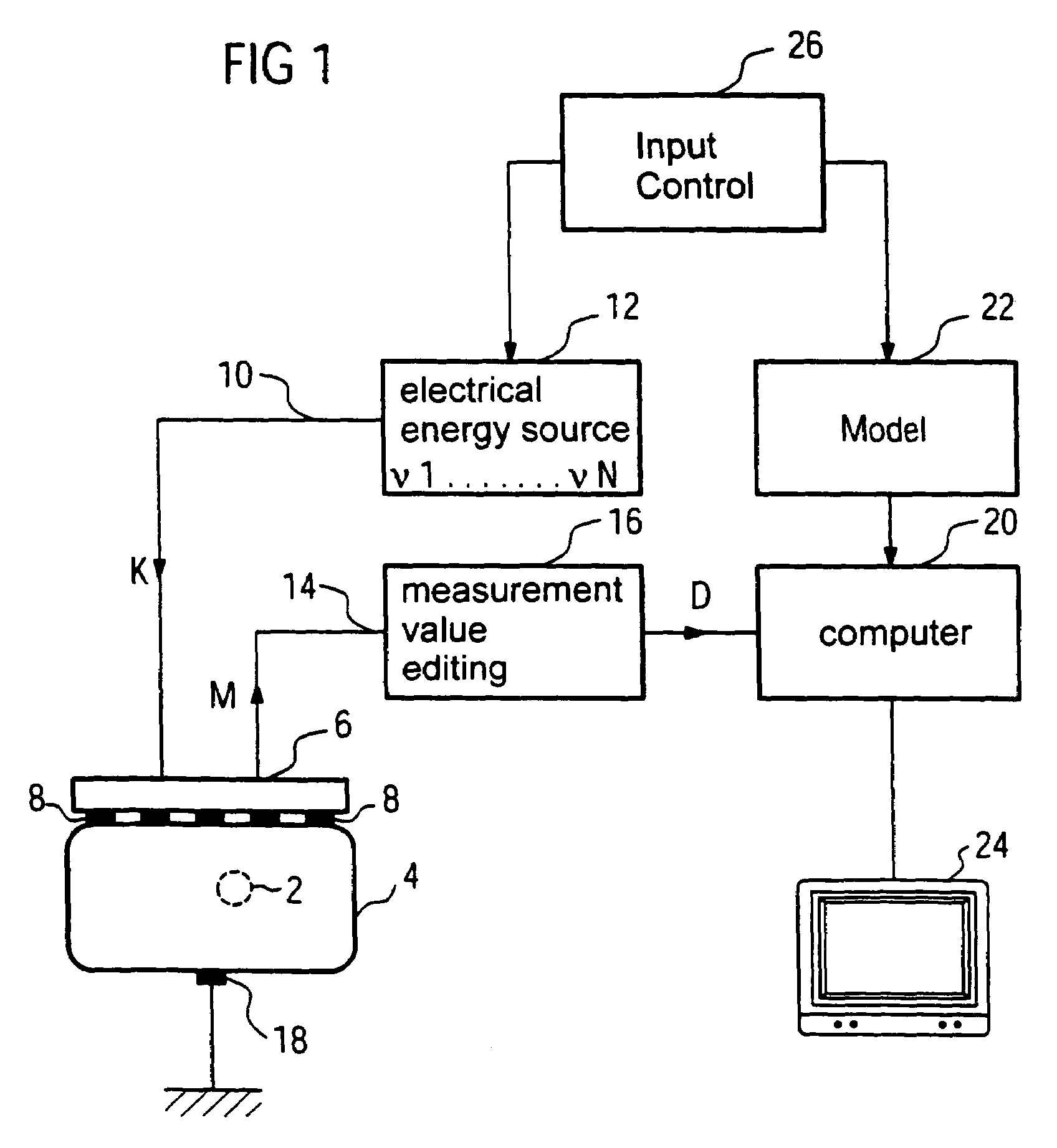
[0016]The overview representation in FIG. 1 shows the basic design of a measurement and evaluation arrangement with which signal activities of a limited spatial area 2 can be localized and identified in a biological tissue section 4. It is thereby assumed that the spatial area 2 possesses an electrical conductivity different from the rest of the tissue section 4, and the rest of the tissue section 4 exhibits an essentially spatially constant electrical conductivity. These assumptions are fulfilled sufficiently well when the biological tissue section 4 is a female breast and the limited spatial area 2 is a tumor.

[0017]The measurement arrangement includes an applicator 6 with a number of spatially distributed, arranged electrodes 8 that are contacted with the surface of the tissue section 4. In FIG. 1, for clarity only five electrodes 8 are shown, however, for a sufficiently precise localization, for example M=256 electrodes 8 should be arranged on a surface of 9×9 cm2.

[0018]The elect...


PUM


Abstract

An apparatus for localizing a focal lesion in a biological tissue section applies electrical excitation signals to the tissue section and measures electrical response signals at a number of measurement locations on a surface of the tissue section that arise there due to the excitation signals. A computer reconstructs a distribution of electrical dipole moments from the response signals, this distribution of dipole moments overall best reproducing the response signals, and supplies the 3D spatial position of the distribution as an output.

Description

BACKGROUND OF THE INVENTION[0001]1. Field of the Invention[0002]The invention concerns an apparatus for localizing a focal lesion in a biological tissue section, the lesion exhibiting an electrical property different from the tissue section, and the electrical property in the tissue section being essentially constant, of the type having means to apply electrical excitation signals to the tissue section and with means to measure electrical response signals at a number of measurement locations on a surface of the tissue section that arise due to the excitation signals.[0003]2. Description of the Prior Art[0004]In imaging by means of electrical impedance, alternating voltages are applied to a tissue section to be examined at one or more locations, and / or electrical alternating currents are applied. Using measurement electrodes that are electrically contacted to the tissue section to be examined at a number of locations, currents (amplitude and phase) are measured that arise due to the ...

Claims


Application Information

Patent Timeline
Patent Type & Authority Patents(United States)
IPC IPC(8): A61B5/06A61B5/04A61B5/053
CPCA61B5/0536
Inventor SCHOLZ, BERNHARD
Owner SIEMENS HEALTHCARE GMBH
Why Eureka
Try Eureka