Vacuum-actuated tissue perforation device for establishing pneumoperitoneum

a tissue perforation device and peritoneum technology, applied in the field of tissue perforation device and method, can solve the problems of significant morbidity and mortality, inexperienced surgeons' injuries are more common, and injuries still occur

Active Publication Date: 2009-09-08
AESCULAP AG
View PDF28 Cites 45 Cited by
  • Summary
  • Abstract
  • Description
  • Claims
  • Application Information

AI Technical Summary

Problems solved by technology

Significant morbidity and mortality occurs each year by iatrogenic injuries during establishment of pneumoperitoneum prior to laparoscopic surgical procedures.
These injuries are more common with inexperienced surgeons, but can occur even in the most experienced hands.
Built-in safety devices exist in the perforation devices themselves, but injuries still occur because of the close proximity of the structures that are intended to be perforated and those to be avoided.

Method used


Image

  • Vacuum-actuated tissue perforation device for establishing pneumoperitoneum
  • Vacuum-actuated tissue perforation device for establishing pneumoperitoneum
  • Vacuum-actuated tissue perforation device for establishing pneumoperitoneum

Examples


example

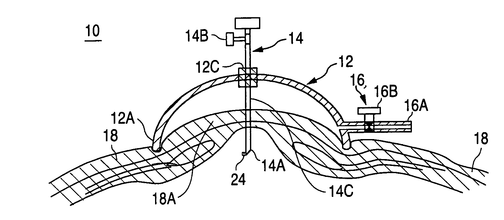
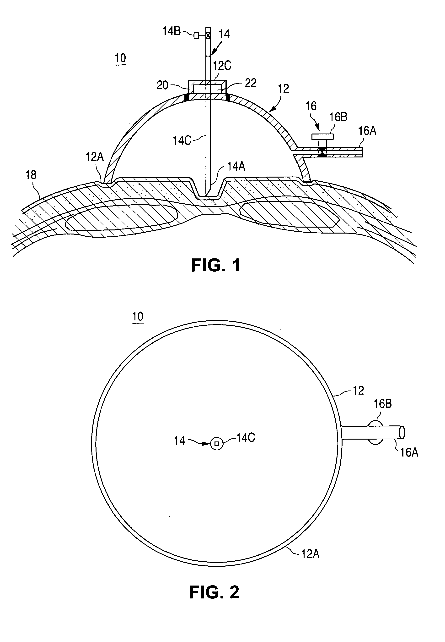
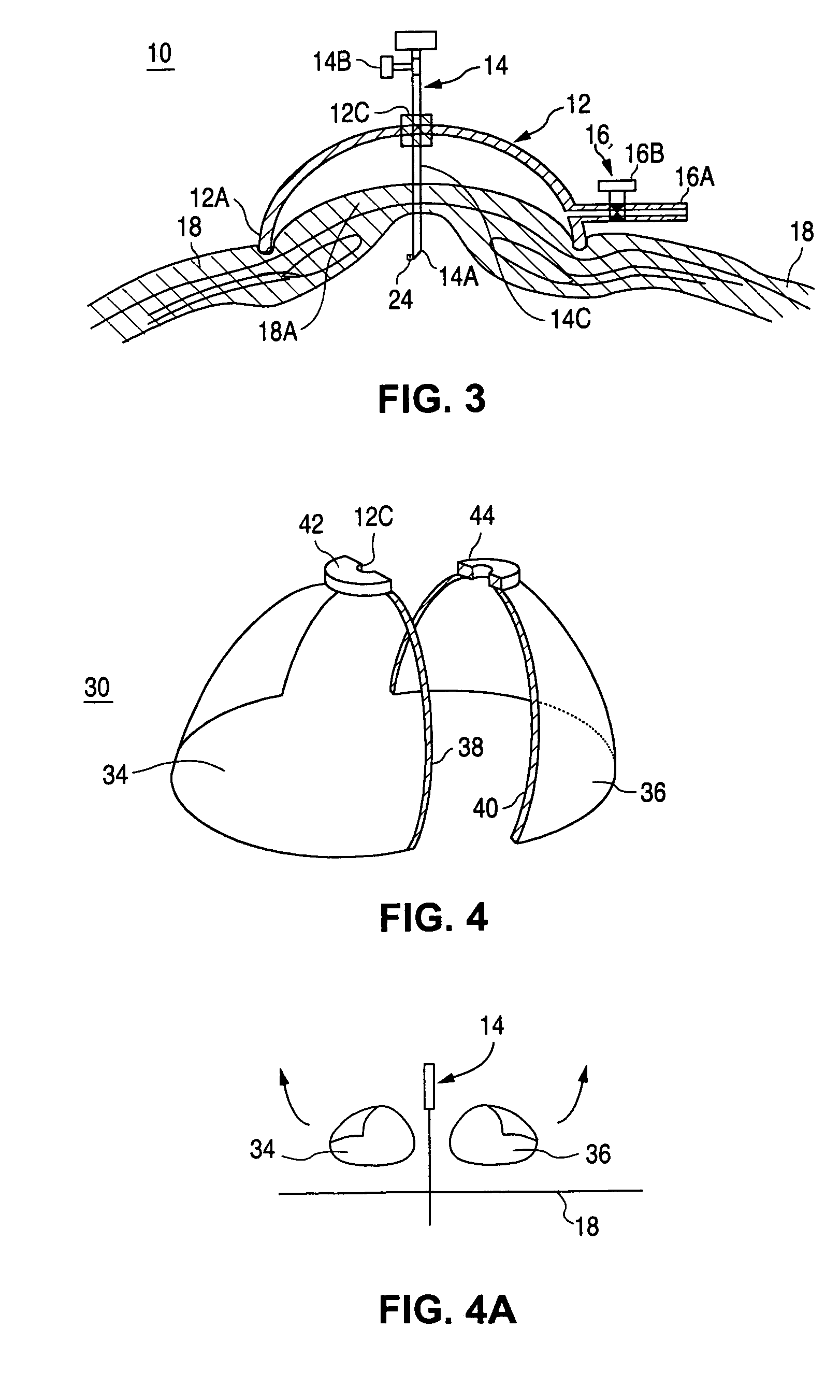
[0066]This example illustrates one embodiment of the present invention shown in FIGS. 1 and 3 in which housing (12) was a TUPPERWARE® bowl having O-ring (22) inserted in the center of the bottom of the bowl. The example is for illustrative purposes only and is not meant to limit the scope of the claims in any way.

[0067]In experimentation with a post mortem female pig, a standard hospital 300 mm Hg vacuum source was used to elevate the external abdominal surface 2-4 inches, depending on the amount of vacuum applied by vacuum system (16). The stationary Veress needle (14C) penetrated the pig's abdomen without difficulty and water was observed to drain upon penetration. CO2 was attached and the abdomen insufflated to 12 mm Hg. Following insufflation, a 10 mm Ethicon Endo-Surgery trocar was installed at the midline 1″ caudal to the xiphoid and a 30° Stortz camera (24) was inserted to verify uninjured tissue below the needle entry site, and to observe the inside abdominal wall. The exper...


PUM


Abstract

A tissue perforation device and method. The device preferably includes a housing having a housing pass-through, a penetrator securely and sealably positioned so that the penetrator device passes through the housing pass-through, and a vacuum system comprising a vacuum source securely and sealably attached through the housing for advancing a patient's tissue onto the penetrator device.

Description

BACKGROUND OF THE INVENTION[0001]1. Field of the Invention[0002]The present invention relates to a tissue perforation device and method. More particularly, the present invention relates to a device, which draws skin and underlying tissue onto a perforation device and away from vulnerable underlying structures.[0003]2. Background of the Invention[0004]Significant morbidity and mortality occurs each year by iatrogenic injuries during establishment of pneumoperitoneum prior to laparoscopic surgical procedures. The main source of these injuries is inadvertent perforation of blood vessels or organ structures within the abdominal cavity when the penetration device (needle, trocar or punch biopsy cutting tool) is advanced too far through the abdominal wall piercing the underlying organs that are located adjacent thereto. These injuries are more common with inexperienced surgeons, but can occur even in the most experienced hands. Built-in safety devices exist in the perforation devices them...

Claims


Application Information

Patent Timeline
Patent Type & Authority Patents(United States)
IPC IPC(8): A61B10/00A61B17/14A61B17/32A61B17/34A61M37/00A61M5/00A61M5/32A61B17/30
CPCA61B17/3403A61B2017/3492A61B2017/306A61B17/3494A61B2017/00561
Inventor NEZHAT, CAMRANMOHR, CATHERINE
Owner AESCULAP AG
Why Eureka
Try Eureka