System and method for enhancing an object of interest in noisy medical images

a noisy, medical image technology, applied in the field of real-time medical viewing systems, can solve the problems of false alarms and detection failures, and the decision about the best alarm is taken by the alarm detection means, so as to minimize matching errors, minimize matching errors, and be more robust to errors

Inactive Publication Date: 2009-11-17
KONINKLIJKE PHILIPS ELECTRONICS NV
View PDF9 Cites 7 Cited by
  • Summary
  • Abstract
  • Description
  • Claims
  • Application Information

AI Technical Summary

Benefits of technology

[0024]An advantage of said temporal tracking means is that they accumulate temporal proofs in favor of an alarm found at time t. Best alarm selection is done by using spatial and temporal criteria and therefore becomes more robust to errors.
[0025]In a preferred embodiment of the invention, said temporal tracking means proceed in an “alarm-centric” way, i.e. said track creation means associate with an alarm at time t that track at time t−1 that minimizes a matching error. In other words, said track creation means start from an alarm at time t and search for the best track ending at time t−1 to be matched with said alarm. This search is thus made in the opposite direction compared with the track formation. Possible associations between said alarm and tracks a time t−1 are evaluated by calculation of matching errors. Only the association that minimizes the matching error for said alarm is able to compete with other associations coming from other alarms to be considered as the best track at time t according to merit criteria. A consequence is to favor the alarms detected at time t to the detriment of the tracks ending at time t−1. In this way, all the alarms are certain to be associated with a track ending at time t−1, whereas some tracks ending at time t−1 may remain unassociated. This is another way of reducing the number of detection failures.

Problems solved by technology

However, unlike in the prior art, no decision about the best alarm is taken by the alarm detection means.
This is a method of compensating for the fact that, in the medical domain, objects of interest usually present a rather low contrast against a noisy background and that they cannot be detected with certainty most of the time, which induces both false alarms and detection failures.

Method used


Image

  • System and method for enhancing an object of interest in noisy medical images
  • System and method for enhancing an object of interest in noisy medical images
  • System and method for enhancing an object of interest in noisy medical images

Examples


Embodiment Construction

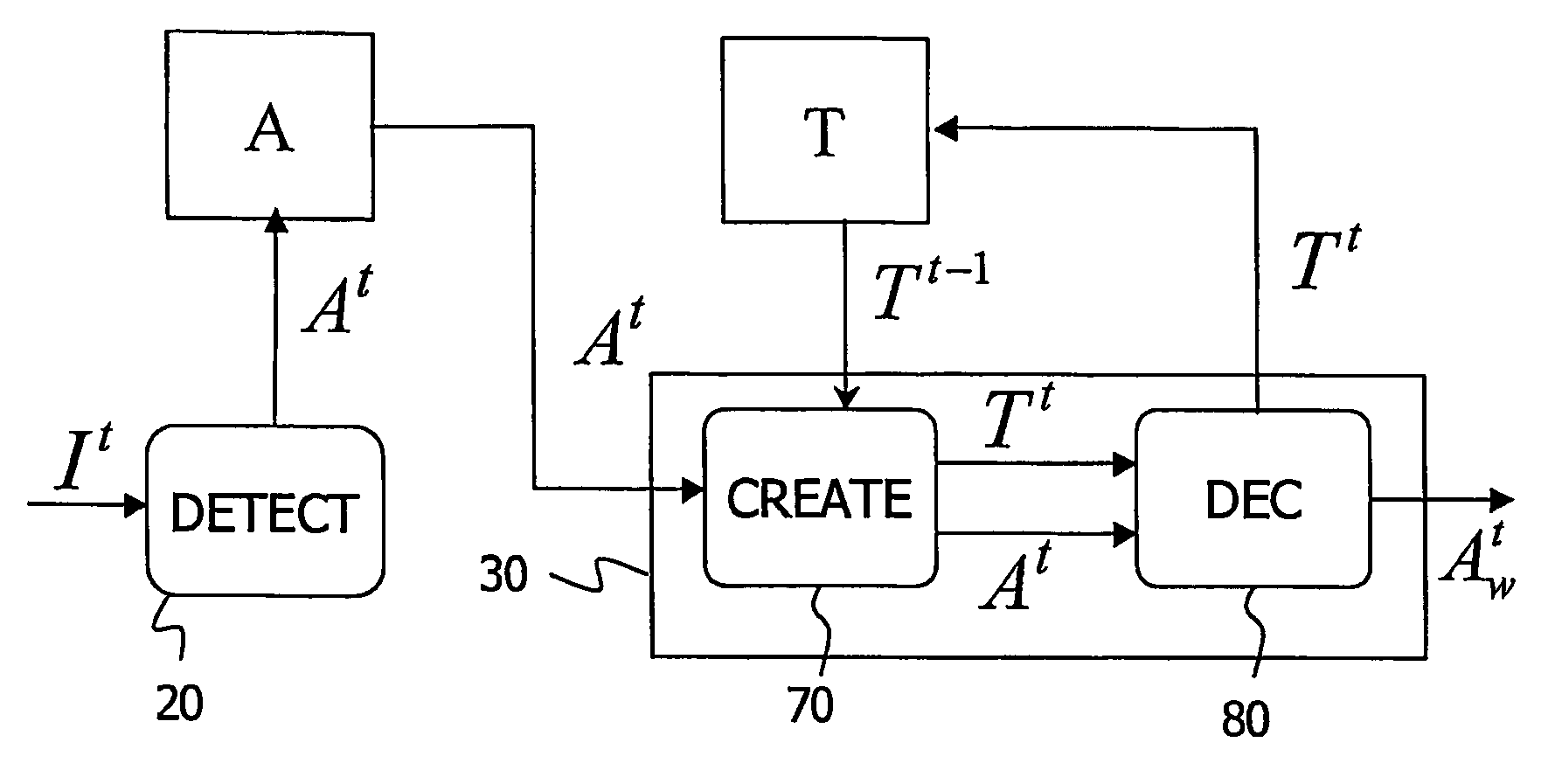
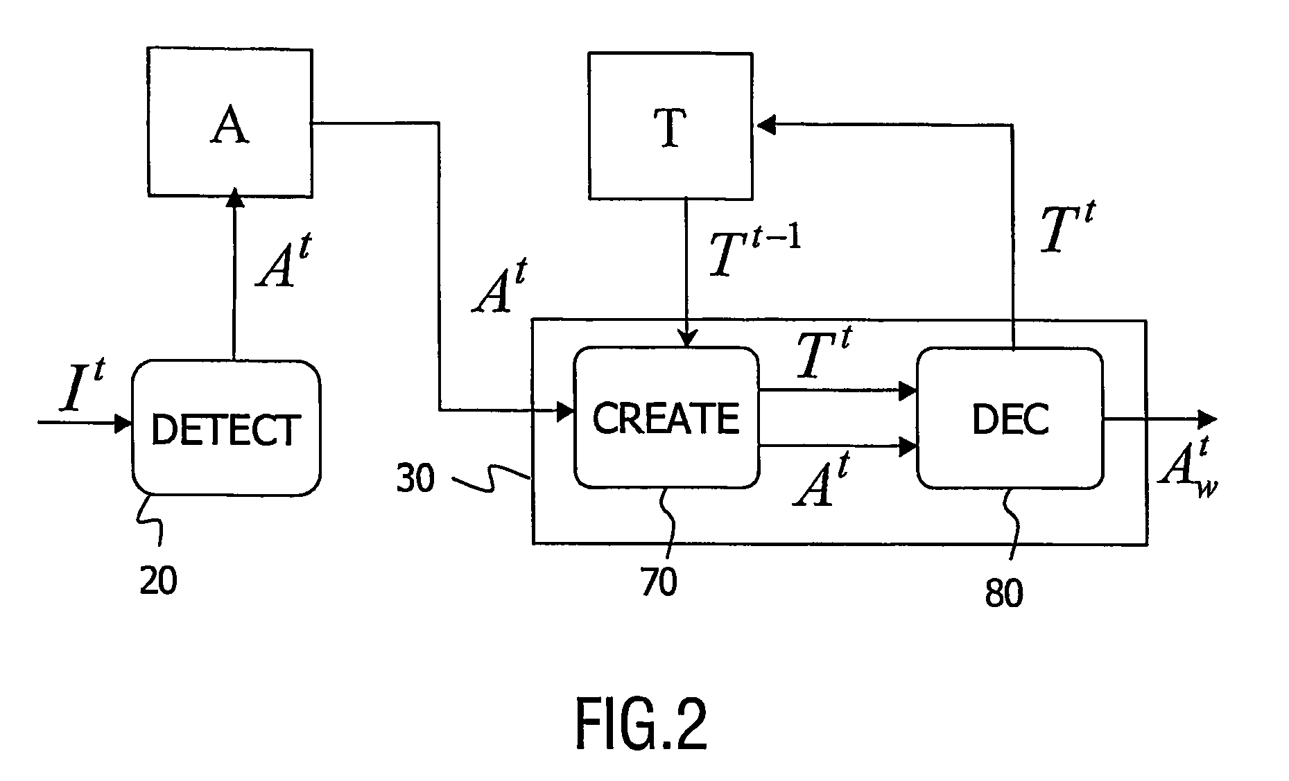
[0042]The invention relates to a viewing system and to a method that is used to actuate the viewing system for detecting an object of interest in real time in a sequence of medical images. The viewing system and the method of the invention are described hereinafter as a example in an application to the medical field of cardiology. In said application, the object of interest is an organ such as an artery or a tool such as a balloon or a stent. These objects are observed during a medical intervention called angioplasty or Percutaneous Transluminal Coronary Angioplasty (PTCA), in a sequence of X-ray fluoroscopic images called angiogram.

[0043]It is to be noted that the system and method may be applied to any object of interest other than a stent, an artery or a catheter in images other than angiograms, for example to the tracking of a biopsy needle in a sequence of breast ultrasound images. In the following, the invention is described in the particular case of a single object of interes...


PUM


Abstract

The invention relates to a system and a method for detecting an object of interest in a sequence of medical images. Said viewing system comprises alarm detection means (20) for detecting a set of alarms of said object of interest in a medical image (I′) at time t, in which an alarms is a set of data describing a possible location of the object. It further comprise temporal tracking means (30) for iteratively creating a set of tracks (T′) at time t by associating alarms of said set of alarms (A′) with tracks (T′1) at time t−1 and for choosing a track from among said set of tracks at time t in accordance with a merit criterion. Said track at time t is ended by an alarm which corresponds to a detection of said object of interest in said medical image at time t. Said viewing system advantageously accumulates temporal proofs for confirming or infirming spatial detections of alarms at time t. Detection of the object of interest is therefore made more robust to errors.

Description

FIELD OF THE INVENTION[0001]The invention relates to a real time medical viewing system for detecting an object of interest in a sequence of medical images. The invention also relates to an image processing method to be used in said system. The invention further relates to a medical examination apparatus coupled to such a system.[0002]The invention finds its application for example in the detection of catheters or stents in a sequence of angiographic images.DOMAIN OF THE INVENTION[0003]Interventional radiology procedures are widely used for the detection and treatment of many diseases and injuries. Catheter-based medical procedures are commonplace and include such medical treatments as balloon angioplasty, stent placement, and laser ablation. In such medical procedures the progress of the catheter or of any object of interest is typically monitored, within a patient's body, by an X-ray fluoroscopic imaging system.[0004]A method for detecting an object of interest like a catheter in ...

Claims


Application Information

Patent Timeline
Patent Type & Authority Patents(United States)
IPC IPC(8): G06K9/00A61B5/06A61B6/12A61B8/08
CPCA61B6/547A61B6/12A61B5/06A61B8/0833
Inventor FLORENT, RAOULNOSJEAN, LUCILELELONG, PIERRE
Owner KONINKLIJKE PHILIPS ELECTRONICS NV
Why Eureka
Try Eureka