Eureka AIR delivers breakthrough ideas for toughest innovation challenges, trusted by R&D personnel around the world.

Magnetic resonance imaging needing a long waiting time between pre-pulses and imaging pulse train

a technology of magnetic resonance imaging and waiting time, applied in the field of magnetic resonance imaging, can solve the problems of deteriorating image quality, difficult to acquire a series of images, and difficult to acquire images in the systole near an r-wave on time, so as to improve the image quality of an mr image, reduce the influence of artifacts, and improve the effect of image quality

Inactive Publication Date: 2009-11-24
TOSHIBA MEDICAL SYST CORP
View PDF8 Cites 11 Cited by
  • Summary
  • Abstract
  • Description
  • Claims
  • Application Information

AI Technical Summary

Benefits of technology

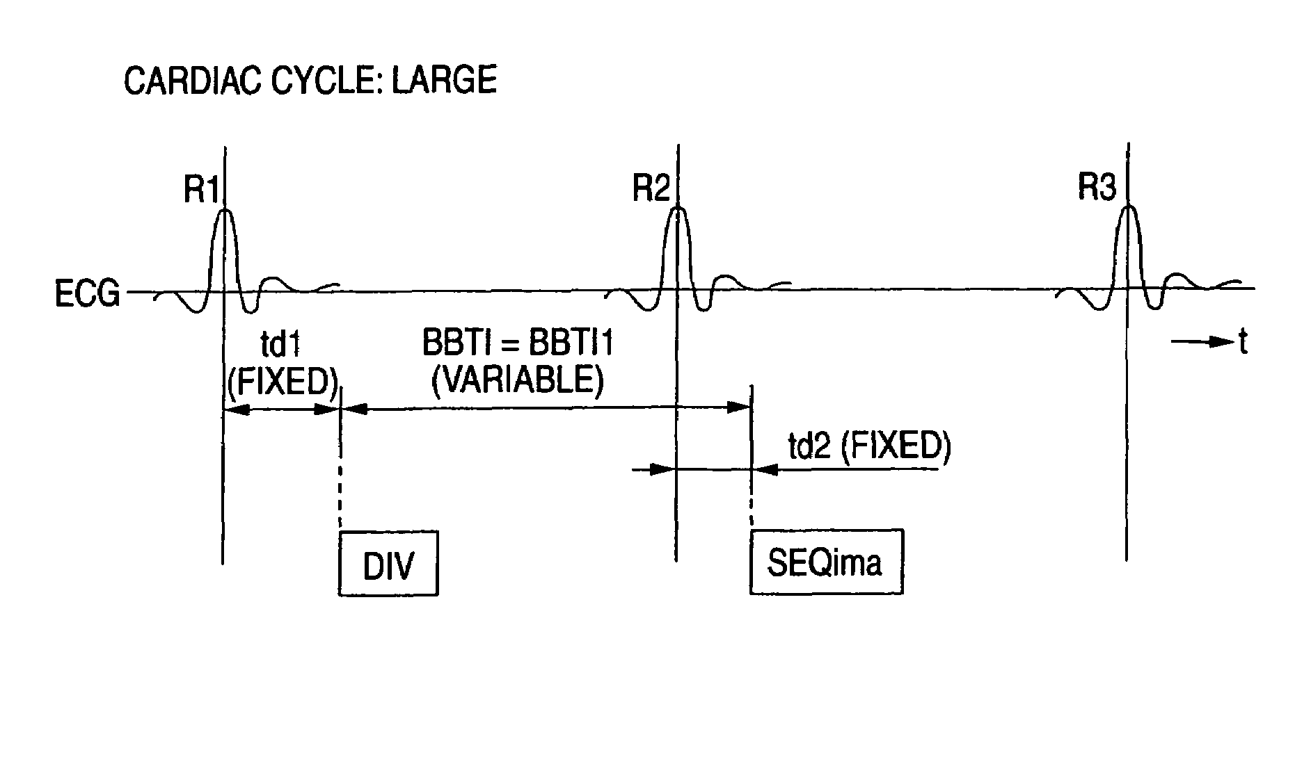
[0024]According to the invention, synchronization can be applied to a pre-pulse and an imaging pulse train that form a pulse sequence in the black blood method or the like respectively with different, specific waveforms (R-waves or the like) of a signal (ECG signal or the like) representing cardiac temporal phases of a subject to be imaged with different delay times. It is thus possible to maintain a regular application timing of an imaging pulse train for acquiring echo signals always even in the presence of a variance of the cardiac cycle. Consequently, in MR imaging using a pulse sequence in which a waiting time between the pre-pulse and the imaging pulse train is long in comparison with the cardiac cycle, images in the systole of the cardiac cycle can be captured in a reliable manner even in the presence of a cycle-to-cycle variance of the cardiac cycle. It is thus possible to improve the image quality of an MR image by reducing the artifacts.
[0025]It should be noted that, in the imaging through the black blood method according to the magnetic resonance imaging of the invention, a time BBTI from the application of the pre-pulse to the application of the imaging pulse train is relatively long in comparison with one cardiac cycle, and a difference of the suppressing effect of a blood signal due to a variance of the BBTI within a range from approximately 400 to 700 ms is so small that the influence on the image quality is almost negligible.

Problems solved by technology

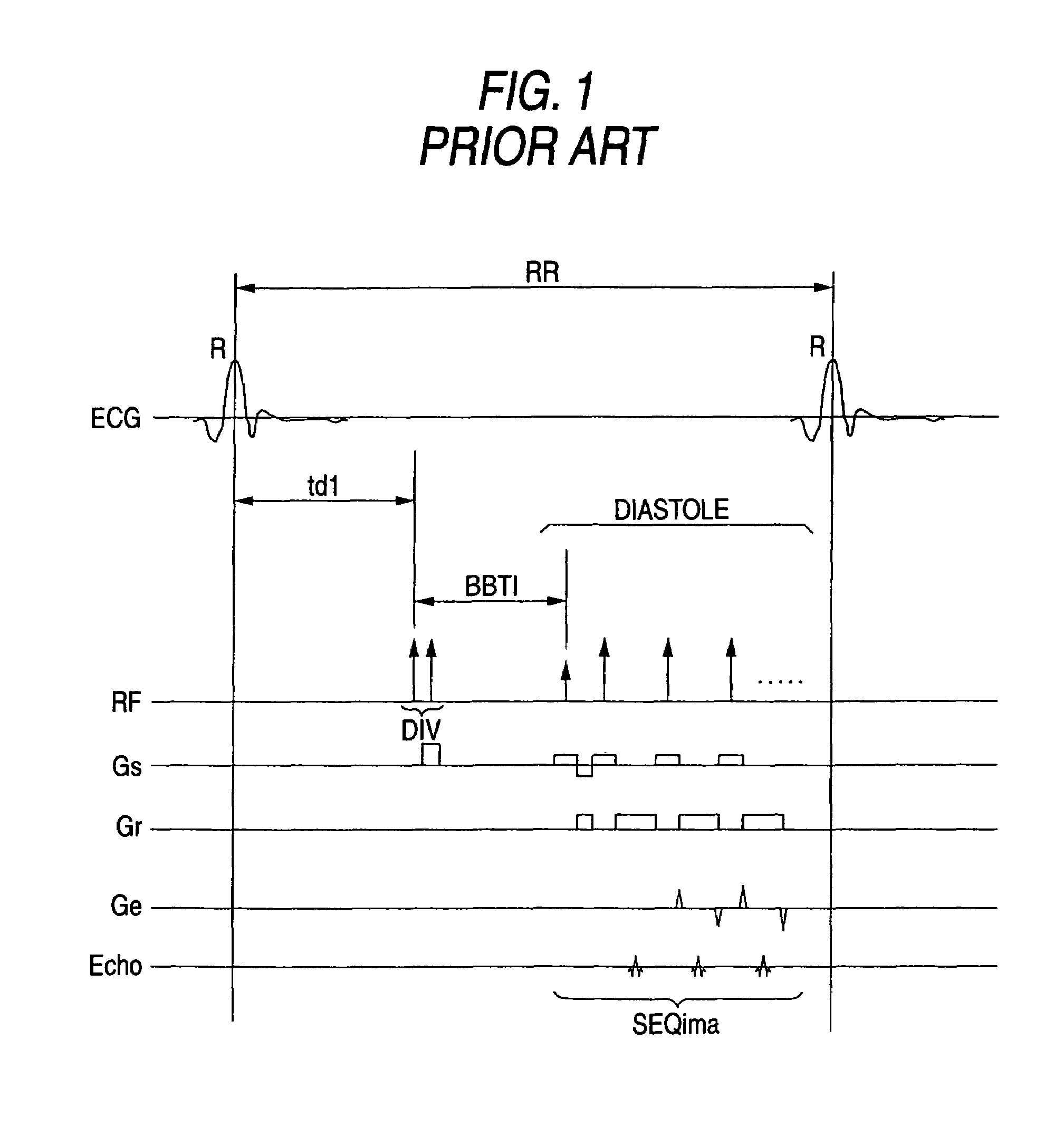
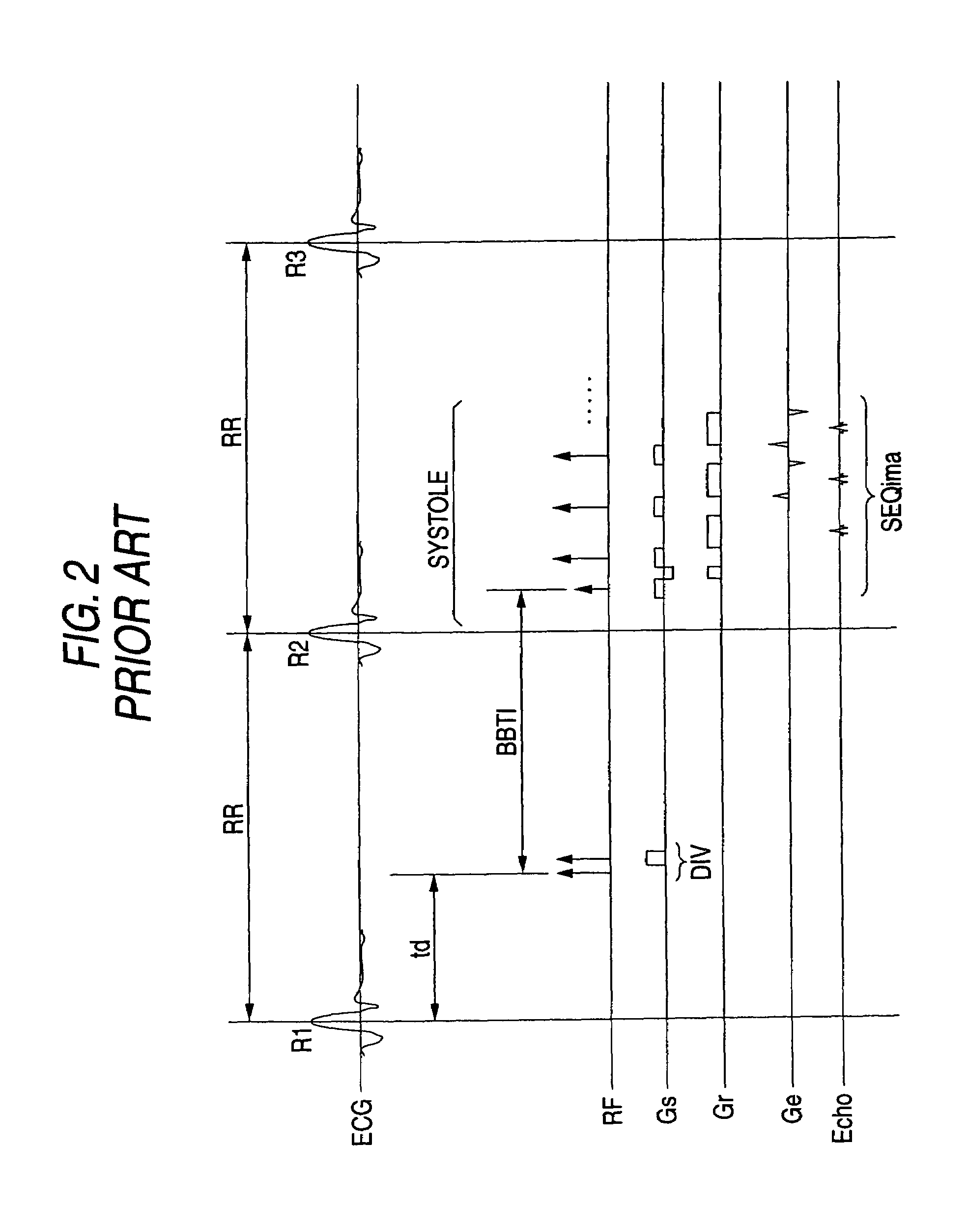
However, when the conventional black blood method is used, there arise problems that images in the systole near an R-wave on time are difficult to acquire, and it is difficult to acquire a series of images having latencies that vary at regular intervals as with a cine mode display.
Hence, even when images are acquired after a certain time “td+BBTI” from an R-wave in the last or earlier cardiac cycle, the position of the cardiac muscle at the time instance of the echo acquisition sways in each shot, which deteriorates the image quality so badly that it is impossible to obtain an image that can be used for a diagnosis. FIG. 3A and FIG. 3B are views schematically showing FIG. 2 divided by two conditions of a long cardiac cycle (FIG. 3A) and a short cardiac cycle (FIG. 3B).

Method used


Image

  • Magnetic resonance imaging needing a long waiting time between pre-pulses and imaging pulse train
  • Magnetic resonance imaging needing a long waiting time between pre-pulses and imaging pulse train
  • Magnetic resonance imaging needing a long waiting time between pre-pulses and imaging pulse train

Examples


Embodiment Construction

[0036]The following description will describe one embodiment of the invention with reference to FIG. 4 through FIG. 6, FIG. 7A, and FIG. 7B.

[0037]FIG. 4 schematically shows an arrangement of a magnetic resonance imaging apparatus of this embodiment.

[0038]The magnetic resonance imaging apparatus includes a patient couch portion on which a subject P to be imaged (a person to be imaged) lies down, a static magnetic field generating portion for generating a static magnetic field, a gradient magnetic field generating portion for appending position information to the static magnetic field, a sending / receiving portion for sending and receiving high-frequency signals, a control and operational portion responsible for the control of an overall system and image reconstruction, an electrocardiogram measuring portion for measuring an ECG (electrocardiogram) signal as a signal representing cardiac temporal phases of the subject P to be imaged, and a breath-hold command portion for directing the ...


PUM


Abstract

In the black blood method using a double inversion pulse, images in the systole of the cardiac cycle can be captured in a reliable manner even in the presence of a cycle-to-cycle variance in the heart beat cycle. A pulse sequence of the black blood method composed of a double inversion pulse DIV and an imaging pulse train SEQima is used. This sequence is applied in sync with an ECG signal of a subject to be imaged, and magnetic resonance imaging is thereby performed. The double inversion DIV is applied in sync with an R-wave:R1 appearing on the ECG signal at a given timing, with a first delay time td1 (fixed value), and the imaging pulse train SEQima is applied in sync with the following R-wave:R2 with a second delay time td2 (fixed value: set in accordance with the systole). A variance of the cardiac cycle is absorbed in an inversion time BBTI.

Description

TECHNICAL FIELD[0001]The present invention relates to magnetic resonance imaging (MRI) for imaging the internal of a subject to be imaged with an imaging sequence containing a pre-pulse, and more particularly to MR imaging using an imaging sequence in which a waiting time until the application of an imaging pulse train is started after a pre-pulse was applied is relatively long in comparison with a cardiac cycle as with the black blood method or the like.BACKGROUND ART[0002]Magnetic resonance imaging is presently used in many cases as one of imaging methods for medical use. Magnetic resonance imaging is an imaging method that gives rise to magnetic excitation of nuclear spins in a subject to be imaged positioned in a static magnetic field with a high frequency signal at the Larmor frequency and then reconstructs an image of the internal of the subject to be imaged using an MR signal induced in association with the excitation. Magnetic resonance imaging includes various types, and th...

Claims


Application Information

Patent Timeline
IPC IPC(8): A61B5/05A61B5/055G01R33/561G01R33/563G01R33/567
CPCA61B5/055A61B5/7285G01R33/563G01R33/567G01R33/56308
Inventor KANAZAWA, HITOSHI
Owner TOSHIBA MEDICAL SYST CORP
Why Eureka
Eureka Blog
Learn More