
Conduit harvesting instrument and method

Inactive Publication Date: 2010-01-12
TYCO HEALTHCARE GRP LP
View PDF154 Cites 47 Cited by
  • Summary
  • Abstract
  • Description
  • Claims
  • Application Information

AI Technical Summary

Benefits of technology

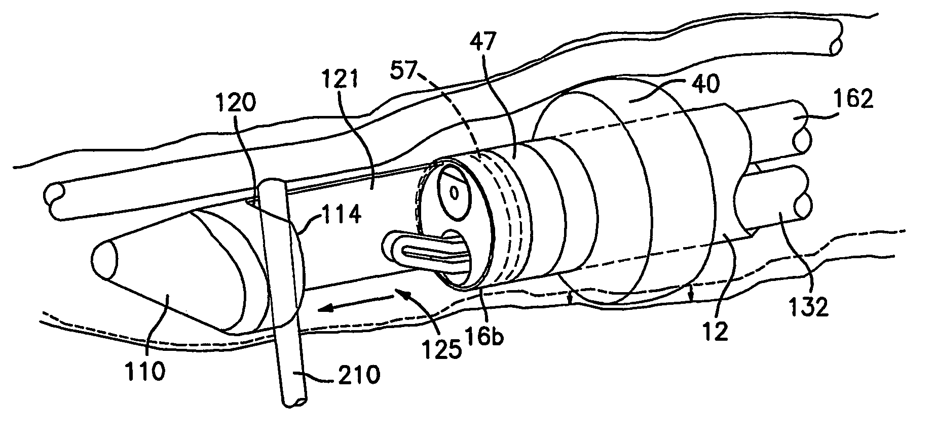
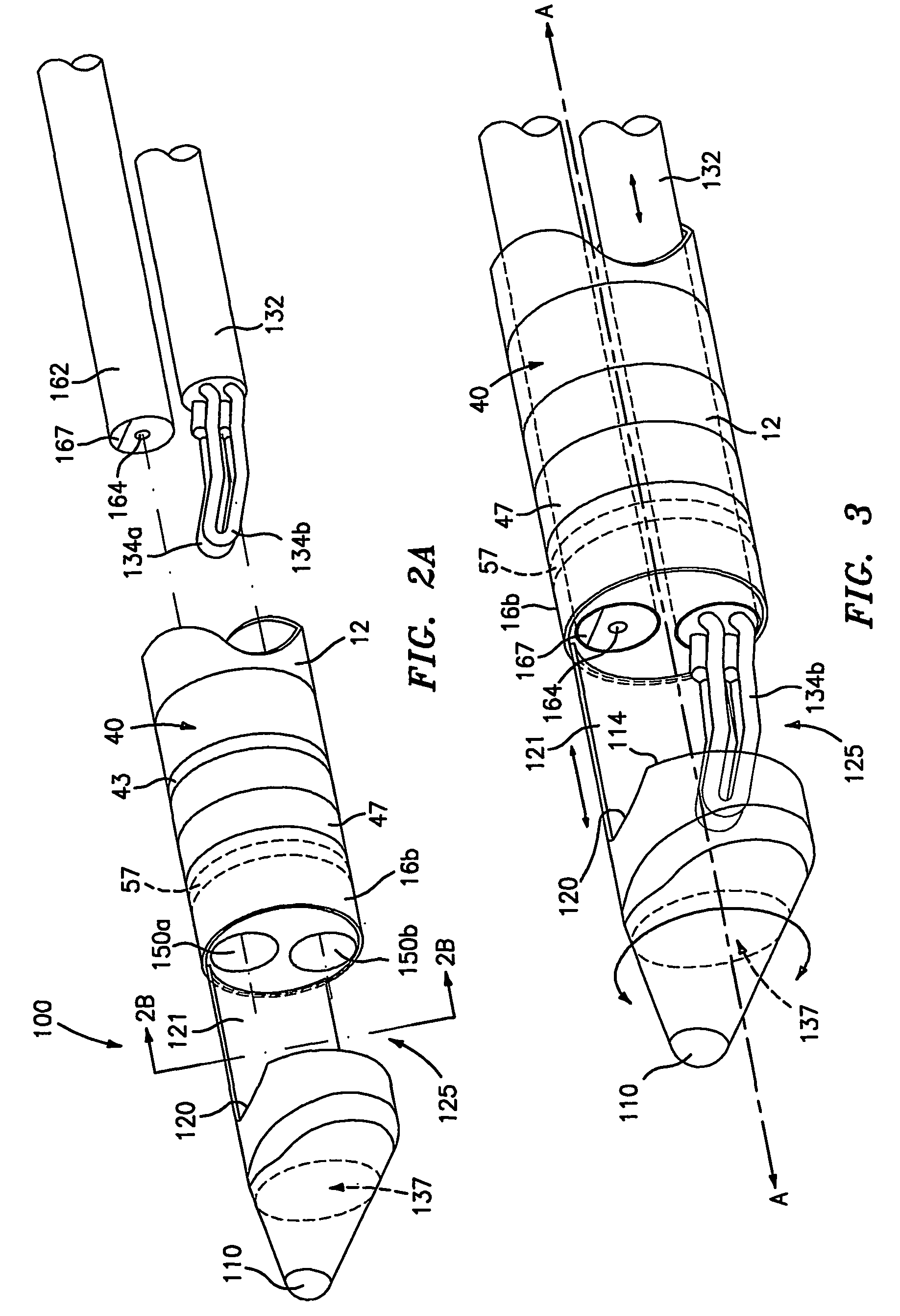
[0019]Preferably, the dissecting portion is transparent and / or conical in dimension to facilitate blunt dissection of surrounding tissue from the vessel. In one embodiment, the tip is extendable along a longitudinal axis defined through the shaft to expose the cradle section. Advantageously, the cradle section includes a notched portion to facilitate manipulation, orientation and positioning and securement of a vessel tributary and to facilitate its ligation and / or separation from the main conduit vessel. The tip and / or the cradle section is preferably rotatable about the endoscope to assist in the orientation of the cradle section for and during manipulation and separation of the vessel tributaries 360° about the vessel. The tip and / or the cradle section can also be selectively rotatable about the axis of the shaft
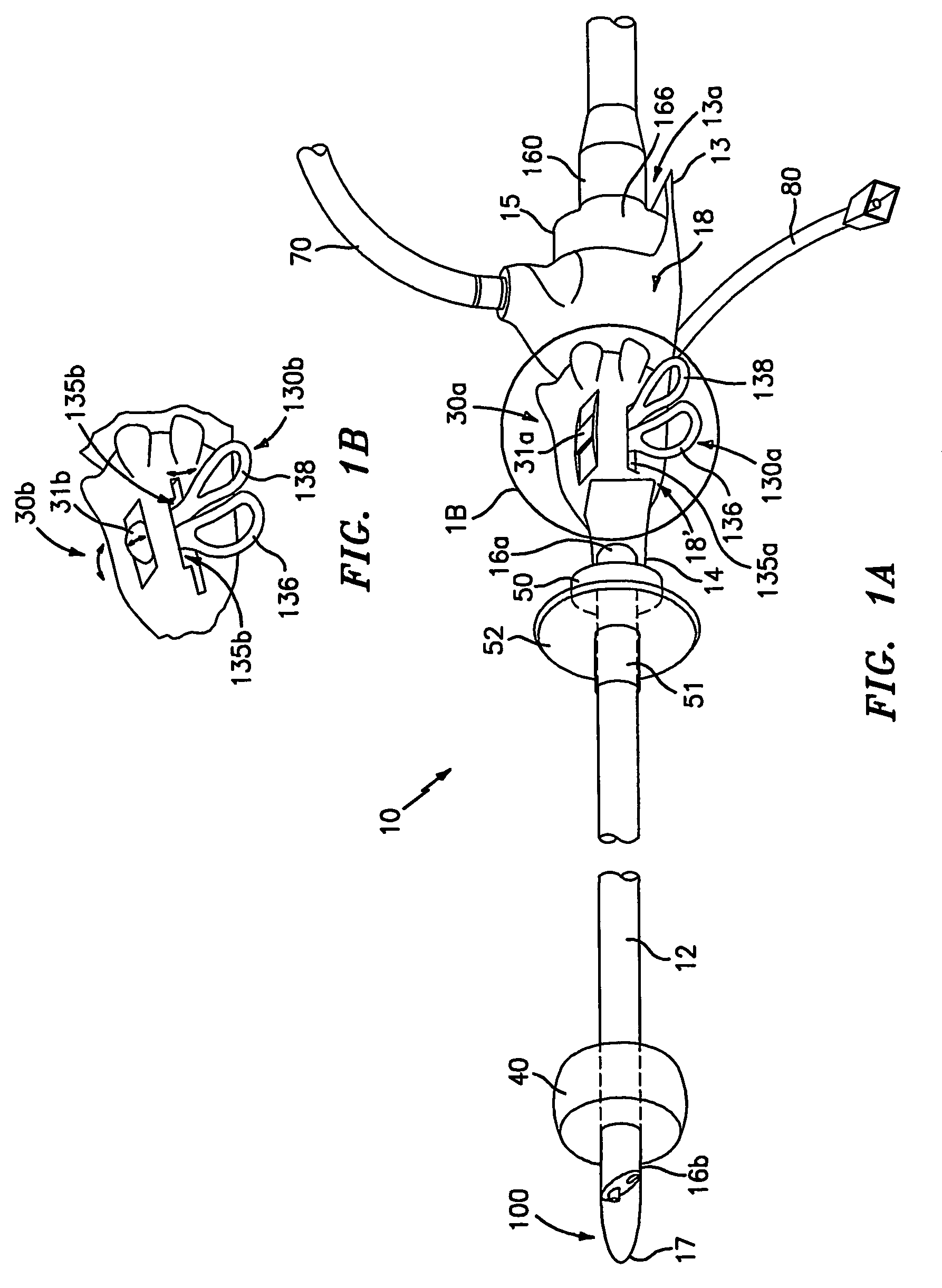
[0020]Additional instruments which can be disposed through one or more of the remaining lumens in the shaft can be selected from the group consisting of: ligating instruments, bipolar shears, ultrasonic shears, clip appliers, coagulating instruments, cutting instruments, vessel sealing instruments, vessel graspers, irrigation instruments, insufflators, suction instruments and combinations of the same. It is envisioned that the additional instruments may be selectively extendable, retractable and / or rotatable relative to the instrument, shaft or endoscope to facilitate operation thereof.
[0022]In another embodiment, the shaft, preferably a distal portion thereof, includes a balloon disposed about the outer periphery thereof and which is selectively inflatable and / or deflatable. The balloon allows the operator to grossly dissect surrounding tissue away from the vessel and create and / or maintain a working space between the vessel and the tissue. The working space may be insufflated as needed during the harvesting procedure to facilitate visualization and removal of the vessel.
[0023]Another embodiment of the present disclosure is a surgical instrument for dissecting a vessel from surrounding tissue which includes a housing and an elongated shaft preferably attached to the housing. The shaft includes a plurality of lumens disposed at least partially therethrough. A blunt tip is disposed at a distal end of the shaft and is selectively movable by an actuator mounted to the housing. The actuator allows the operator to extend the tip from a first dissecting position wherein the tip is positioned proximate the distal end of the shaft (i.e., positioned to separate surrounding tissue from the vessel) to at least one additional position distally further from the distal end of the shaft to expose a cradle section. An endoscope is disposed in at least one of the lumens for visualization purposes and one or more additional surgical instrument(s) (preferably selected from the list mentioned above) is disposed in one or more of the remaining lumens.
[0030]The method can also include the steps of: inserting the instrument through an incision in the body; advancing the instrument through the incision and along the vessel utilizing the endoscope to view and / or the blunt tip to dissect surrounding tissue from the vessel; selectively extending the blunt tip to expose the cradle section and position a vessel tributary; sealing and separating a portion of the tributary from the vessel; repeating the advancing, extending and treating steps as needed to clear surrounding tissue from the vessel and / or seal or separate additional vessel tributaries; and removing the vessel from the body. Preferably, the advancing step is effected with the blunt tip retracted to reduce exposure of the cradle section. After the extending step, the method can further include the step of: rotating the cradle section to position tributaries for treatment.

Problems solved by technology

Coronary bypass surgery is required when these methods of treatment cannot be used or have failed to clear the blocked artery.
As can be appreciated, harvesting these conduits from the body often requires enormous skill and precision due to the delicate nature of the tissue structure.
As can be appreciated, ligating and separating these tributaries from the vein requires a high degree of skill and accuracy and is typically a very tedious procedure.
As can be appreciated from the above descriptions, the harvesting of vessels can be very traumatic and is often the most troublesome part of the bypass operation.
Moreover, the incisions, especially the long ones, created in the leg or arm to harvest the vessel tend to heal slowly and are often very painful.
As can be appreciated, this simply adds to the overall complexity of the operation since it requires the repeated exchange of surgical instruments through the working lumen to perform the different tasks associated with blunt dissection and removal of the vessel tributaries.

Method used


Image

  • Conduit harvesting instrument and method
  • Conduit harvesting instrument and method
  • Conduit harvesting instrument and method

Examples


Embodiment Construction

[0070]Referring now to FIGS. 1A-4, there is disclosed one embodiment of an endoscopic vessel harvesting instrument 10 that can be employed for harvesting vessels 200 (FIG. 5) for use, e.g., in bypass procedures, in particular, coronary bypass procedures. Harvesting instrument 10 includes a proximal end 15, a distal end 17 and an elongated shaft 12 disposed therebetween, but not necessarily in direct communication with such ends 15 and 17. For the purposes herein, it is understood that FIG. 1A discloses one embodiment of the presently disclosed instrument. As described in more detail below, it is envisioned that the instrument may include a base housing which is integral with or attachable to a shaft housing and a shaft which is integral with or attachable to the shaft housing. In such instances, what is commonly termed the “proximal end” may change depending upon whether a single, integral, base housing is utilized or a combination base housing and shaft housing is utilized.

[0071]El...


PUM


Abstract

A surgical instrument for harvesting vessels from the body includes an elongated shaft (12) having distal and proximal ends and a plurality of lumens (150a, 150b) disposed therethrough. The shaft also includes a tip having a dissecting portion disposed at a distal end thereof and a cradle section (114). The tip is movable from a first position proximate the distal end of the shaft to at least one additional position distally further from the distal end of the shaft to expose the cradle section. The instrument also includes an endoscope (162) disposed in one of the plurality of lumens and at least one additional surgical instrument (132) disposed in one of the remaining lumens. Methods are disclosed for utilizing the surgical instrument.

Description

CROSS REFERENCE TO RELATED APPLICATION[0001]This application claims the benefit of and priority to U.S. Provisional Patent Application Ser. No. 60 / 301,059 entitled “CONDUIT HARVESTING INSTRUMENT AND METHOD” filed on Jun. 26, 2001 by Hanspeter R. Bayer, the entire contents of which is hereby incorporated by reference herein.BACKGROUND[0002]1. Field of the Disclosure[0003]This present disclosure relates to instruments and methods for performing minimally invasive, laparoscopic or endoscopic surgical procedures. More particularly, the present disclosure relates to instruments and methods that are especially suitable for procedures that require or benefit from minimally invasive access to anatomical conduits or vessels for harvesting the same. The instrument is suitable for harvesting vessels from surrounding tissue for use in bypass procedures including, but not limited to, coronary artery bypass grafting (CABG) or reverse or in-situ femoral-popliteal or femoral-tibia peripheral bypass...

Claims


Application Information

Patent Timeline
IPC IPC(8): A61B17/22A61B17/00A61B17/02A61B17/12A61B17/125A61B17/32A61B18/00A61B18/04A61B18/14A61B19/00
CPCA61B17/00008A61B18/1445A61B18/1482A61B2090/306A61B2017/00969A61B2017/22051A61B2017/2945A61B2017/320048A61B2018/0022A61B2018/00232A61B2018/00404A61B2018/00601A61B2018/00982A61B2018/1432A61B2090/3614A61B17/32
Inventor BAYER, HANSPETER ROBERT
Owner TYCO HEALTHCARE GRP LP
Learn More