Computerized detection of breast cancer on digital tomosynthesis mammograms

a technology of digital tomography and breast cancer, applied in the field of breast cancer detection, can solve the problems of increasing reducing the detection accuracy of breast cancer, so as to improve the detection accuracy and increase the time required for interpretation.

Inactive Publication Date: 2010-01-12
RGT UNIV OF MICHIGAN
View PDF7 Cites 43 Cited by
  • Summary
  • Abstract
  • Description
  • Claims
  • Application Information

AI Technical Summary

Benefits of technology

[0008]Digital tomosynthesis mammography (DTM) is a promising new modality that has the potential to improve breast cancer detection, especially in dense breasts. However, the number of slices per breast may range from 30 to over 80, thus increasing the time required for interpretation. A computer-aided diagnosis (CAD) method and a system is disclosed that provides a second opinion to radiologists during their interpretation of DTMs. The disclosed CAD system uses a combination of computer vision techniques, including image enhancement, 3D gradient field analysis, 3D object segmentation, feature extraction, feature classification, to detect breast lesions on mammograms and to characterize it as malignant or benign. This is the first CAD system that uses 3D image processing techniques to detect and classify lesions in DTMs.

Problems solved by technology

However, mammographic sensitivity is often limited by the presence of overlapping dense fibroglandular tissue in the breast.
A drawback of conventional tomography is that each tomogram can only image one plane at a selected depth in relatively sharp focus.
If the exact depth of interest is not known in advance or the abnormality encompasses a range of depths, a tomogram at each depth will have to be acquired at separate imaging, thus costing additional dose and examination time.

Method used


Image

  • Computerized detection of breast cancer on digital tomosynthesis mammograms
  • Computerized detection of breast cancer on digital tomosynthesis mammograms
  • Computerized detection of breast cancer on digital tomosynthesis mammograms

Examples


Embodiment Construction

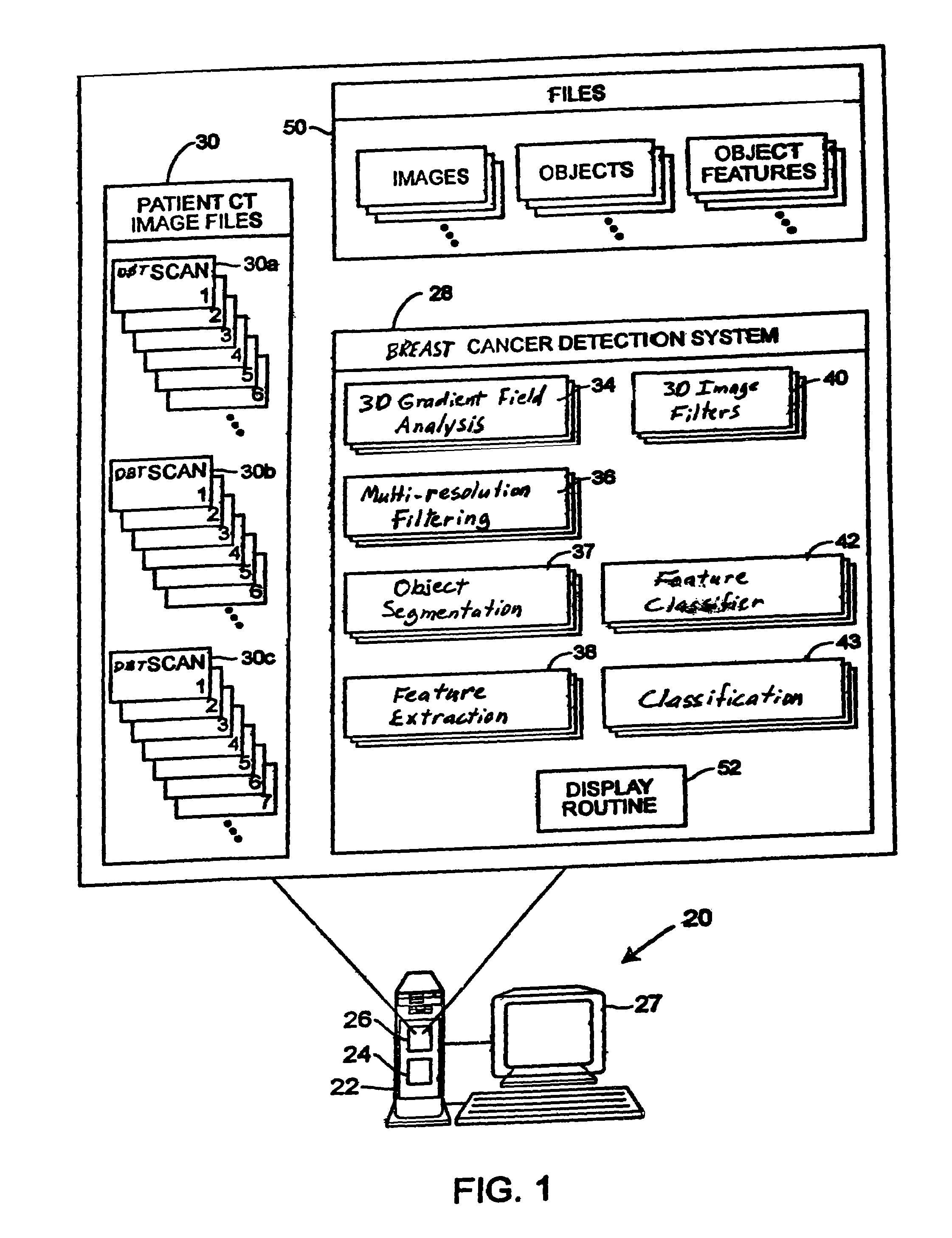
[0021]Referring to FIG. 1, a computer aided diagnosis (CAD) system 20 that may be used to detect and diagnose breast cancers includes a computer 22 having a processor 24 and a memory 26 therein and having a display screen 27 associated therewith. As illustrated in an expanded view of the memory 26, a breast cancer detection and diagnostic system 28 in the form of, for example, a program written in computer implementable instructions or code, is stored in the memory 26 and is adapted to be executed on the processor 24 to perform processing on one or more sets of digital tomosynthesis mammography (DTM) images 30, which may also be stored in the computer memory 26. The DTM images 30 may include DTM images for any number of patients and may be entered into or delivered to the system 20 using any desired importation technique. Generally speaking, any number of sets of images 30a, 30b, 30c, etc. (called image files) can be stored in the memory 26 wherein each of the image files 30a, 30b, ...


PUM


Abstract

A method for using computer-aided diagnosis (CAD) for digital tomosynthesis mammograms (DTM) including retrieving a DTM image file having a plurality of DTM image slices; applying a three-dimensional gradient field analysis to the DTM image file to detect lesion candidates; identifying a volume of interest and locating its center at a location of high gradient convergence; segmenting the volume of interest by a three dimensional region growing method; extracting one or more three dimensional object characteristics from the object corresponding to the volume of interest, the three dimensional object characteristics being one of a morphological feature, a gray level feature, or a texture feature; and invoking a classifier to determine if the object corresponding to the volume of interest is a breast cancer lesion or normal breast tissue.

Description

CROSS-REFERENCE TO RELATED PATENT APPLICATIONS[0001]This application claims benefit of U.S. Provisional Application Ser. No. 60 / 650,923, entitled “Computerized Detection Of Masses On digital Tomosynthesis Mammograms” filed Feb. 8, 2005, the disclosure of which is hereby incorporated by reference in its entirety for all purposes.STATEMENT REGARDING FEDERALLY SPONSORED RESEARCH OR DEVELOPMENT[0002]This invention was made with government support under Grant numbers CA091713 and CA095153 awarded by the National Institutes of Health (Department of Health and Human Services) and Grant number DAMD17-02-1-0214 awarded by the Army Medical Research and Materiel Command. The government has certain rights in the invention.FIELD OF TECHNOLOGY[0003]This relates generally to breast cancer detection and, more particularly, to a system and method for using computer-aided diagnosis (CAD) for Digital Tomosynthesis Mammograms (DTMs) for detection and characterization of breast lesions.DESCRIPTION OF TH...

Claims


Application Information

Patent Timeline
Patent Type & Authority Patents(United States)
IPC IPC(8): G06K9/00
CPCG06K9/00G06T7/0012G06T7/403G06K2209/05G06T2207/10081G06T2207/30068G06T7/44G06V2201/03G06F18/00
Inventor CHAN, HEANG-PINGWEI, JUNBERKMAN, SAHINERHADJIISKI, LUBOMIR M.
Owner RGT UNIV OF MICHIGAN
Why Eureka
Try Eureka