Gynecological ablation procedure and system

a pelvic tumor and ablation technology, applied in the field of gynecological disorders, can solve the problems of pelvic and/or abdominal pressure or pain, increased abdominal girth, irregular bleeding, etc., and achieve the effect of effective ablation of pelvic tumors, rapid recovery time and significant cost savings

Active Publication Date: 2010-03-16
ACESSA HEALTH INC +1
View PDF126 Cites 139 Cited by
  • Summary
  • Abstract
  • Description
  • Claims
  • Application Information

AI Technical Summary

Benefits of technology

[0008]The present invention, also referred to as “the Halt procedure,” is an innovative, outpatient procedure that utilizes electromagnetic energy to effectively ablate pelvic tumors. The invention employs an ablation device that uses radio-frequency (RF) energy to treat pelvic tumors, while sparing the surrounding normal tissue. Although the ablation device utilized in the present invention has FDA approval for ablation of soft tissue tumors, no known reports exist in the medical literature of the ablation device's application to uterine leiomyomata or other pelvic tumors. In addition, current results indicate that, compared to other conservative therapies, the present method is very effective. Thus far, the present invention has provided relief from most types of symptoms caused by uterine leiomyomata. Furthermore, the present invention is versatile, safe, and well-accepted by patients. Advantages of the present invention include a quick recovery time, typically no more than a week, and significant cost savings. More importantly, the present invention provides a practical and efficient way to achieve uterine conservation on an out-patient basis.

Problems solved by technology

Although uterine leiomyomata rarely (0.1%) progress to cancer, these tumors can cause excessive menstrual bleeding, irregular bleeding, pregnancy loss, infertility, urinary frequency and retention, increased abdominal girth, and pelvic and / or abdominal pressure or pain with sexual activity, menses, or daily activities.
These methods necessarily destroy normal overlying and surrounding tissue in order to treat the tumor.
As a result, the integrity of the uterus is compromised, and harmful scar tissue (e.g., adhesions) may occur.

Method used


Image

  • Gynecological ablation procedure and system
  • Gynecological ablation procedure and system
  • Gynecological ablation procedure and system

Examples


Embodiment Construction

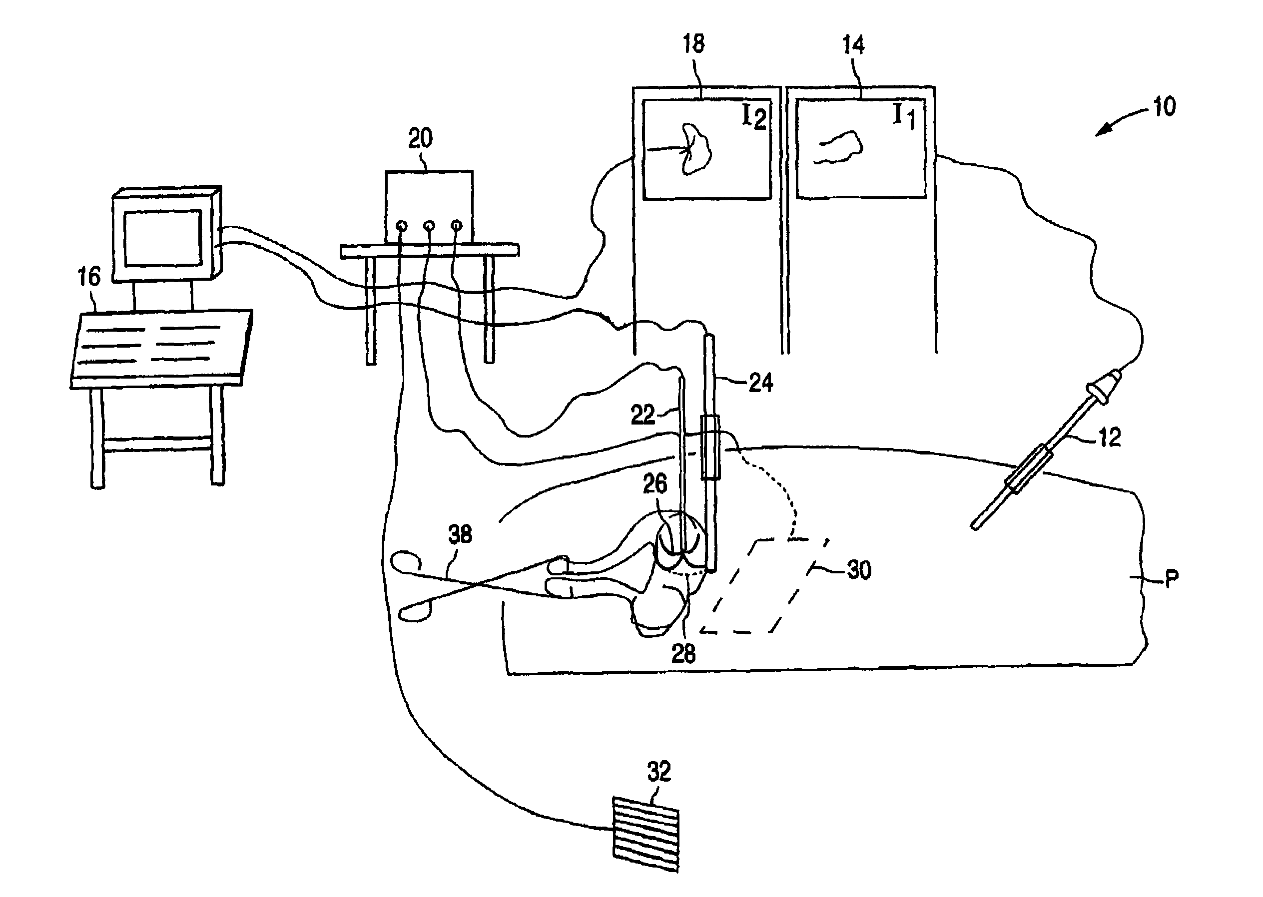
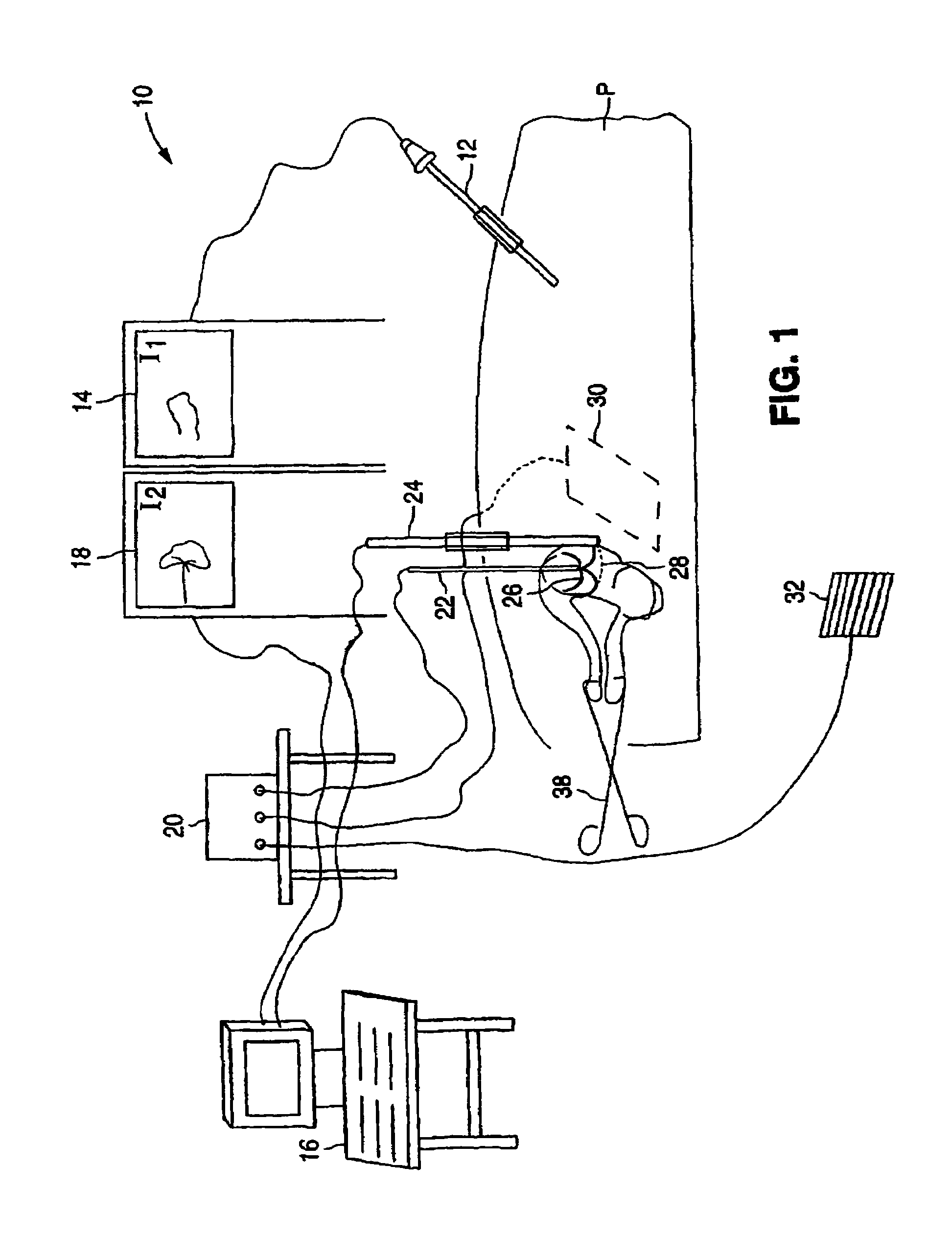
[0019]Referring first to FIG. 1, a surgical system 10 for ablating pelvic tumors includes a laparoscope 12, a video monitor 14 associated with laparoscope 12, an imaging device 16, a video monitor 18 associated with imaging device 16, an energy source 20, and an ablation device 22.

[0020]Laparoscope 12, which is inserted into a patient P, is connected to a light source and to a video monitor 14 for displaying an image from laparoscope 12. As will be explained in greater detail below, laparoscope 12 enables a surgeon to view the structures in the pelvis and abdomen and the insertion and placement of ablation device 22 into a pelvic or abdominal region of the patient. Standard recording devices such as a VCR, DVD, or CD recorders may be utilized to record and document laparoscopic images.

[0021]Imaging device 16 is connected to a video monitor 18 to provide images of the patient's pelvic region in one example. These images, which are displayed on video monitor 18, enable the surgeon to ...


PUM


Abstract

A method for treating tumors, such as uterine leiomyomata, includes inserting an ablation apparatus into a pelvic region and positioning the ablation apparatus into a pelvic tumor. The method further includes using a laparoscope and an imaging device, such as an ultrasound machine, to confirm the location of the pelvic tumor and placement of the ablation apparatus. The method further includes delivering electromagnetic energy or other energy through the ablation apparatus to the pelvic tumor to directly ablate the tumor. A surgical system for ablating pelvic tumors is also provided.

Description

RELATED APPLICATIONS[0001]This application is a continuation-in-part of U.S. patent application Ser. No. 09 / 920,425, filed Jul. 31, 2001, now U.S. Pat. No. 6,840,935, which claims priority to U.S. Provisional Patent Application No. 60 / 224,191, filed Aug. 9, 2000. This application is related to U.S. patent application Ser. No. 10 / 853,599, filed May 24, 2004. These prior patent applications are incorporated by reference herein for all purposes.BACKGROUND OF THE INVENTION[0002]1. Field of Invention[0003]The present invention relates to a procedure and system for treating gynecological disorders. More particularly, the present invention relates to the treatment of abdomino-pelvic tumors.[0004]2. Discussion of the Related Art[0005]Benign and malignant tumors can occur in the abdomen and pelvis. For example, uterine leiomyomata are muscle cell tumors that occur in 77% of women in the reproductive years. Although uterine leiomyomata rarely (0.1%) progress to cancer, these tumors can cause ...

Claims


Application Information

Patent Timeline
Patent Type & Authority Patents(United States)
IPC IPC(8): A61B18/04A61B18/18A61B8/12A61B17/42A61B18/00A61B18/12A61B19/00
CPCA61B17/42A61B18/148A61B18/1485A61B19/52A61B18/00A61B19/5212A61B19/5244A61B2017/4216A61B2018/00559A61B2019/5276A61B90/36A61B34/20A61B90/361A61B2090/378A61B17/3205A61B18/12A61B18/14
Inventor LEE, BRUCE B.
Owner ACESSA HEALTH INC
Why Eureka
Try Eureka