Method and apparatus for calculating 3D volume of cerebral hemorrhage

a hemorrhage and 3d technology, applied in the field of methods and apparatus for calculating 3d volume of cerebral hemorrhage, can solve the problems of insufficient reproducibility by a single specialist, affecting segmentation accuracy, and requiring labor and time, and achieve the effect of stable results

Active Publication Date: 2010-03-23
GE MEDICAL SYST GLOBAL TECH CO LLC
View PDF7 Cites 7 Cited by
  • Summary
  • Abstract
  • Description
  • Claims
  • Application Information

AI Technical Summary

Benefits of technology

[0008]Therefore, an object of the present invention is to provide a method and apparatus for calculating the 3D volume of cerebral hemorrhage lesion in an appropriate manner. Another object of the present invention is to provide a method and apparatus for calculating the 3D volume of cerebral hemorrhage lesion, which allows stable results to be obtained without the dependency on the technical skill of user.
[0009]Additionally, still another object of the present invention is to provide a method and apparatus for calculating 3D volume of cerebral hemorrhage lesion, which facilitate the user-friendly operation. Yet another object of the present invention is to provide a method and apparatus for calculation 3D volume of cerebral hemorrhage lesion, which accommodate to lower-, middle-, and upper-cerebral sections, and overcome the problems of partial volume effect and CT value shift among a plurality of images.
[0037]In accordance with the aspects described above of the present invention, the present invention allows to provide a method and apparatus for calculating 3D volume of cerebral hemorrhage lesion, which may obtain stable results without the dependency to the technical skill of the user. In addition, in accordance with the aspects described above of the present invention, the present invention allows to provide a method and apparatus for calculating 3D volume of cerebral hemorrhage lesion, which facilitate the user-friendly operation.
[0038]In accordance with the aspects described above of the present invention, the present invention allows to provide a method and apparatus for calculating 3D volume of cerebral hemorrhage lesion, which may accommodate to lower-, middle-, and upper-cerebral sections, and overcome the problems of partial volume effect and CT value shift among a plurality of images.

Problems solved by technology

In a semi-automatic method, a specialist is involved in the segmentation of cerebral hemorrhage lesion, therefore requiring labor and time.
Since the accuracy of the segmentation largely depends on the technical skill of the specialist, the result may be different among specialists and the reproducibility by a single specialist is not sufficient.
In contrast, fully automatic method is economic but has a problem in the segmentation accuracy, and does not yet provide a sufficient result.
Moreover, there may be cases that the section that has no cerebral hemorrhage is segmented, and that the cerebral hemorrhage lesion is not sufficiently segmented.
In both methods, the calculation ignores the partial volume effect and the CT value shift among a plurality of images so that the calculation result must be inaccurate.

Method used


Image

  • Method and apparatus for calculating 3D volume of cerebral hemorrhage
  • Method and apparatus for calculating 3D volume of cerebral hemorrhage
  • Method and apparatus for calculating 3D volume of cerebral hemorrhage

Examples


Embodiment Construction

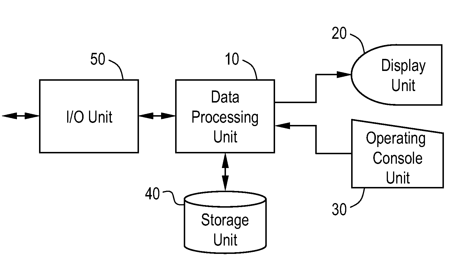
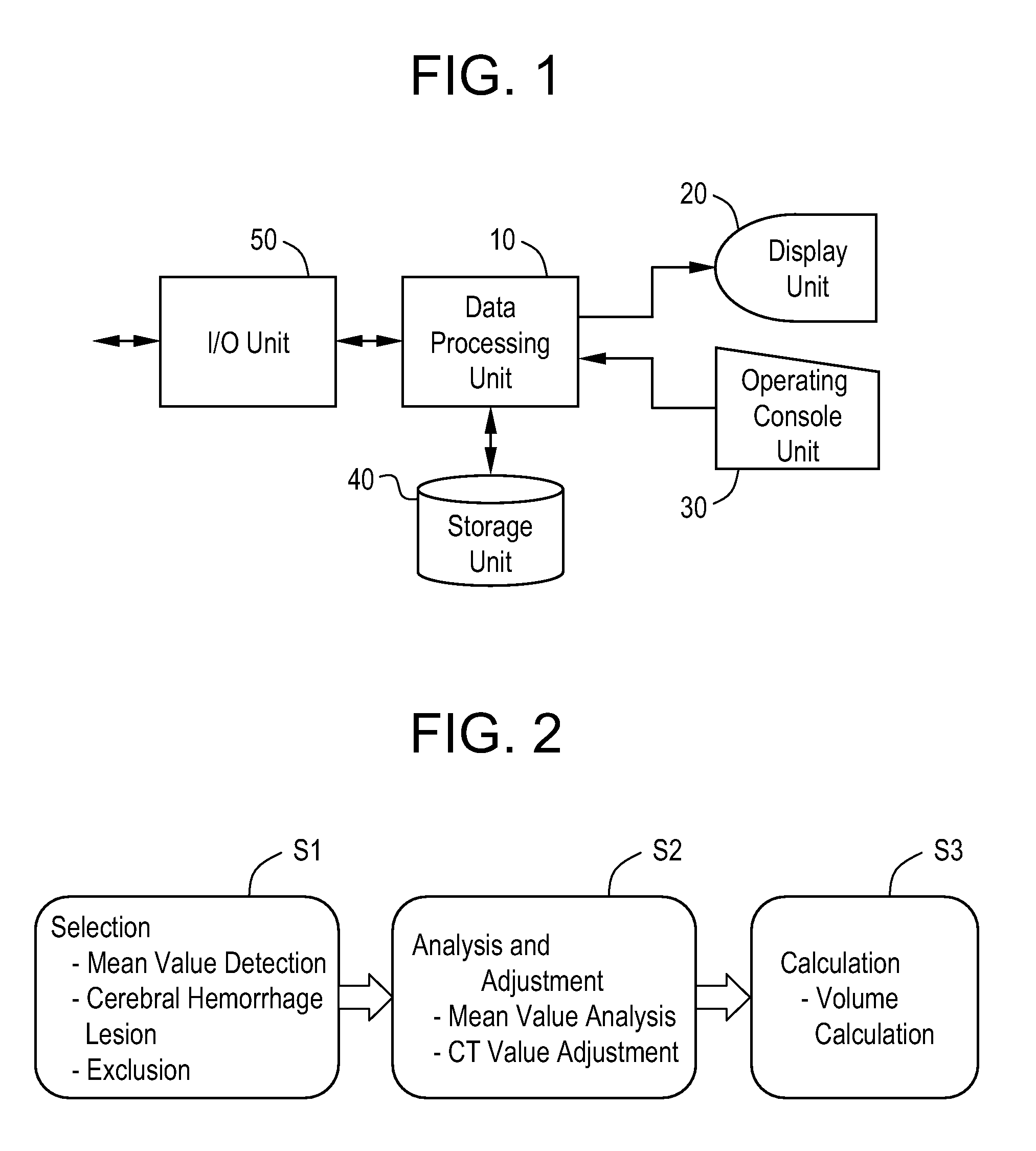
[0048]A best mode for carrying out the invention will be described in greater details herein below with reference to accompanying drawings. It is to be understood that the present invention is not limited to the best mode for carrying out the invention. FIG. 1 shows a schematic block diagram illustrating an image processing apparatus.

[0049]The apparatus shown is an exemplary embodiment of the best mode for carrying out the invention. The structure of the apparatus indicates an exemplary embodiment of the best mode for carrying out the invention of the apparatus for calculating 3D volume of cerebral hemorrhage lesion. The operation of the apparatus indicates an exemplary embodiment of the best mode for carrying out the invention of the method for calculating 3D volume of cerebral hemorrhage lesion.

[0050]As shown in FIG. 1, the apparatus includes a data processing unit 10, a display unit 20, an operating console unit 30, a storage unit 40, and an I / O unit 50. The data processing unit ...


PUM


Abstract

The present invention provides a method and apparatus for appropriately calculating 3D volume of cerebral hemorrhage lesion. The present invention sets a ROI for normal cerebral parenchymal region and a ROI for cerebral hemorrhage lesion respectively for a plurality of continuous slice images obtained by imaging the head having cerebral hemorrhage onset with an X-ray CT device, determines a mean value of the CT values of pixels within the ROI of normal cerebral parenchymal region for each of a plurality of images, then determines an amount of shift of mean values of images based on the mean value of one image, adjusts the CT value of pixels in the ROI of cerebral hemorrhage lesion for each image by using the amount of shift for each image, specifies the maximum value of the adjusted CT value through a plurality of images, and then calculates the 3D volume of the cerebral hemorrhage lesion.

Description

CROSS REFERENCE TO RELATED APPLICATIONS[0001]This application claims the benefit of Chinese Application No. 200510137354.1 filed Dec. 1, 2005.BACKGROUND OF THE INVENTION[0002]The present invention is related to method and apparatus for calculating 3D volume of cerebral hemorrhage, in particular to method and apparatus for calculating 3D volume of cerebral hemorrhage based on X-ray CT (computed tomography) images of the head in which the intracerebral hemorrhage has been affected.[0003]In order to diagnose and treat cerebral hemorrhage, 3D volume calculation of cerebral hemorrhage lesion is performed based on the X-ray CT images of the affected head. The 3D volume calculation of cerebral hemorrhage lesion is performed by identifying the location of cerebral hemorrhage on the image, then performing 3D volume calculation of identified lesion. The calculation methods consist of semi-automatic method (see for example patent reference 1) in which a specialist is involved in the segmentati...

Claims


Application Information

Patent Timeline
Patent Type & Authority Patents(United States)
IPC IPC(8): G06K9/00A61B5/05
CPCA61B6/032A61B6/501
Inventor WANG, XUELI
Owner GE MEDICAL SYST GLOBAL TECH CO LLC
Why Eureka
Try Eureka