Systems and methods for segmenting object of interest from medical image

a technology of medical images and object of interest, applied in the field of image processing, can solve the problems of difficult to establish a criterion to identify the precise location of liver tumors, difficult to precisely segment liver tumors from liver anatomy,

Inactive Publication Date: 2010-04-20
SIEMENS MEDICAL SOLUTIONS USA INC
View PDF4 Cites 52 Cited by
  • Summary
  • Abstract
  • Description
  • Claims
  • Application Information

AI Technical Summary

Problems solved by technology

It is not an easy task to precisely segment the liver tumor from the liver anatomy, except possibly in cases when the liver tumor is well isolated from adjacent anatomical structures.
A large number of liver tumors reside next to anatomical structures with similar intensity distribution in CT images, which may result in boundary blurriness between the liver tumors and the neighboring anatomy structures.
It is very difficult to establish a criterion to identify the precise location of liver tumor boundary when such blurriness exists.
Given that there are large variations in the intensity characteristics in liver CT images, neighboring anatomical structures, and intra-tumor intensity distribution, it is not easy to design a liver segmentation algorithm that produces a segmentation result that is robust to user interactions.
Liver tumor segmentation may be handled as a modified region-growing problem, which in most cases does not produce an acceptable segmentation result, Moreover, this approach requires extensive user editing, which is not desirable in tumor segmentation.
This technique may work reasonably well on some isolated tumors, but does not handle the blurred boundary cases.
This technique enables more informative constraints to be incorporated in the segmentation algorithm but does not handle the blurred boundary cases, and it requires a significant amount of user interactions, which is not desirable in 3-D tumor segmentation.

Method used


Image

  • Systems and methods for segmenting object of interest from medical image
  • Systems and methods for segmenting object of interest from medical image
  • Systems and methods for segmenting object of interest from medical image

Examples


Embodiment Construction

[0021]Hereinafter, exemplary embodiments of the present invention will be described in detail with reference to the accompanying drawings.

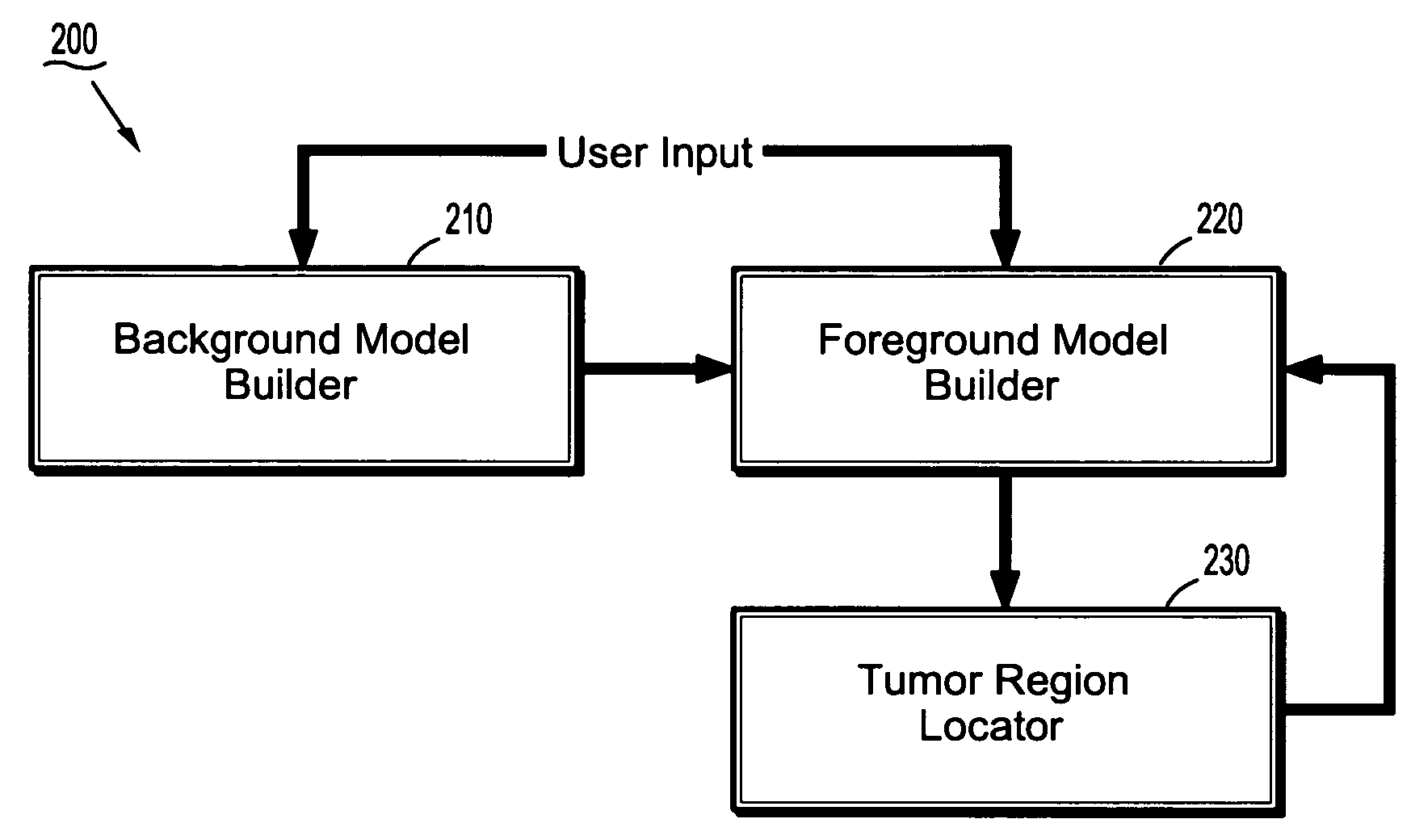
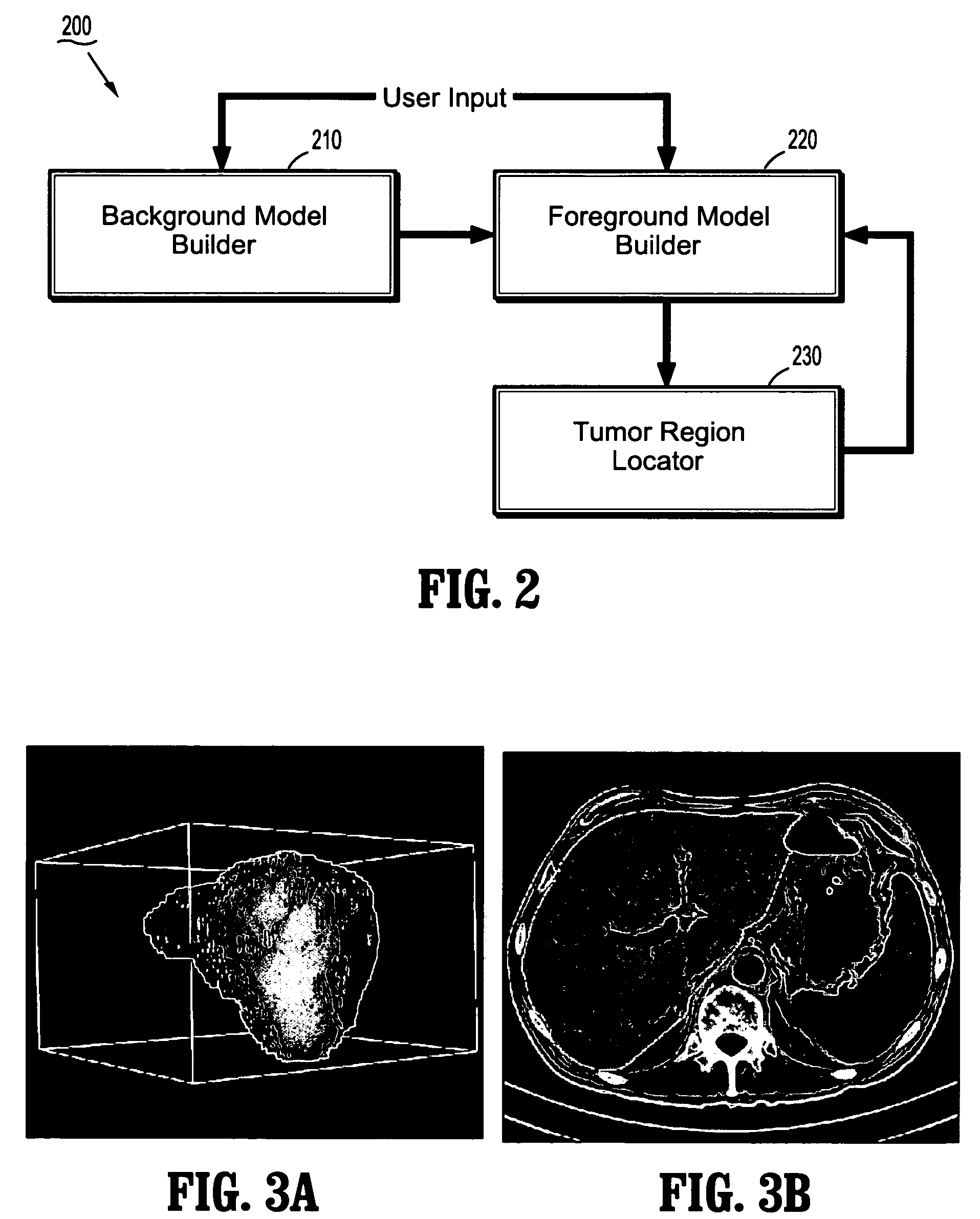
[0022]FIG. 2 shows a block diagram of system for segmenting a target organ tumor from an image, according to an exemplary embodiment of the present invention. Images may be obtained using one or more imaging modalities. Examples of images include X-ray images, positron emission tomography (PET) images, computed tomography (CT) images, magnetic resonance imaging (MRI) images, single-photon emission computed tomography (SPECT) images, etc. An image may comprise 2-D image data, 3-D image data and / or higher-dimensional image data. As used herein, “organ” refers to cells, tissues, organs and / or organ systems. Examples of organs include a liver, a pancreas, a lung or a kidney

[0023]Referring to FIG. 2, the tumor segmentation system 200 comprises a background model builder 210, a foreground model builder 220, and a tumor region locator 230. In an exemplar...


PUM


Abstract

A system for segmenting a target organ tumor from an image includes a background model builder, a foreground model builder and a tumor region locator. The background model builder uses an intensity distribution estimate of voxels in an organ region in an image to build a background model. The foreground model builder uses an intensity distribution estimate of voxels in a target organ tumor to build a first foreground model. The tumor region locator uses the background model and the first foreground model to segment the target organ tumor to obtain a first segmentation result.

Description

CROSS-REFERENCE TO RELATED APPLICATION[0001]This application claims the benefit of U.S. Provisional Application Ser. No. 60 / 724,445, filed Oct. 7, 2005 and entitled “A Novel Interactive Segmentation Algorithm for Liver Tumor Assessment”, the content of which is herein incorporated by reference in its entirety.BACKGROUND OF THE INVENTION[0002]1. Technical Field[0003]The present disclosure relates to image processing, and more particularly to systems and methods for segmenting an object of interest from a medical image.[0004]2. Discussion of Related Art[0005]In recent years, medical imaging has experienced an explosive growth due to advances in imaging modalities such as X-rays, computed tomography (CT), magnetic resonance imaging (MRI) and ultrasound. Image segmentation plays an important role in computer-based medical applications for diagnosis and analysis of anatomical data by enabling automatic or semi-automatic extraction of an anatomical organ or object of interest from a datas...

Claims


Application Information

Patent Timeline
Patent Type & Authority Patents(United States)
IPC IPC(8): G06K9/34G06V10/28
CPCG06K9/38G06T7/0081G06T7/0087G06K2209/053G06T2207/30056G06T2207/20104G06T2207/20144G06T2207/30096G06T2207/10081G06T7/11G06T7/143G06T7/194G06V10/28G06V2201/032
Inventor HONG, LINSHEN, HONG
Owner SIEMENS MEDICAL SOLUTIONS USA INC
Why Eureka
Try Eureka