System and method for automatically registering three dimensional cardiac images with electro-anatomical cardiac mapping data

a technology of cardiac imaging and cardiac mapping data, applied in the field of system and method for automatically registering three dimensional cardiac images with electroanatomical cardiac mapping data, can solve problems such as difficult navigation

Inactive Publication Date: 2010-05-11
SIEMENS MEDICAL SOLUTIONS USA INC +1
View PDF15 Cites 88 Cited by
  • Summary
  • Abstract
  • Description
  • Claims
  • Application Information

AI Technical Summary

Problems solved by technology

However, fluoroscopic images are two dimensional and do not have good resolution thereby making navigation difficult.

Method used


Image

  • System and method for automatically registering three dimensional cardiac images with electro-anatomical cardiac mapping data
  • System and method for automatically registering three dimensional cardiac images with electro-anatomical cardiac mapping data
  • System and method for automatically registering three dimensional cardiac images with electro-anatomical cardiac mapping data

Examples


Embodiment Construction

[0012]The present invention is directed to a system and method for automatically registering pre-operative high-resolution three dimensional (3D) cardiac images with corresponding intra-operative electrophysiological (EP) points of 3D electro-anatomical (EA) maps. The 3D images can be obtained using either a Computed Tomography (CT) imaging system or a Magnetic Resonance (MR) imaging system. Registration of left atrial (LA) high-resolution CT and MR images with a cardiac mapping system can provide precise cardiac anatomical information, along with real-time cardiac electrical activation information, catheter tracking and 3D location, and lesion position.

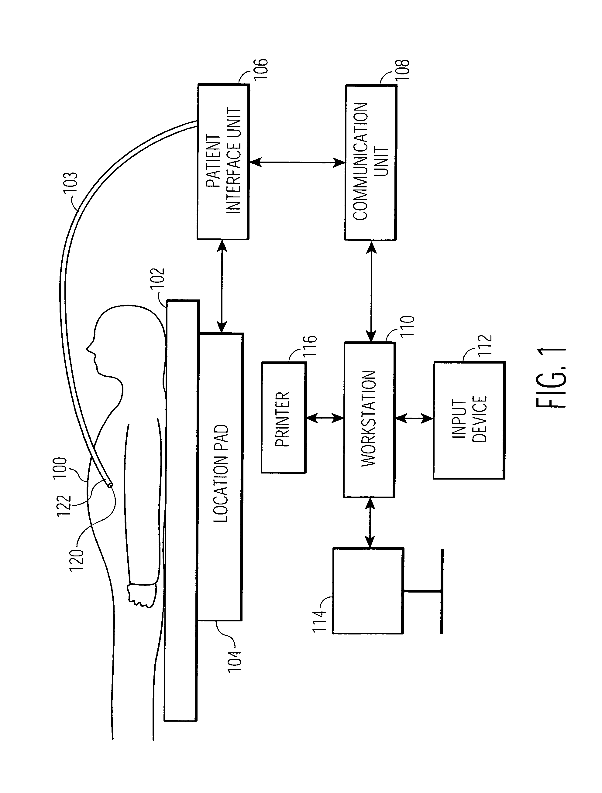
[0013]An exemplary cardiac mapping and navigation system is illustrated in FIG. 1. The cardiac mapping system comprises a miniature passive magnetic field sensor 120 which is located at a tip of a catheter 122 which is inserted in a chamber of a patient's heart. The patient 100 is placed on a patient table 102. A location pad 104 is ...


PUM


Abstract

A system and method for automatically registering a three dimensional (3D) pre-operative image of an anatomical structure with intra-operative electrophysiological (EP) points of a 3D electro-anatomical (EA) image map of the anatomical structure is disclosed. The pre-operative image is displayed in a first supporting view. The intra-operative EA image map is displayed in a second supporting view. An alignment of the pre-operative image with the intra-operative map is performed by identifying at least one corresponding point on each image. The view of the pre-operative image is integrated with the EA map based on the alignment.

Description

CROSS REFERENCE TO RELATED APPLICATION[0001]This application claims the benefit of U.S. Provisional Application Ser. No. 60 / 644,817 filed on Jan. 18, 2005, which is incorporated by reference in its entirety.FIELD OF THE INVENTION[0002]The present invention is directed to a system and method for automatically registering three dimensional cardiac images with electro-anatomical cardiac mapping data, and more particularly, to a system and method for automatically registering pre-operative three dimensional atrial images with intra-operative electro-anatomical cardiac mapping data.BACKGROUND OF THE INVENTION[0003]Cardiovascular disease is one of the leading causes of death in the United States as well as in many other countries. Cardiovascular disease includes a number of conditions affecting the structures or functions of the heart. Arrhythmias can be an indication of a number of heart problems. An Electrophysiology (EP) test can be performed to record the electrical activity of the he...

Claims


Application Information

Patent Timeline
Patent Type & Authority Patents(United States)
IPC IPC(8): G06K9/00A61B5/05G06K9/32
CPCA61B5/0035A61B5/042G06T7/0028G06T2207/30048A61B6/03A61B5/055A61B5/283G06T7/33
Inventor SUN, YIYONGAZAR, FRED S.XU, CHENYANGSAUER, FRANKRAHN, NORBERT
Owner SIEMENS MEDICAL SOLUTIONS USA INC
Why Eureka
Try Eureka