Ultrasound simulator for craniosynostosis screening

a craniosynostosis and ultrasound simulator technology, applied in the field of anatomically correct medical models, can solve the problems of not readily available medical training models that can be used to train radiologists and technicians to assess the skull sutures of children with abnormal head shape using ultrasound, and require sedation of young children, etc., to achieve uniform feel and appearance.

Inactive Publication Date: 2010-06-08
UNIV OF WASHINGTON
View PDF4 Cites 13 Cited by
  • Summary
  • Abstract
  • Description
  • Claims
  • Application Information

AI Technical Summary

Benefits of technology

[0017]Each opening corresponding to a simulated patent skull suture is preferably filled with a hypoechoic material. The echogenicity of the hypoechoic material enables each opening corresponding to a simulated patent skull suture to be readily distinguishable in an ultrasound image of the model, as long as the material from which the model is formed has a different echogenicity than the fill material. Preferably, the echogenicity of the fill material is lower than the echogenicity of the model material, so that in an ultrasound image of the model, the fill material appears darker than the model material.
[0018]The method for making a model may also include the step of covering the scalp of the model with a layer of the fill material, so that the scalp of the model has a uniform feel and appearance.

Problems solved by technology

CT suffers from the disadvantages of relying on ionizing radiation (exposure to which in children is to be avoided when possible), frequently requiring the sedation of young children, and being expensive.
Medical training models that can be used to train radiologists and technicians to assess the skull sutures in children with abnormally shaped heads using ultrasound simply are not readily available.

Method used


Image

  • Ultrasound simulator for craniosynostosis screening
  • Ultrasound simulator for craniosynostosis screening
  • Ultrasound simulator for craniosynostosis screening

Examples


Embodiment Construction

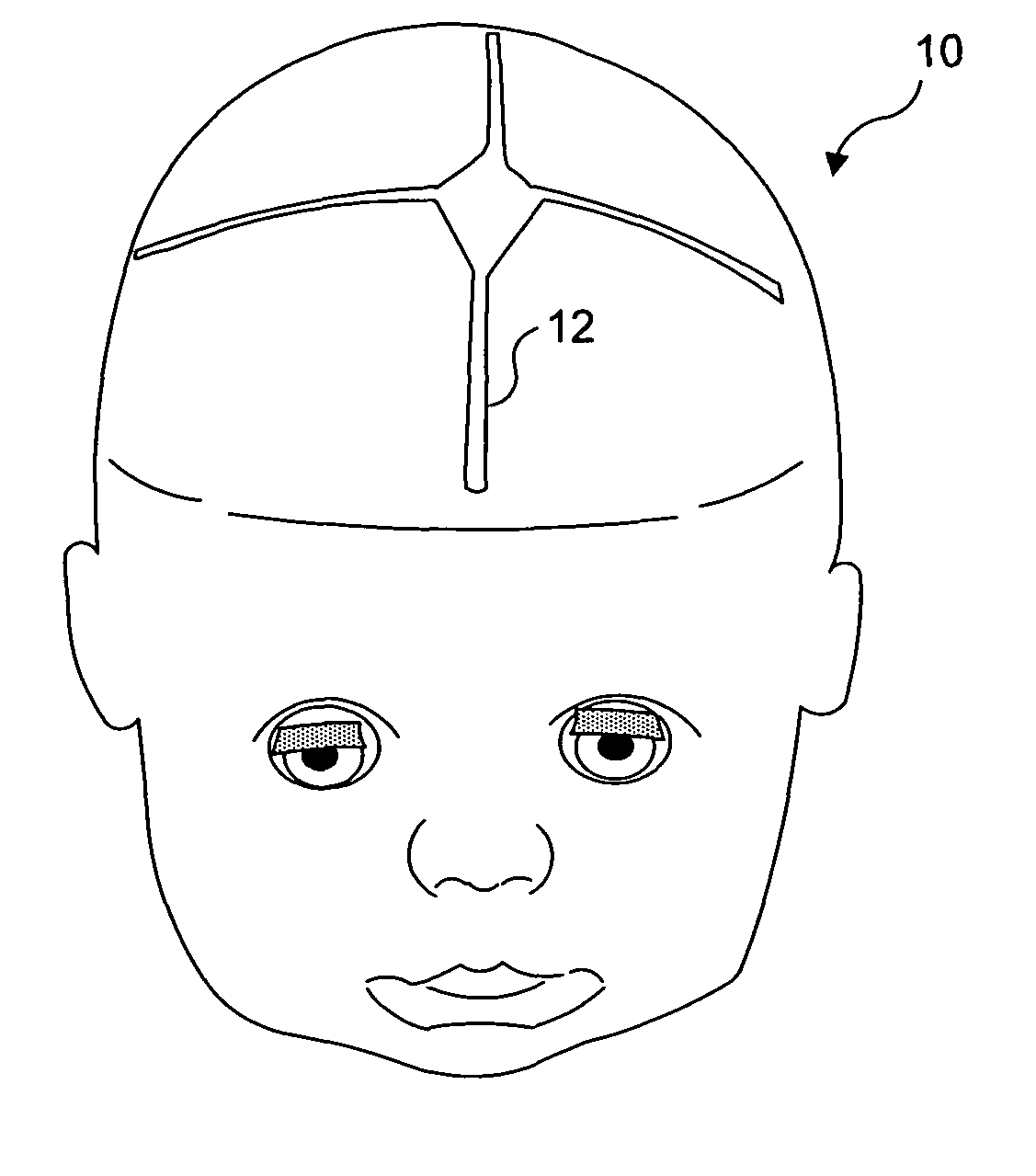
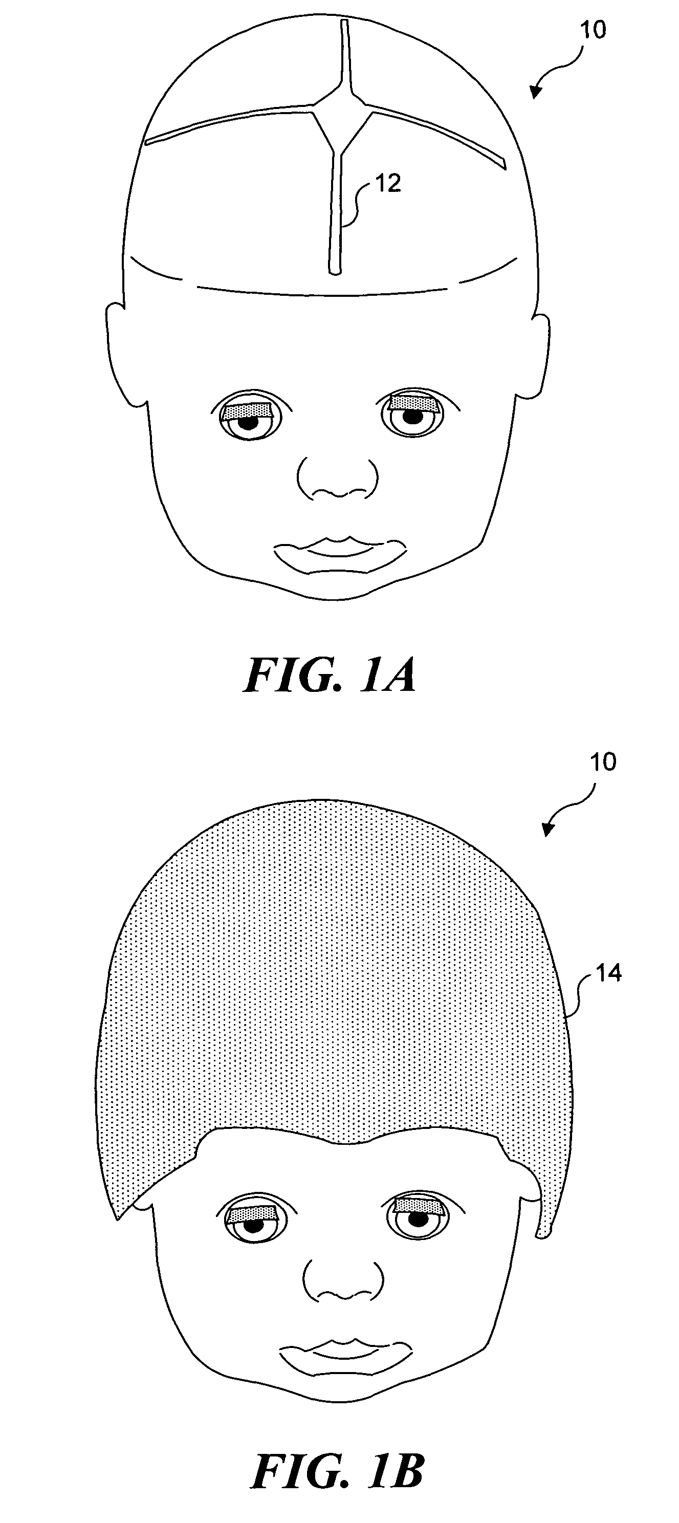
[0038]The present invention is an ultrasound simulator that can be used to train radiologists and ultrasound technicians in the anatomy of pediatric cranial sutures and in techniques for using ultrasound as a screening tool to detect craniosynotosis. A life sized model of a human head includes simulated skull sutures that can be detected using ultrasound. Additional aspects of the present invention relate to methods for making and using such a model.

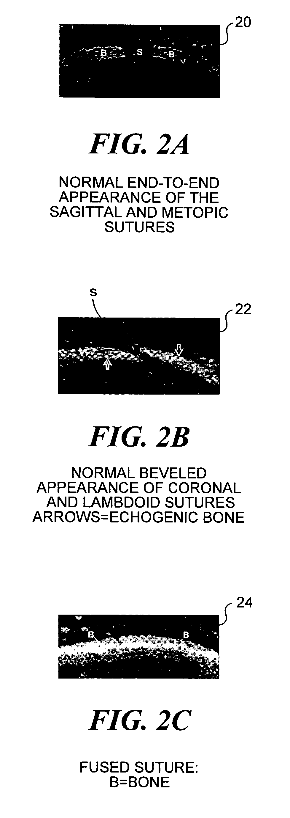
[0039]In the following description and the claims that follow, it will be understood that the term “patent” means a skull suture in which a gap exists between adjacent portions of the skull. Normal skull sutures have such a gap. Abnormal skull sutures are closed, and in the following description and the claims that follow, it will be understood that the term “fused” is employed to refer to such abnormal, or closed, skull sutures. The term “life size” is meant to refer to a model that simulates a portion of human anatomy such that the mod...


PUM


Abstract

An ultrasound simulator to train radiologists and technologists to locate and recognize patent and fused cranial sutures. The model is formed, for example, using specially fabricated heads or from life-sized plastic doll heads. Simulated suture lines are cut in the heads in anatomically correct positions. The typical end-to-end morphology of the sagittal and metopic sutures and the typical beveled appearance of the paired coronal and lambdoid sutures are created by angling the cutting blade. The hypoechoic appearance of patent sutures in ultrasound images is simulated by filling the gaps that were formed by cutting with a hypoechoic material. Fused echoic sutures are simulated by leaving that portion of the doll's head uncut, or by filling the openings with an echoic material. When imaged using ultrasound, the portions cut and filled with a hypoechoic material are readily distinguishable from uncut portions, and from portions cut and filled with an echoic material.

Description

RELATED APPLICATIONS[0001]This application is based on a prior copending provisional application Ser. No. 60 / 431,279, filed on Dec. 5, 2002, the benefit of the filing date of which is hereby claimed under 35 U.S.C. §119(e).FIELD OF THE INVENTION[0002]The present invention generally relates to anatomically correct medical models, and more specifically pertains to a medical model that simulates the anatomical position of skull sutures in children to the extent that ultrasound images from such a model closely correspond to ultrasound images from actual patients, and to a method for using such a medical model for training radiologists and ultrasound technologists in the detection and examination of skull sutures in children.BACKGROUND OF THE INVENTION[0003]Abnormal head shapes in children can be caused by either positional molding (a change in head shape due to a child's preferential head position during sleep), or a more serious condition referred to as craniosynostosis (the premature ...

Claims


Application Information

Patent Timeline
Patent Type & Authority Patents(United States)
IPC IPC(8): G09B23/28
CPCG09B23/286
Inventor SZE, RAYMOND W.NGO, ANH-VU
Owner UNIV OF WASHINGTON
Why Eureka
Try Eureka