

Systems and methods of classification utilizing intensity and spatial data
a technology of intensity and spatial data and classification methods, applied in image data processing, image enhancement, instruments, etc., can solve the problems of relying on manual or automated segmentation, unable to achieve the effect of effective classification of new subjects
- Summary
- Abstract
- Description
- Claims
- Application Information
AI Technical Summary
Benefits of technology
Problems solved by technology
Method used
Image

Examples
Embodiment Construction
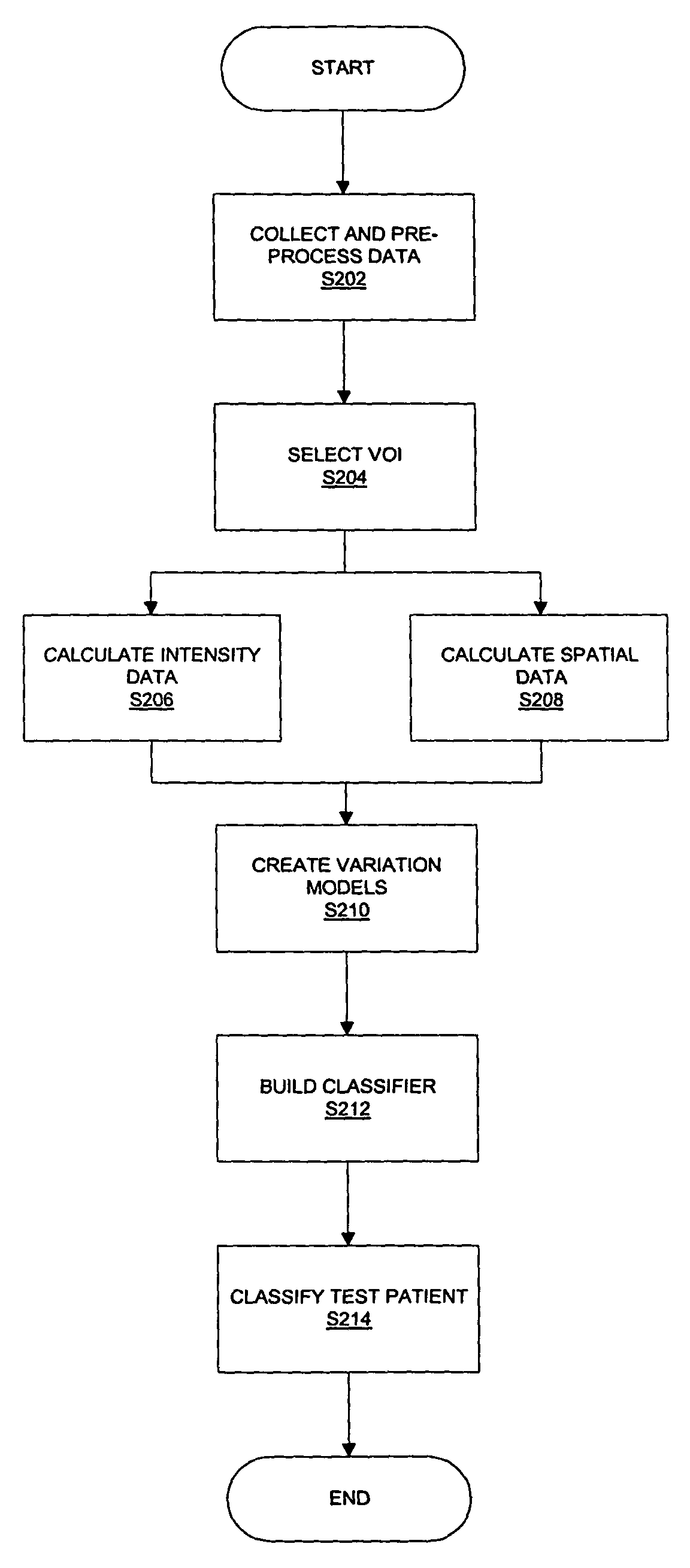
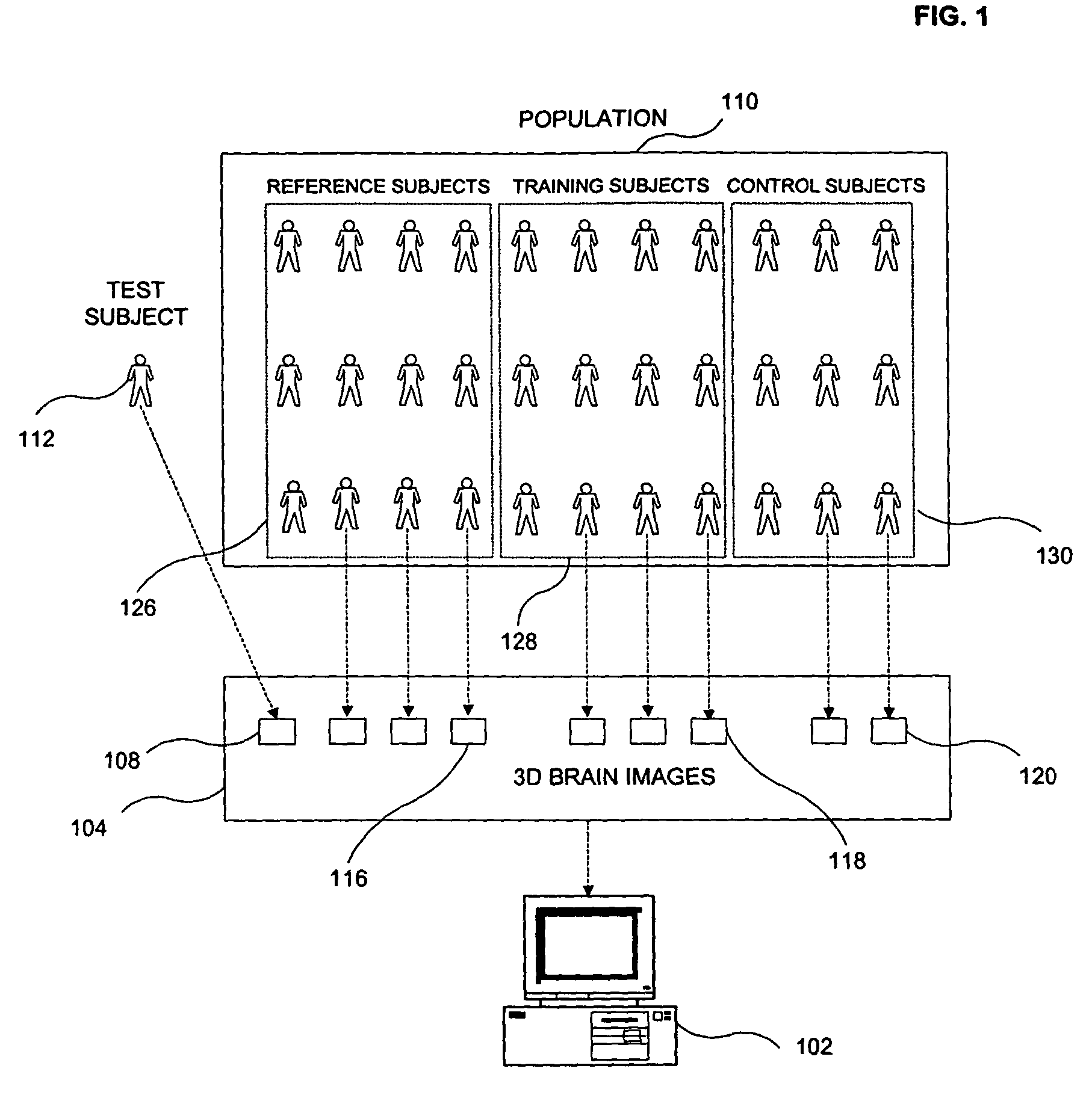
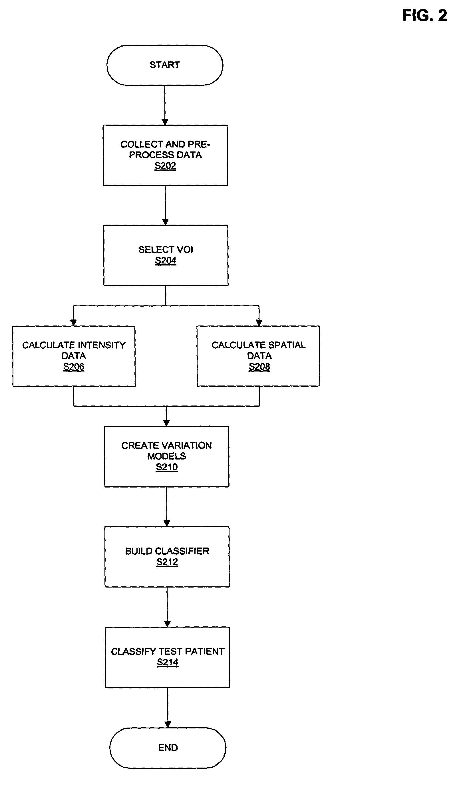
[0029]A schematic illustration of the design of an automated classification system 102 in manners exemplary of the present invention is shown in FIG. 1. The example automated classification system 102 determines a classification and diagnosis of the neurological disease state of a given test subject 112, based on 3D image data of the brain 104. The image data 104 may be mono-modal or multi-modal. Possible types of images that may be acquired include, but are not limited to images based on: T1w MRI, T2w MRI, PD MRI, PET, SPECT, and CT.
[0030]As illustrated, example automated classification system 102 is determined using a general purpose computing device, executing software exemplary of the aspects of the present invention. The computing device may have any suitable combination of dynamic and persistent storage memory. To classify the disease state of a test subject 112, a plurality of 3D images 104 is first collected from subjects in population 110 (in some embodiments, typically onl...
PUM
 Abstract
Description
Claims
Application Information