Method for deriving a treatment plan for orthognatic surgery and devices therefor

a treatment plan and orthognatic technology, applied in the field of orthognatic surgery treatment plan derivation and the field of orthognatic surgery treatment plan, can solve the problems of inability to plot on a three-dimensional view the exact position of teeth to obtain the bite, and the currently available 3d surface representation algorithm does not provide a suitable framework comprising anatomically relevant references

Active Publication Date: 2010-09-07
MEDICIM
View PDF6 Cites 29 Cited by
  • Summary
  • Abstract
  • Description
  • Claims
  • Application Information

AI Technical Summary

Benefits of technology

[0013]The present invention aims to provide a method for performing a cephalometric and / or anthropometric analysis. In a second object it aims to provide a method for deriving a treatment plan for orthognatic surgery, comprising said analysis method. In a further object it aims to provide devices suitable therefor.

Problems solved by technology

Further, this simulation is only done in two dimensions based on a plane profile view of the skull.
However, the currently available 3D surface representations algorithms do not provide a suitable framework comprising anatomically relevant references, which allow the clinician to easily and reliably reposition a bone fragment in the virtual 3D environment.
A second problem is associated with the use of 3D surface representations derived from CT-scans of patients having amalgam dental fillings.
Using these CT images as such, it is impossible to plot on a three-dimensional view the exact position of the teeth to obtain the bite.

Method used


Image

  • Method for deriving a treatment plan for orthognatic surgery and devices therefor
  • Method for deriving a treatment plan for orthognatic surgery and devices therefor
  • Method for deriving a treatment plan for orthognatic surgery and devices therefor

Examples


Embodiment Construction

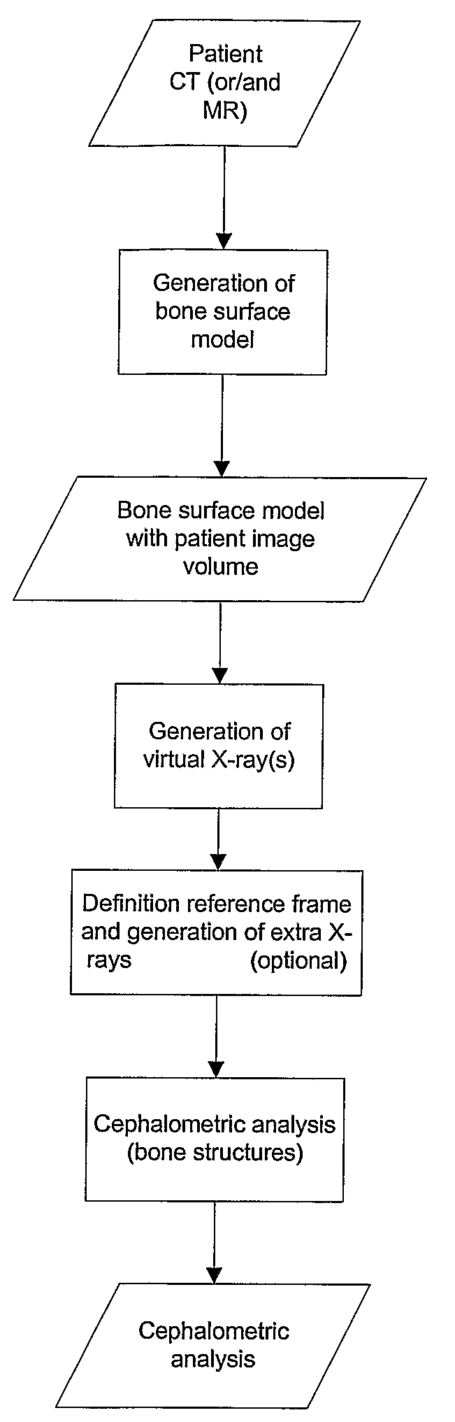
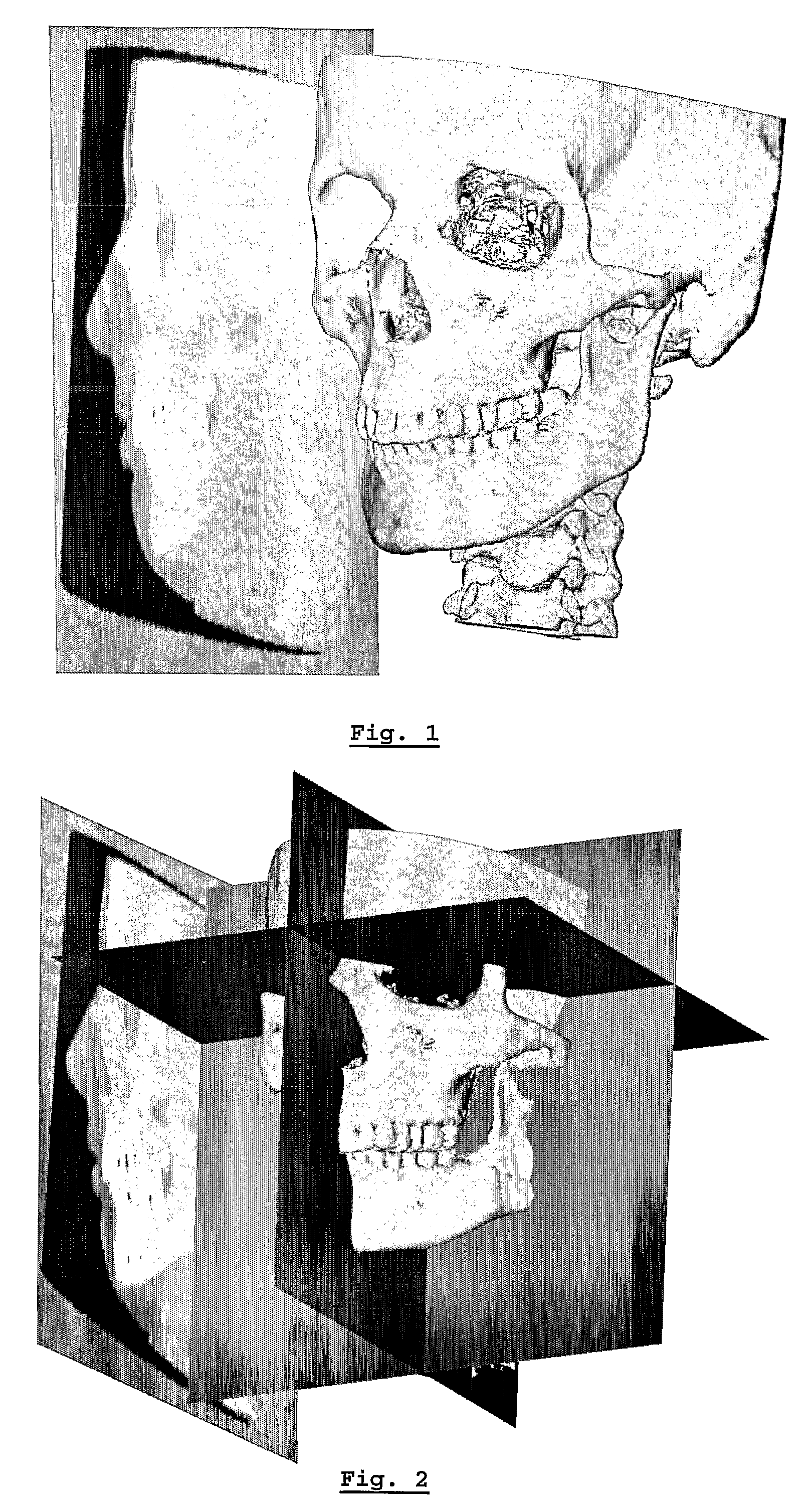
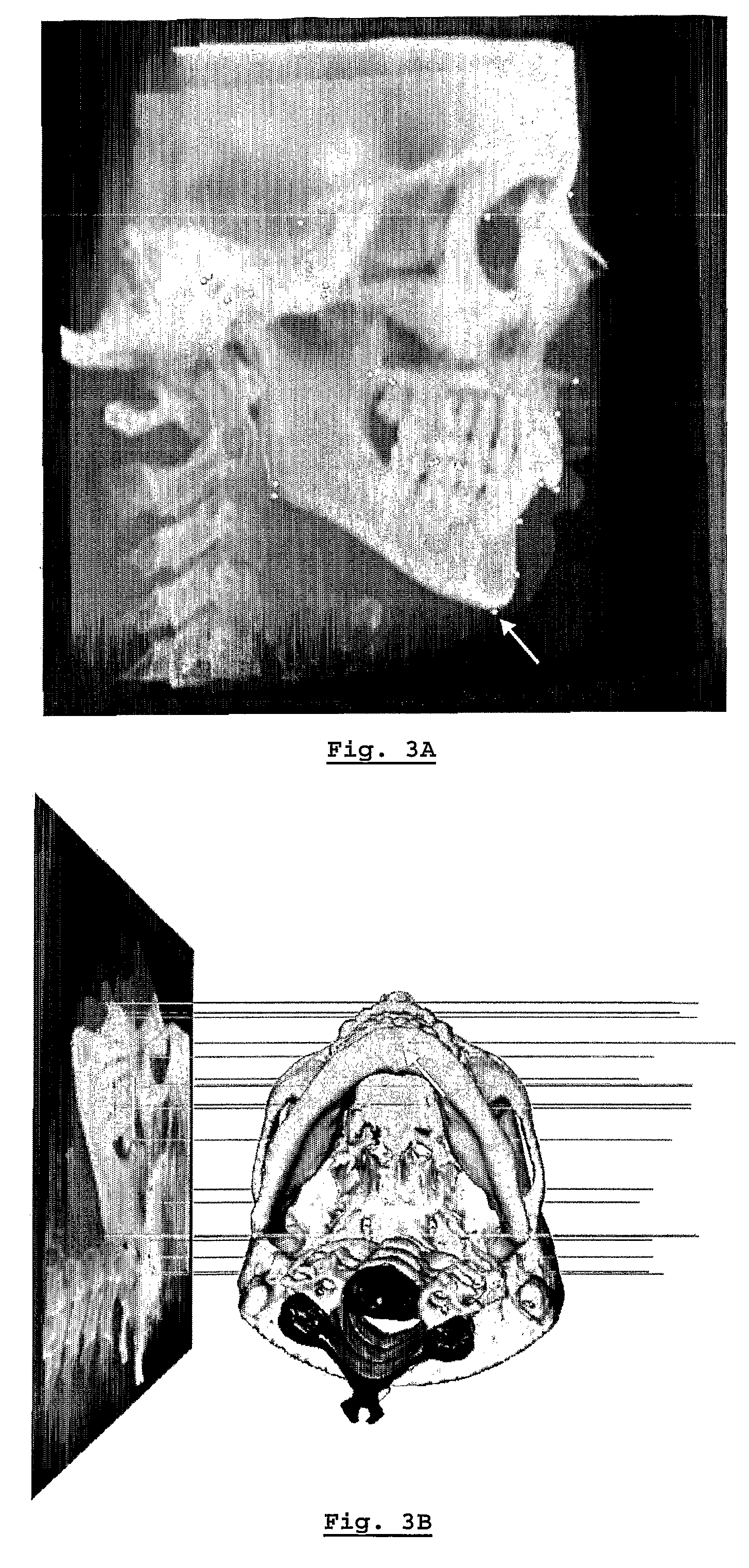
[0051]In order to perform an adequate 3D cephalometric analysis of bone tissue and / or soft tissues, the ability to indicate the relevant points on the 3D structures alone does not suffice. Points required for adequate 3D cephalometric tracing that are not well defined on the 3D structure are available on a 2D representation and vice-versa. The present invention describes a computerised system that solves this problem.

[0052]The present invention provides a method to redefine the framework of a 3D surface representation into an anatomically relevant framework. The anatomically relevant framework allows a clinician to perform an accurate cephalometric and / or anthropometric analysis in an intuitive manner. Moreover, a 3D surface representation comprising an anatomically relevant framework has the advantage that it allows the virtual repositioning of bone fragments in relation to anatomically relevant landmarks, making it particularly suited for the planning of surgical interventions. Th...


PUM


Abstract

The present invention is related to a method for performing a cephalometric or anthropometric analysis comprising the steps of: acquiring a 3D scan of a person's head using a 3D medical image modality, generating a 3D surface model using data from the 3D scan, generating from the 3D scan at least one 2D cephalogram geometrically linked to the 3D surface model, indicating anatomical landmarks on the at least one 2D cephalogram and / or on the 3D surface model, performing the analysis using the anatomical landmarks.

Description

CROSS-REFERENCE TO RELATED PATENT APPLICATIONS[0001]This patent application is the National Stage of International Application No. PCT / BE2005 / 000100 filed Jun. 27, 2005, that claims the benefit of Great Britain Application No. 0414277.4 filed Jun. 25, 2004.FIELD OF THE INVENTION[0002]The present invention relates to methods for assessing the shape of the skull and soft tissues and for determining a treatment plan for maxillofacial surgery and more particularly for orthognatic surgery and devices used in such surgery or in the preparation thereof.STATE OF THE ART[0003]In maxillofacial surgery, the skull and dentition is surgically remodelled or restored. This surgical discipline encompasses surgical interventions of repair, in particular, of a mis-positioning of the jaws with respect to one another, called orthognatic surgery. Typically, orthognatic surgery involves osteotomies of the maxilla and / or mandible to reposition these bone fragments correctly with respect to the rest of the...

Claims


Application Information

Patent Timeline
Patent Type & Authority Patents(United States)
IPC IPC(8): G06K9/00A61B19/00G06T17/40
CPCG06T7/0012G06T7/60G06T19/00G06T2207/30008G06T2210/41G06T2207/00B33Y80/00B33Y50/00
Inventor SCHUTYSER, FILIP
Owner MEDICIM
Why Eureka
Try Eureka