Protein synthesis monitoring (PSM)

a protein synthesis and monitoring technology, applied in the field of ribosome protein synthesis monitoring, can solve the problems of missing entire protein families, difficult and slow, expensive, etc., and achieves the effects of high quantitation accuracy, high sensitivity, and high sensitivity

Active Publication Date: 2010-10-05
ANIMA CELL METROLOGY
View PDF7 Cites 4 Cited by
  • Summary
  • Abstract
  • Description
  • Claims
  • Application Information

AI Technical Summary

Benefits of technology

[0031]Protein synthesis information can optionally be tabulated to correlate with chemical or environmental effects applied to the cells being studied. In this way the present invention is able to decipher the functionality of chemical compounds; elucidate cellular proteomic mechanisms; control protein production systems; and help study the factors affecting protein synthesis.
[0107]Another important point is photobleaching of the fluorophores. When a fluorescent dye is excited it is susceptible to oxidation or photobleaching. With standard fluorophores such as naturally fluorescent proteins or small organic dyes, bleaching can be minimized both by eliminating as far as possible the sample exposure to oxygen, and on the other hand employing an enzymatic oxygen scavenger system. Singlet oxygen is presumed to be the main culprit in photobleaching, and in some cases oxygen removal has a considerable effect on reducing photobleaching (cf. T. Ha, METHODS 25, 78-86(2001)). If the system in use is two-photon microscopy, than photobleaching is reduced considerably compared to confocal microscopy, especially because of the significant. or even near complete reduction in background excitation. Another configuration that helps lengthen the signal generation time is the use of quantum dots, which are practically immune to bleaching. When quantum dots are used as donors, and if the excitation radiation is well outside the excitation spectrum of the acceptor, very long monitoring times can be expected.

Problems solved by technology

Current technologies fail at several key points: they can miss entire protein families; fail to identify protein pathways; focus on a-single protein at a time; and they are expensive, difficult and slow.
Importantly, no current technology provides information on protein dynamics.
In fact, results of current large-scale and high-throughput protein analysis are often delayed by days or weeks following an experiment, and are usually restricted to the form of a catalogue, tabulating those proteins of a database that have been putatively identified from the analyzed sample.
Unfortunately, proteins are also so much more complex and difficult to study than DNA.
Proteins are more difficult to study than DNA, since protein extraction, separation and identification are difficult; there is no amplification technique that parallels PCR; three-dimensional protein structure is hard to obtain and use; protein expression has high dynamic range; protein modifications, cleavages and interactions are to a large extent unknown; and, finally, both as cause and effect, protein databases are thin and sparsely populated, encompassing a small fraction of all theoretical proteins, especially in higher organisms, such as Homo sapiens.
Unfortunately, first for genomics and then for proteomics, it became abundantly clear that though genomic and proteomic data is extremely valuable, it is far from sufficient for achieving the breakthrough that was hoped for (Miklos, G. L. and Maleszka, R., Protein functions and biological contexts.
The extraction of proteins from bacterial or cell culture invariably involves lysis (and therefore death) of the cells.
Protein separation with two-dimensional gels requires at least 24 hours and an expert human operator; their analysis is often much more difficult, even with modern software (Smilansky, Z.
Even worse, two-dimensional gel technology is not applicable to very acidic or very basic proteins, to many membranal proteins, and most importantly, to proteins that are expressed in low amounts.
However, all three are hopelessly inaccurate.
Moreover, quantitation at best means quantity of protein in the extracted, processed sample, such as in a gel spot or in a chromatographic fraction; estimation of protein copies in a cell at any given time is not even attempted today.
All in all, the standard technique for identifying proteins in a cell culture takes from weeks to months, is suitable for only a small part of the proteome, does a bad job of quantitating protein amounts, and provides no clue as to proteome dynamics.
The procedure requires a full day and provides no quantitative or dynamic information.
However, they are still difficult and expensive to carry out, require cell lysis, take days for complete analysis, and provide no dynamic information.
All of these may aid protein analysis in some way, but none of them provides the data that the disclosed method provides.
It is known to generate a large number of false-positives and also cannot generate dynamic information.
However, cell based screening suffers from certain disadvantages.
These disadvantages include the need to engineer a specific cell line with the required reporting capability, and the lack of information about the would-be protein target.
This of course limits the scope of information obtainable, and the entire cascade of events following administration of the compound under analysis remains hidden from the researcher.
More discouraging yet is the fact that while advances in high-throughput screening, chemical compound library design and bioinformatics have helped multiply the number of “hits” in HTS assays, the number of “leads” has not increased at all.
Thus, the pharmaceutical pipeline today has an abundance of targets on the one side and an abundance of candidate compounds on the other, but attempts to combine this information has yielded little.
While the process is long and very expensive, it is narrow in the sense that relatively little is known about the target protein, its function, its isoforms and look-alikes, its roles in disease and in health.
Thus, information that may indicate that a compound cannot become a suitable drug candidate is revealed only at later stages and at a high cost—sometimes only after being distributed on the market.

Method used


Image

  • Protein synthesis monitoring (PSM)
  • Protein synthesis monitoring (PSM)
  • Protein synthesis monitoring (PSM)

Examples


Embodiment Construction

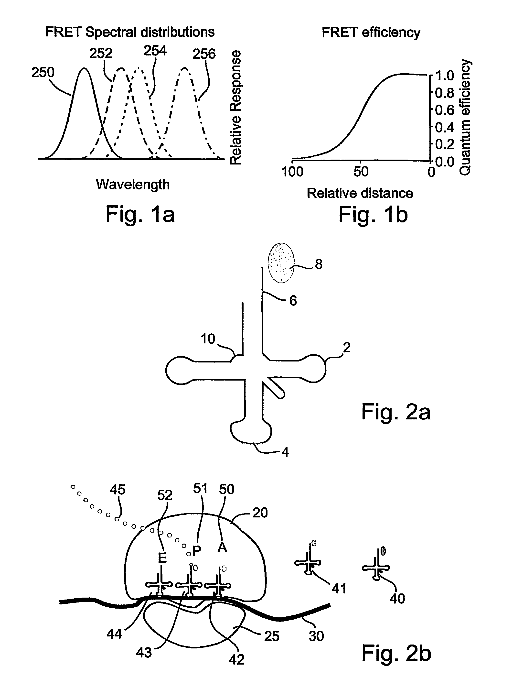
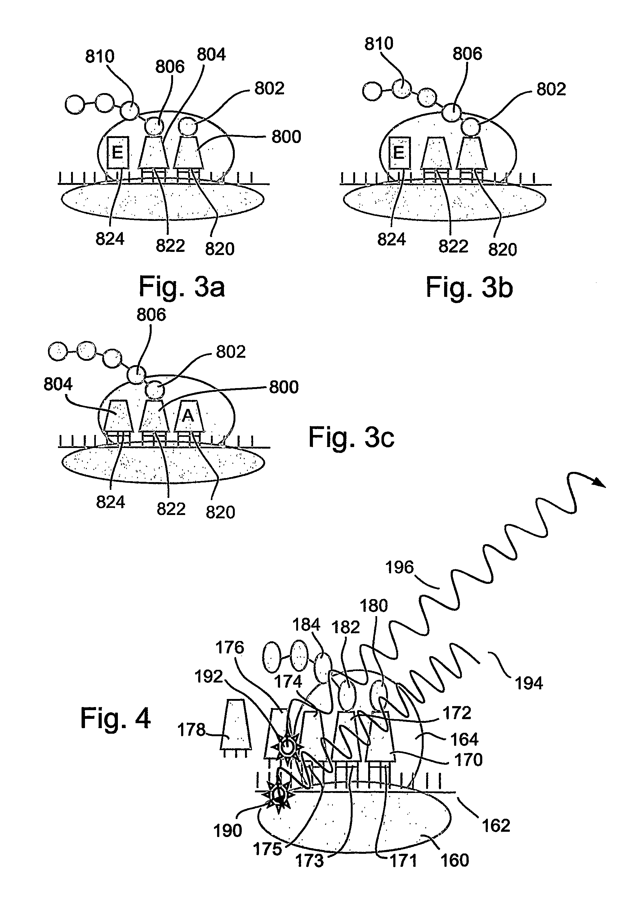
[0173]The present invention is of a system and method for monitoring protein synthesis in a protein synthesis system, by using a marker for protein synthesis in the system, which preferably causes electromagnetic radiation to be emitted. The protein synthesis system may optionally comprise a single ribosome, or alternatively a plurality of ribosomes. The marker optionally comprises at least one photo-active component.

[0174]The emitted electromagnetic radiation is then detected and can be analyzed to monitor protein synthesis. The present invention may optionally be performed qualitatively, but is preferably performed quantitatively. As used herein, “monitoring” may also optionally include at least the initial detection of a protein synthetic act or process, such as an interaction between a tRNA and a ribosome for example, preferably in real time. Optionally and preferably, monitoring includes identification of the tRNA or tRNA species, amino acid or amino acid species, codon or codo...


PUM

PropertyMeasurementUnit
distancesaaaaaaaaaa
volumeaaaaaaaaaa
thicknessaaaaaaaaaa

Abstract

A method and a device are disclosed for monitoring the synthesis of proteins by the ribosome in real time, in vivo as well as in in-vitro. The ribosome is engineered to carry a donor fluorophore, and tRNA and / or amino acids and / or some other part of the ribosome are either engineered to carry acceptor fluorophores or else their natural fluorescent properties are utilized as acceptors. As the ribosomes mechanism processed the mRNA and tRNA molecules and synthesizes a polypeptide chain, a light source illuminates the ribosome, exciting the donor fluorophores and thereby the acceptor fluorophores whenever these are in sufficient proximity to a donor. The resulting signals are detected and used as a key for real-time database searching and identification of the protein being synthesized. The resulting data can be tabulated and interpreted in different ways. FIG. (1) describes the properties of a FRET pair and the dependence of FRET on pair distance.

Description

CROSS REFERENCE TO RELATED APPLICATIONS[0001]This application is a 371 filing of PCT International application no. PCT / IL2003 / 001011 filed Nov. 27, 2003 and published in English as WO 2004 / 050825 on Jun. 17, 2004 which claims the priority of U.S. application No. 60 / 429,532 filed Nov. 29, 2002, the disclosures of which are hereby incorporated by reference in their entirety.FIELD OF THE INVENTION[0002]The present invention relates to the monitoring of protein synthesis by ribosomes, and in particular to such monitoring being performed in real time.BACKGROUND OF THE INVENTION[0003]The study of proteins is a key endeavor of current biological research, as well as a focus of pharmaceutical research and development. The information revealed by sequenced genomes increases the pace and activity of protein research, for example for the development of a cell-based assay, analysis of a pathway, study of a single receptor, or the application of proteomics. Current technologies fail at several k...

Claims


Application Information

Patent Timeline
Patent Type & Authority Patents(United States)
IPC IPC(8): C12Q1/68G01N33/554G01N33/53G01N33/48C12Q1/37C12Q1/02G01N33/542G01N33/68
CPCB82Y5/00B82Y10/00G01N33/542G01N33/6803G01N33/6842
Inventor SMILANSKY, ZEEV
Owner ANIMA CELL METROLOGY
Why Eureka
Try Eureka