
Targeted dual-axes confocal imaging apparatus with vertical scanning capabilities

a confocal imaging and vertical scanning technology, applied in the field of tissue microscopy, can solve the problems of terminal experiments, lower efficiency, distorting pulse profiles, etc., and achieve the effect of improving fluorescence detection efficiency and overcoming blurring

Active Publication Date: 2018-03-20
RGT UNIV OF MICHIGAN
View PDF26 Cites 11 Cited by
  • Summary
  • Abstract
  • Description
  • Claims
  • Application Information

AI Technical Summary

Benefits of technology

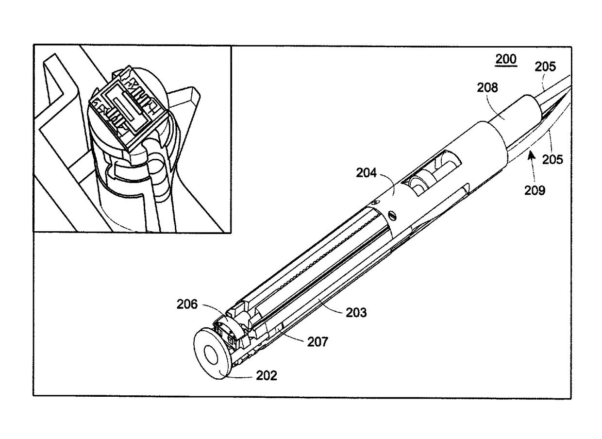
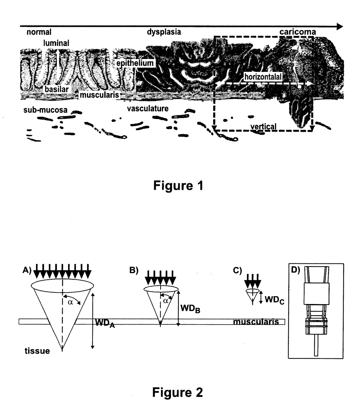
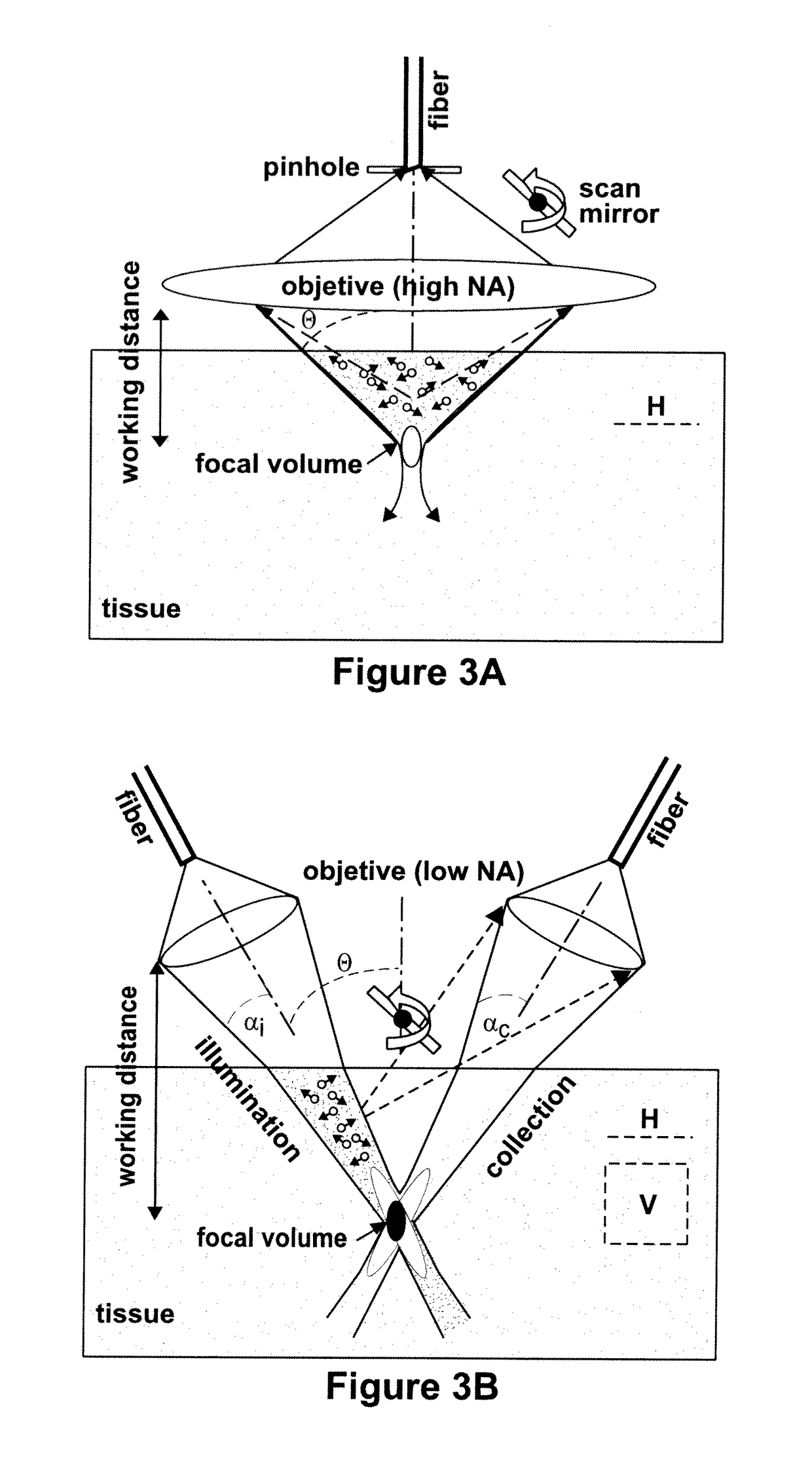
[0019]To address the foregoing, the present application describes an optical device that may be used as a microscope system for real-time, three-dimensional optical imaging at depths substantially greater than existing intra-vital imaging technologies. The optical device comprises a probe that can be used as part of an integrated molecular imaging strategy using fluorescence-labeled peptides to detect cell surface targets that are up-regulated by the epithelium and / or endothelium of colon and breast tumors in small animal models of cancer. A miniature, fiber optic, intra-vital probe microscope that uses the dual-axes confocal architecture has been developed to detect expressions of molecular targets through vertical cross-sectional imaging. The innovative approach uses off-axis illumination and collection of light to achieve sub-cellular resolution (e.g., <5 μm) with deep tissue penetration (e.g., >500 μm). Moreover, the design geometry can overcome blurring caused by tissue scattering to achieve superior dynamic range for fluorescence detection. The design is highly sensitive to the subtle changes in normal vertical patterns of tissue differentiation that can reveal the presence of early cancer biomarkers.
[0020]The present dual-axes design has advantages over single-axis two-photon microscopy, which is a commonly used method for performing intra-vital imaging studies. The two-photon effect occurs when two lower energy (longer wavelength) photons arrive at a biomolecule simultaneously to excite fluorescence. The probability of absorbing two photons increases with the square of the intensity, thus a high numerical objective in the single axis configuration is used to maximize the intensity at the focus. Because of this physical principle, there is less sensitivity to tissue scattering and reduced photobleaching in comparison to that for single photon fluorescence. Moreover, the longer excitation wavelengths used provide deeper tissue penetration. However, most two-photon instruments use a large, bulk optic objective that requires a large region of exposure to image the target organs, resulting in terminal experiments. Recently, fiber-optic two-photon instruments have been developed that use a hollow-core (photonic bandgap) fiber. Near-infrared femtosecond pulses are focused by tiny gradient index (GRIN) micro-lens, and can collect images with a transverse and axial resolution of 1 and 8 μm, respectively. Because the pulse lengths are ultrashort, the pulses propagating through the fiber are sensitive to group velocity dispersion and self-phase modulation at high peak powers. These distort the pulse profile, lower efficiency, and reduce sensitivity. Furthermore, expensive light sources (lasers) are needed to provide the high peak powers and ultrashort pulses required to perform real time in vivo imaging. Also, these lasers need to be tuned to produce different excitation bands, thus multi-color images are collected sequentially rather than simultaneously. This limits the ability of two-photon microscopy to study ligand-receptor interactions and cell tracking behavior.
[0021]The present techniques, as will be described further, are able to employ a low NA objective lens (e.g., <0.3). The techniques offer many of the same advantages of two-photon microscopy in terms of reduced tissue scattering, deeper tissue penetration, and less photobleaching. In addition, however, the present dual-axes design provides a significant improvement in dynamic range by collecting light off-axis of the illumination beam so that vertical cross-sectional images can be achieved. This orientation is valuable to viewing the vertical epithelial differentiation patterns in tissue such as the colon and breast to better understand the molecular mechanisms of cancer biology. Also, the dual-axes architecture can be implemented with a relatively inexpensive light source (semiconductor lasers) that cost only a tiny fraction of that for ultra-fast (femtosecond) lasers. In addition, as discussed below, in some examples multiple light sources can be used at the same time (multiplexed) to perform multi-color excitation where the fluorescence images are collected simultaneously using the same optical fibers and objectives. This powerful feature can be used to observe ligand-receptor interactions and cell tracking behavior, both very important biological phenomena.
[0023]In the examples illustrated herein below, the proposed dual-axes architecture is used to form an optical probe capable of targeted imaging of molecular changes that occur in transformed cells and tissues that progress to cancer in the colon and breast provide critical imaging targets. These improvements in imaging technology, for identifying and localizing the presence of these cancer biomarkers, may dramatically improve our ability to perform early detection, risk stratification, and therapeutic monitoring of cancer. Generally speaking, molecular probes have demonstrated tremendous potential for detecting early neoplastic changes in small animal models by their high binding specificity to pre-malignant targets. As used herein, “specificity” means that the molecular probe can identify, bind and / or interact with one target with a higher affinity and / or avidity compared to all other targets. For example, the use of monoclonal antibodies to bind cell surface targets in tumors in the colon and breast for diagnosis and therapy has been demonstrated. In addition, proteases are proteolytic enzymes that are important targets that play an important role in cell proliferation, invasion, apoptosis, angiogenesis, and metastasis. Examples include cathepsin B and matrix metalloproteinases. Molecular probes that bind to these targets can be radiolabeled for detection on whole body imaging, such as with PET and SPECT, and can be fluorescence labeled for detection on endoscopy and microscopy.
[0024]The dual-axes architectures described herein provide an imaging geometry capable of overcoming tissue scattering issues of conventional imaging modalities, while providing superior dynamic range, allowing for the collection of vertical cross-sectional images. This orientation is valuable for visualizing the epithelium (and other tissue) because this tissue differentiates in the plane perpendicular to the tissue surface. No other molecular imaging modality can visualize the epithelium in this orientation. The present application, however, describes dual-axes microscopy devices that are capable of sub-cellular resolution and fast imaging speeds that can be formed into the small package dimensions desired for intra-vital microscopy. This provides for better study of cell-protein and protein-protein interactions. Moreover, because this novel architecture can be scaled down in size to millimeter dimensions, the dual-axes architectures may be generalized for imaging the epithelium of hollow organs, such as the bladder, colon, esophagus, lung, and stomach, via a medical endoscope. Thus, this instrument can be used to guide tissue biopsy, increase the yield of disease detection, and reduce the risk of bleeding.
[0025]In some applications, the dual-axes architecture can be used to form optical probes that address an important need for a miniature intra-vital microscope and that provide sub-cellular resolution with deep tissue penetration in vertical cross-sections to visualize the perpendicular differentiation patterns of the epithelium and sub-epithelium in small animal models of cancer. The dual-axes confocal architecture provides a novel and powerful optical configuration that meets the demanding environment of live animal imaging with use of low numerical aperture objectives and post-objective scanning to achieve high dynamic range and scalability.

Problems solved by technology

However, most two-photon instruments use a large, bulk optic objective that requires a large region of exposure to image the target organs, resulting in terminal experiments.
These distort the pulse profile, lower efficiency, and reduce sensitivity.
Furthermore, expensive light sources (lasers) are needed to provide the high peak powers and ultrashort pulses required to perform real time in vivo imaging.
This limits the ability of two-photon microscopy to study ligand-receptor interactions and cell tracking behavior.
Thus, off-axis light collection and post-objective scanning cannot be performed with present two-photon techniques; and the level of performance for in vivo imaging has been limited so far to horizontal cross-sectional images.

Method used


Image

  • Targeted dual-axes confocal imaging apparatus with vertical scanning capabilities
  • Targeted dual-axes confocal imaging apparatus with vertical scanning capabilities
  • Targeted dual-axes confocal imaging apparatus with vertical scanning capabilities

Examples


example 1

[0129]Peptides have demonstrated tremendous potential for use as molecular probes to identify cell surface targets in vivo. In addition to high clonal diversity, small size, low immunogenicity and compatibility with fluorescence dyes, peptides exhibit rapid binding kinetics and high binding specificity. Targeting peptides have been developed using techniques of phage display, a powerful combinatorial method that uses recombinant DNA technology to generate a complex library of peptides for selection by preferential binding to cell surface targets. The protein coat of bacteriophage, such as the filamentous M13 or T7, is genetically engineered to express a very large number (>109) of different peptides with unique sequences to achieve high affinity binding (New England Biolabs, Inc. Ph.D.7™ phage display peptide library kit. Instruction manual 2006; ver 2.7). Selection is then performed by biopanning the phage library against cultured cells that overexpress the target (Zurita et al., C...

example 2

[0132]Small animal models play an important role in the study of colon cancer biology because the natural history of these tumors develop over an abbreviated time course (between 6 to 12 months) compared to that of humans.

[0133]A genetically engineered mouse model has been developed that spontaneously expresses polyps in the distal colon and rectum (Hinoi et al., Cancer Res. 2007; 67:9721-30; Akyol et al., Nat Methods 2008; 5:231-3). This small animal model can be used to demonstrate the imaging performance of the novel dual-axes confocal microscope and to evaluate affinity binding of the fluorescence-labeled peptides. The model is based on mutations in the adenomatous polyposis coli (APC) gene to initiate cancer development in the distal colon. Previous mouse models based on the APC gene expressed tumor mainly in the small intestine where it would be extremely difficult to access using techniques of intra-vital microscopy in a non-terminal experiment. A new mouse model of colon can...

example 3

[0138]Chemokine receptor CXCR7 was recently identified as a highly promising new molecular target of angiogenesis in breast cancer.

[0139]On immunohistochemistry, 97% of human primary breast tumors expressed CXCR7 in the tumor vascular endothelium, while the receptor was undetectable in normal breast tissue, as shown in FIG. 26C (Miao et al., Proc Natl Acad Sci USA 2007; 104:15735-15740). CXCR7 expression in tumor vasculature can be demonstrated on immunohistochemistry using the mouse monoclonal antibody 11G8 (Burns et al., J Exp Med 2006; 203:2201-2213). Invasive ductal carcinoma of the breast reveals CXCR7 expression in the tumor vasculature (arrow), magnification 10×, shown in FIG. 26A. Endothelial staining of a blood vessel (arrow) from a) is shown in FIG. 26B, magnification 40×. Normal breast tissue vasculature shows no CXCR7 expression, as shown in FIG. 26C, magnification 10×. CXCR7 negative blood vessels from c) are shown in FIG. 26D, magnification 40×. In addition, CXCR7 imag...


PUM

PropertyMeasurementUnit
depthaaaaaaaaaa
depthaaaaaaaaaa
diameteraaaaaaaaaa

Abstract

An optical device is described that may be used as a microscope system for real-time, three-dimensional optical imaging. The device includes a miniature, fiber optic, intra-vital probe microscope that uses a dual-axes confocal architecture to allow for vertical scanning perpendicular to a surface of the sample (e.g., a tissue surface). The optical device can use off-axis illumination and collection of light to achieve sub-cellular resolution with deep tissue penetration. The optical device may be used as part of an integrated molecular imaging strategy using fluorescence-labeled peptides to detect cell surface targets that are up-regulated by the epithelium and / or endothelium of colon and breast tumors in small animal models of cancer.

Description

CROSS REFERENCE TO RELATED APPLICATIONS[0001]This application claims the benefit of U.S. Provisional Application No. 61 / 256,937, filed Oct. 30, 2009, the entirety of which is expressly incorporated herein by reference.STATEMENT OF GOVERNMENT INTEREST[0002]This invention was made with government support under CA136429 and CA093990 awarded by the National Institutes of Health. The government has certain rights in the invention.BACKGROUND OF THE DISCLOSURE[0003]Field of the Disclosure[0004]The disclosure relates generally to microscope imaging of tissue and, more particularly, to devices for dual-axes confocal microscopy.[0005]Brief Description of Related Technology[0006]While tremendous advances have been made in technologies for whole body molecular imaging, including PET, SPECT, MRI, and ultrasound, techniques for visualizing biological phenomenon with sub-cellular resolution in optically thick tissue are lacking. In addition, these conventional techniques lack the ability to obtain...

Claims


Application Information

Patent Timeline
Patent Type & Authority Patents(United States)
IPC IPC(8): A61B5/00G02B26/10G01N21/64G02B21/00G02B26/08
CPCG02B26/101A61B5/0068A61B5/0073A61B5/4255G02B21/0028G02B21/0036G02B21/0076G02B26/0841G01N21/6458A61B2503/40
Inventor WANG, THOMAS D.KURABAYASHI, KATSUOOLDHAM, KENNQIU, ZHEN
Owner RGT UNIV OF MICHIGAN
Learn More