
Image measuring apparatus and method, and image measuring system for glomerular filtration rate

a technology of image measurement and glomerular filtration rate, applied in the field of image processing, can solve the problems of insufficient precision, low efficiency, and insufficient accuracy of existing measuring methods, and achieve the effects of improving efficiency, ensuring the computing speed of characteristic data, and improving precision of characteristic data

Active Publication Date: 2010-10-12
NEUSOFT MEDICAL SYST CO LTD
View PDF5 Cites 9 Cited by
  • Summary
  • Abstract
  • Description
  • Claims
  • Application Information

AI Technical Summary

Benefits of technology

The solution significantly enhances the precision and efficiency of characteristic data measurement, enabling high-precision, high-efficiency computation of CT values, volumes, and glomerular filtration rates, making it suitable for clinical applications.

Problems solved by technology

However, the measuring method for diagnostic reference values of CT images in the prior art depends more on manual work, resulting in a low efficiency and an insufficient precision.
However, to obtain some preciser and complexer characteristic data, it is needed to collect the basic characteristic data such as CT value and volume precisely and quickly and to have a large amount of complex computation.
At this point, the existing measuring method becomes insufficient.
This method often creates a big error.
Although the volume of the part under test can be obtained by the above methods, due to the complex shape of the part under test on the medical image, it is difficult to obtain an accurate value and sufficient parameters.
As a result, the characteristic data obtained such as volume have a low precision and cannot be applied to cases with high precision requirement.
Moreover, because the above methods depend more on personal experience and manual operations, the efficiency is usually very low.
However, due to the fact that the distribution of the characteristic data in the part under test is not always uniform, the precision of the average value of the characteristic data obtained by the above method is usually low.
In order to improve the precision, the operator need to determine and collect lots of representative points to obtain a more accurate result, at the cost of much more time and cost and a lower efficiency.
Consequently, by use of the prior art image measuring methods, the resulting precision of the characteristic data of the part under test is relatively insufficient, or the speed and efficiency of the measuring process are very low (that is, considerable time and cost must be taken to improve the precision).

Method used


Image

  • Image measuring apparatus and method, and image measuring system for glomerular filtration rate
  • Image measuring apparatus and method, and image measuring system for glomerular filtration rate
  • Image measuring apparatus and method, and image measuring system for glomerular filtration rate

Examples


Embodiment Construction

[0068]The above objects, characteristics and advantages of the present invention will be further understood by the following detailed description of the present invention in conjunction with the drawings and the preferred embodiments.

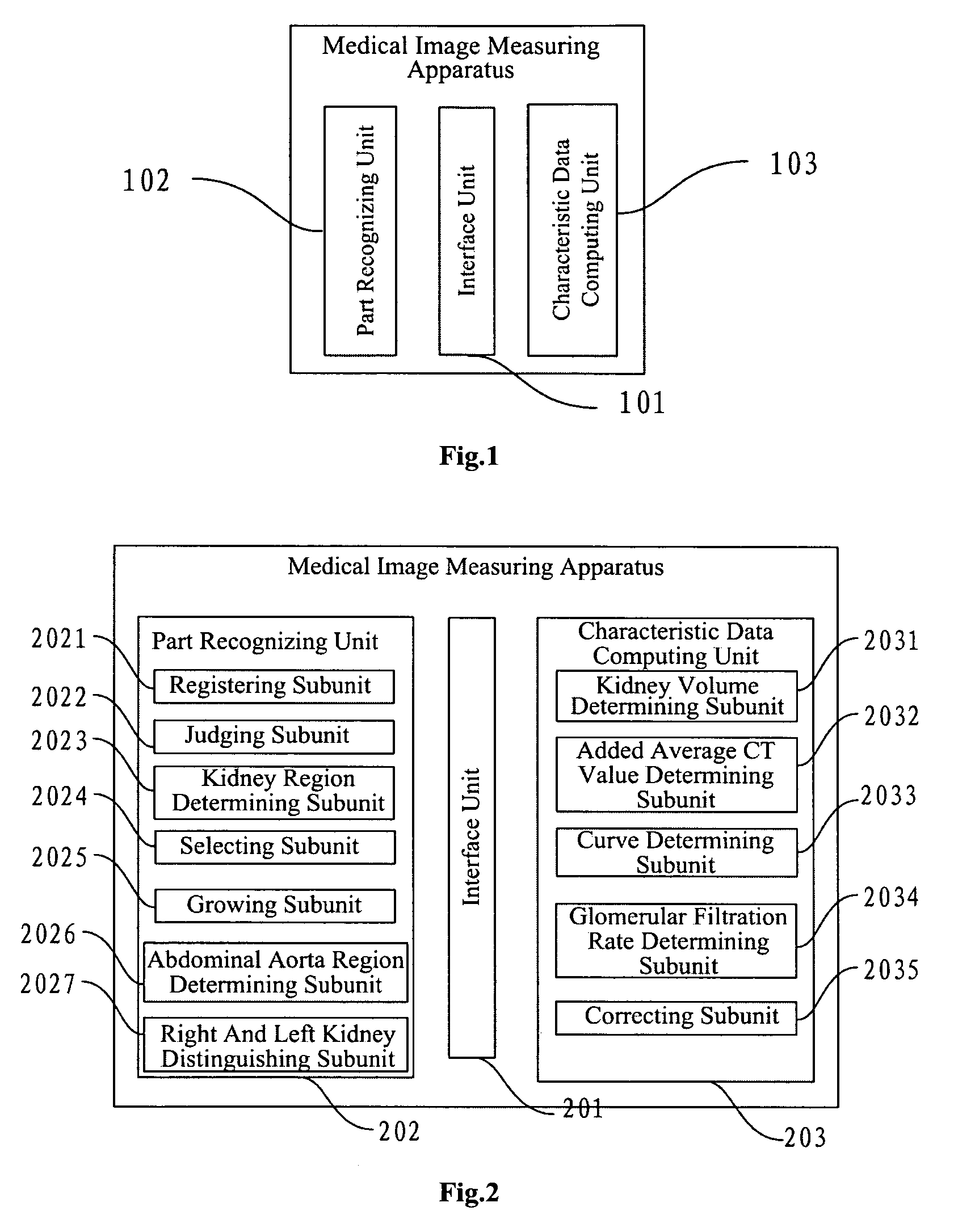
[0069]Refer to FIG. 1, which shows a structure diagram of an abdominal CT image measuring apparatus according to an embodiment of the present invention, including:

[0070]an interface unit 101, for obtaining two-phase or multi-phase scan images of a part under test, wherein the scanned images include plain scan image series and enhanced scan image series, and the images especially refer to abdominal CT images in the embodiments of the present invention;

[0071]a part recognizing unit 102, for recognizing the points representing the part under test on the scan images; and

[0072]a characteristic data computing unit 103, for extracting the CT values of the points representing the part under test, and computing the required characteristic data of the part under ...


PUM


Abstract

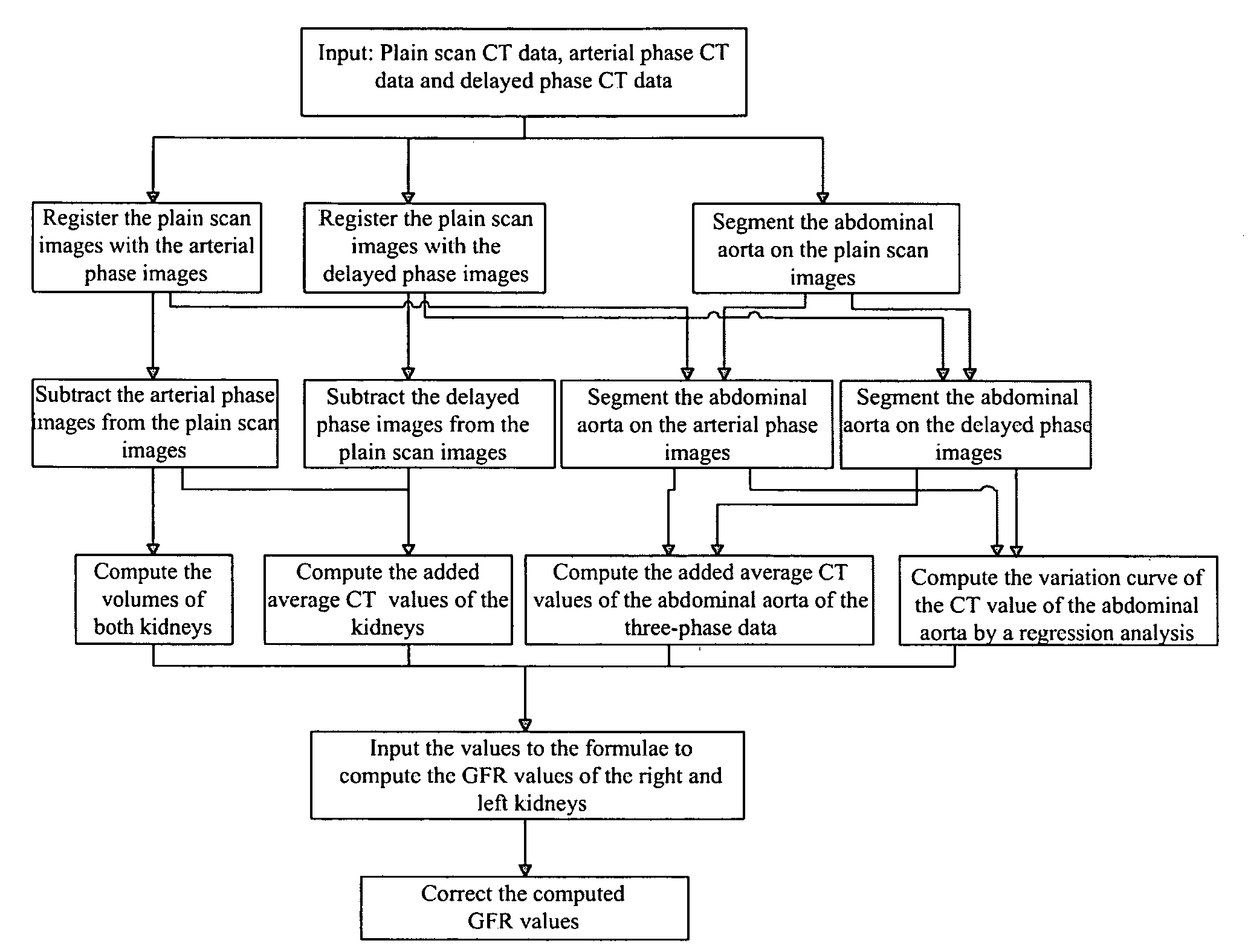
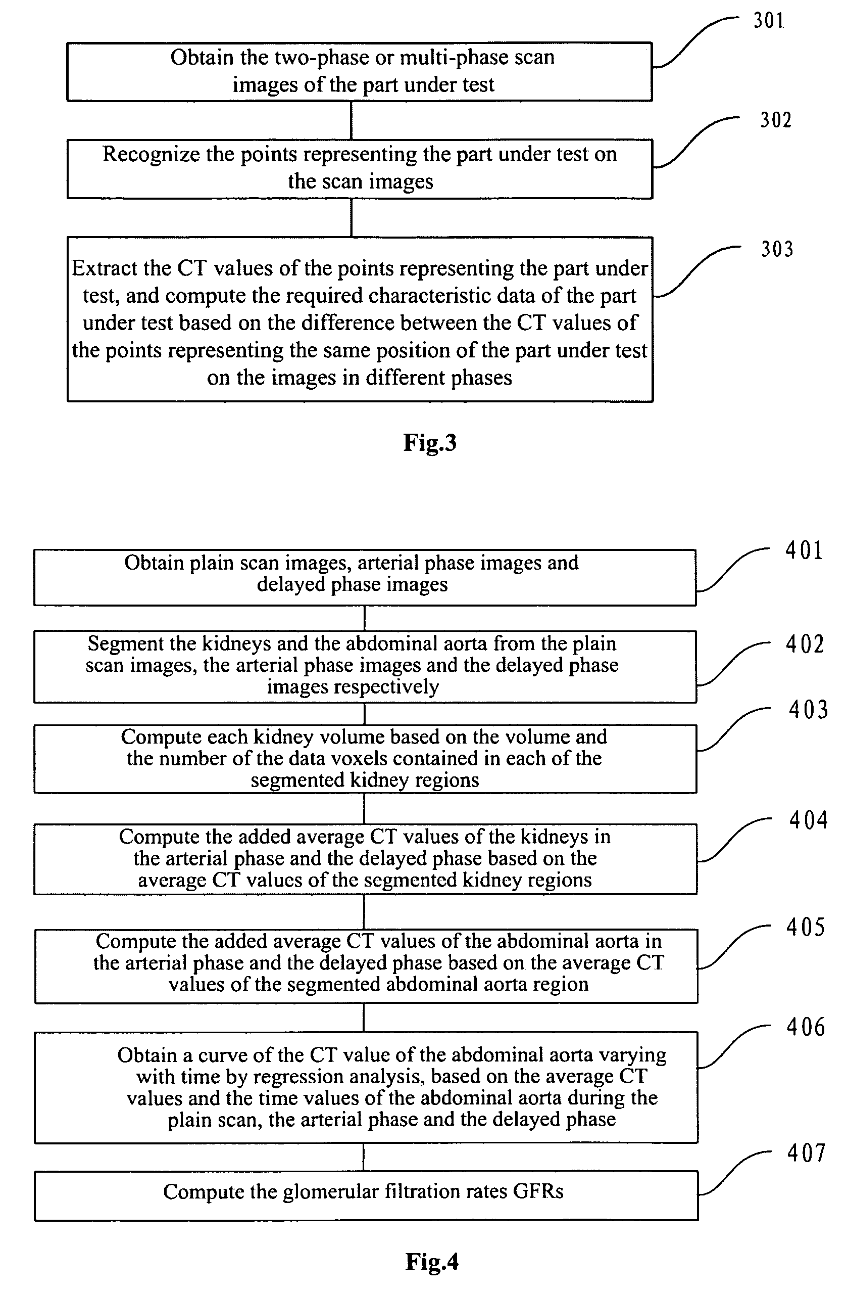
The present invention discloses an abdominal CT image measuring apparatus and method. The abdominal CT image measuring apparatus includes: an interface unit; a part recognizing unit and a characteristic data computing unit. The present invention can determine the specific region of the part under test with a little amount of computation, by registration and subtraction operation on the two-phase scan images. This is easy to be carried out in computers, thus the computing speed of the characteristic data can be guaranteed and the efficiency can be improved. By the recognizing of the kidney regions and the abdominal aorta region in the present invention, the glomerular filtration rates obtained by applying the key concept of the present invention to the image measuring of glomerular filtration rate can meet the clinical application requirements in both precision and speed.

Description

FIELD OF THE INVENTION[0001]The present invention relates to the field of image processing, and more particularly, to a method and an apparatus for measuring diagnostic reference values based on abdominal CT images, and an image measuring system for glomerular filtration rate.BACKGROUND OF THE INVENTION[0002]Computer Tomography (CT) uses computer-controlled X-rays to scan human in stratum. It can accurately display an anatomical structure by comparing the different densities of human body tissues presented under X-rays. Medical images obtained by CT are usually inspected and measured by physicians or the like to obtain required characteristic data as diagnostic basis. However, the measuring method for diagnostic reference values of CT images in the prior art depends more on manual work, resulting in a low efficiency and an insufficient precision.[0003]For example, Chinese patent application No. 03136006.8 discloses a medical image processing apparatus for processing medical image(s)...

Claims


Application Information

Patent Timeline
Patent Type & Authority Patents(United States)
IPC IPC(8): G06K9/00G06V10/26
CPCG06K9/34G06T7/0012G06T7/0081A61B6/50G06K2209/05G06T2207/10081G06T2207/30084G06T7/11G06V10/26G06V2201/03
Inventor MA, HONGFENGDI, QIANGWU, LEIFU, HONGLEIMEI, LONGWEITIAN, LIANGQUAN, SONGMINZHANG, JUNKANG, YAN
Owner NEUSOFT MEDICAL SYST CO LTD
Learn More