Endoscopic anchoring device and associated method

a technology of endoscopic anchoring and associated methods, which is applied in the field of endoscopic anchoring devices and surgical anchors, can solve the problems of no reliable method for securing clips, staples, or other surgical fasteners inside a patient, and no such devices currently available, and achieve the effect of reducing the risk of surgical anchoring

Active Publication Date: 2010-11-16
GRANIT MEDICAL INNOVATION
View PDF44 Cites 55 Cited by
  • Summary
  • Abstract
  • Description
  • Claims
  • Application Information

AI Technical Summary

Benefits of technology

[0018]In one embodiment of the invention, the anchors include anchor spines, which are curved at their distal ends for anchoring into tissue, and are sharply pointed at their distal ends for the purpose of easily penetrating the tissue. Two corresponding spines are formed from one thin metal rod that is bent at a medial aspect thereof. Several such metal rods are thusly bent, and coupled with a crimping mechanism such as, for example, a metal ring or collar. The crimping mechanism is placed proximate the bight portion created by the bend in the rods, forming a proximal loop.

Problems solved by technology

Currently, there is no reliable method for securing clips, staples, or other surgical fasteners inside a patient's body in conjunction with a flexible endoscope.
Secondly, a flexible endoscope bends along with curvatures of the internal body lumens, requiring similar flexibility of a fastener delivery and deployment device to enable such a journey.
Both of these challenges have not yet been surmounted; hence there are no such devices currently available.
Suturing operations have similar limitations, and as such physicians have been unable to perform surgical procedures via natural body orifices using a flexible endoscope.
Consequently, a staple may be displaced, or slip out of the delivery device's jaws.
The problem with the embodiment of the '249 patent is the following: bowel wall thickness, for example, is approximately 0.5 cm, and its consistency is slightly firmer than that of a calf's liver.

Method used


Image

  • Endoscopic anchoring device and associated method
  • Endoscopic anchoring device and associated method
  • Endoscopic anchoring device and associated method

Examples


Embodiment Construction

Definitions

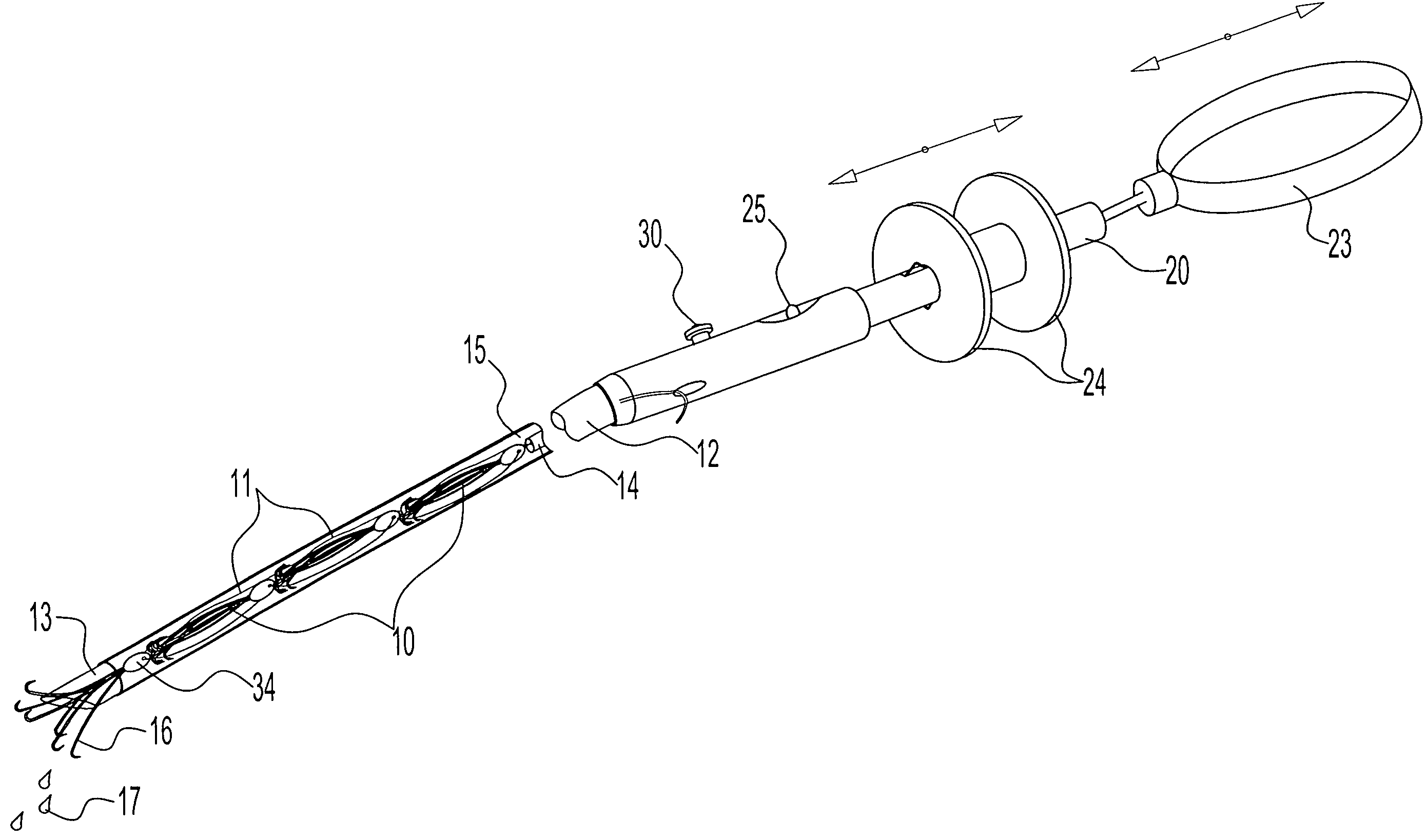
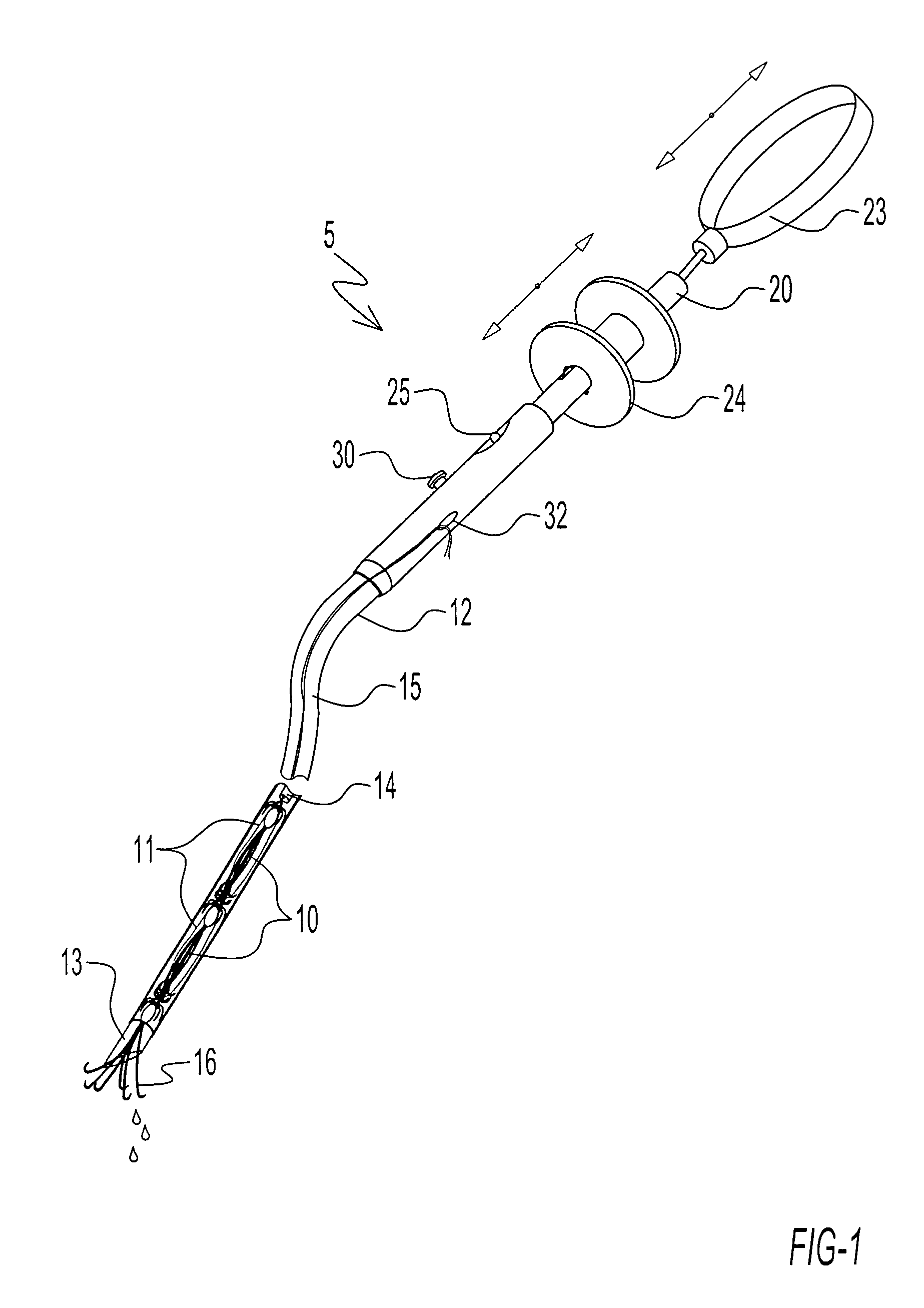
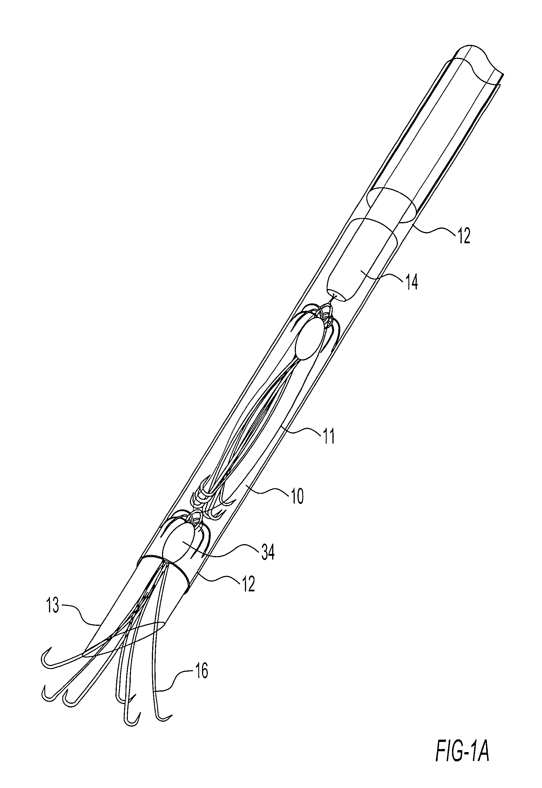
[0042]The term “endoscopic” is used herein to designate any of a variety of minimally invasive surgical procedures wherein optical elements are used to view internal spaces and tissues of a patient through relatively small surgically created openings or natural orifices. Concomitantly, the term “endoscope” as used herein refers to any optical or tubular instrument inserted through such openings or orifices for purposes of enabling visualization of and / or access to internal tissues during a minimally invasive procedure.

[0043]During a laparoscopic procedure, for example, an optical element may be inserted through one small incision, while one or more cannulas would be inserted through one or more separate incisions. The surgical instruments inserted through the cannulas are visualized by means of the first optical element. During a flexible endoscopic procedure on the other hand, a flexible endoscope may include, for example, both the optical element and one or more channel...


PUM


Abstract

An endoscopic fastening assembly having a diameter sufficiently small so that the fastening assembly is slidably insertable into a working channel of a flexible or rigid endoscope for performing a fastening operation, comprises one or more surgical anchors within an elongated tubular member configured for delivery and deployment of said anchors. One or more anchors being disposed in a substantially closed pre-deployment configuration inside the tubular body are configured for being delivered and deployed into a target tissue while remaining in a substantially closed configuration, and assuming its open anchoring properties only once embedded in tissue.

Description

RELATED APPLICATIONS[0001]This application claims the benefit of the priority of U.S. Provisional Application No. 60 / 563,534, filed on Apr. 19, 2004, which application is incorporated herein by reference in its entirety.FIELD OF THE INVENTION[0002]The present invention relates to an endoscopic anchoring device and surgical anchors to be used in conjunction with flexible or rigid endoscopy, or during open surgery. Anchoring is performed on internal body tissues as part of a surgical diagnostic or therapeutic procedure using one or more surgical anchors and an associated anchor delivery and deployment assembly designed for delivering one or more anchors.BACKGROUND OF THE INVENTION[0003]Conventional surgery often necessitates the use of clips, staples, or other surgical fasteners. Such procedures generally require that an extensive incision be made (open surgery), or that a series of small incisions be created, through which several cannulas are placed for providing access to a body ca...

Claims


Application Information

Patent Timeline
Patent Type & Authority Patents(United States)
IPC IPC(8): A61B17/08A61B17/04A61D1/00A61B17/00
CPCA61B17/0401A61B17/0469A61B2017/00004A61B2017/00862A61B2017/00867A61B2017/0412A61B2017/0414A61B2017/0437A61B2017/0464
Inventor NAKAO, NAOMI L.
Owner GRANIT MEDICAL INNOVATION
Why Eureka
Try Eureka