Dedicated mobile high resolution prostate PET imager with an insertable transrectal probe

a high-resolution, mobile technology, applied in tomography, x/gamma/cosmic radiation measurement, instruments, etc., can solve the problems of insufficient accuracy, bulky standard pet imagers, and existing mobile pet imagers that do not meet the special combined requirements of size, resolution and sensitivity, and achieve fast data replay

Active Publication Date: 2010-12-28
JEFFERSON SCI ASSOCS LLC
View PDF10 Cites 4 Cited by
  • Summary
  • Abstract
  • Description
  • Claims
  • Application Information

AI Technical Summary

Benefits of technology

[0013]Several advantages are achieved with the dedicated prostate PET imager system with insertable probe of the present invention, including:
[0014](1) The imager will provide a large active field of view including torso-wide (40 cm or more) by at least 10 cm wide.
[0015](2) The outside imager includes detector modules placed in front of the patient and behind the patient to improve reconstruction resolution of the prostate region.
[0016](3) The outside imager will provide a 3D reconstruction resolution of 1.5-2.0 mm (over body slice involved).
[0017](4) A reconstruction resolution of 1.0-1.5 mm 2D will be provided with the probe in coincidence with the outside imager (over the small field of view of the probe).
[0018](5) A rotating open geometry gantry will enable 360 deg angular sampling in the 3D imaging mode with the outside imager.
[0019](6) The transrectal probe of the current invention will image a larger portion of the tissue surrounding the prostate as compared to prior art probes.
[0020](7) The prostate imager includes a multi-channel fast data acquisition and processing system capable of recording data with at least 200 kHz trigger rate in a list mode to enable prompt limited data analysis and fast data replay and image reconstruction during the same scan session.

Problems solved by technology

In the current state of the art, standard PET imagers are bulky devices that are placed in dedicated imaging rooms and require the patient to be transported to the imager.
These standard imagers have poor spatial resolution that is inadequate for accurate imaging of small organs such as the prostate.
Existing mobile PET imagers do not satisfy the special combined requirements of size, resolution and sensitivity for prostate imaging tests.
In this approach, the limited detector size and limited angular sampling of the imaging procedure does not allow for full scale all-angle 3D tomographic imaging of the prostate region and of surrounding organs.
Unfortunately, this approach has limited angular sampling of the imaged organ.
There are practical implementation issues related to the use of insertable transrectal probes.
They cannot be treated as non-invasive, with the related patient safety issues involving active probes.
There are limits to the useful detection volume, probe positioning relative to prostate, and non-uniformity of sensitivity for different parts of prostate.
Insertable probes, while being close to parts of the prostate, will not image enough surrounding tissue.

Method used


Image

  • Dedicated mobile high resolution prostate PET imager with an insertable transrectal probe
  • Dedicated mobile high resolution prostate PET imager with an insertable transrectal probe
  • Dedicated mobile high resolution prostate PET imager with an insertable transrectal probe

Examples


Embodiment Construction

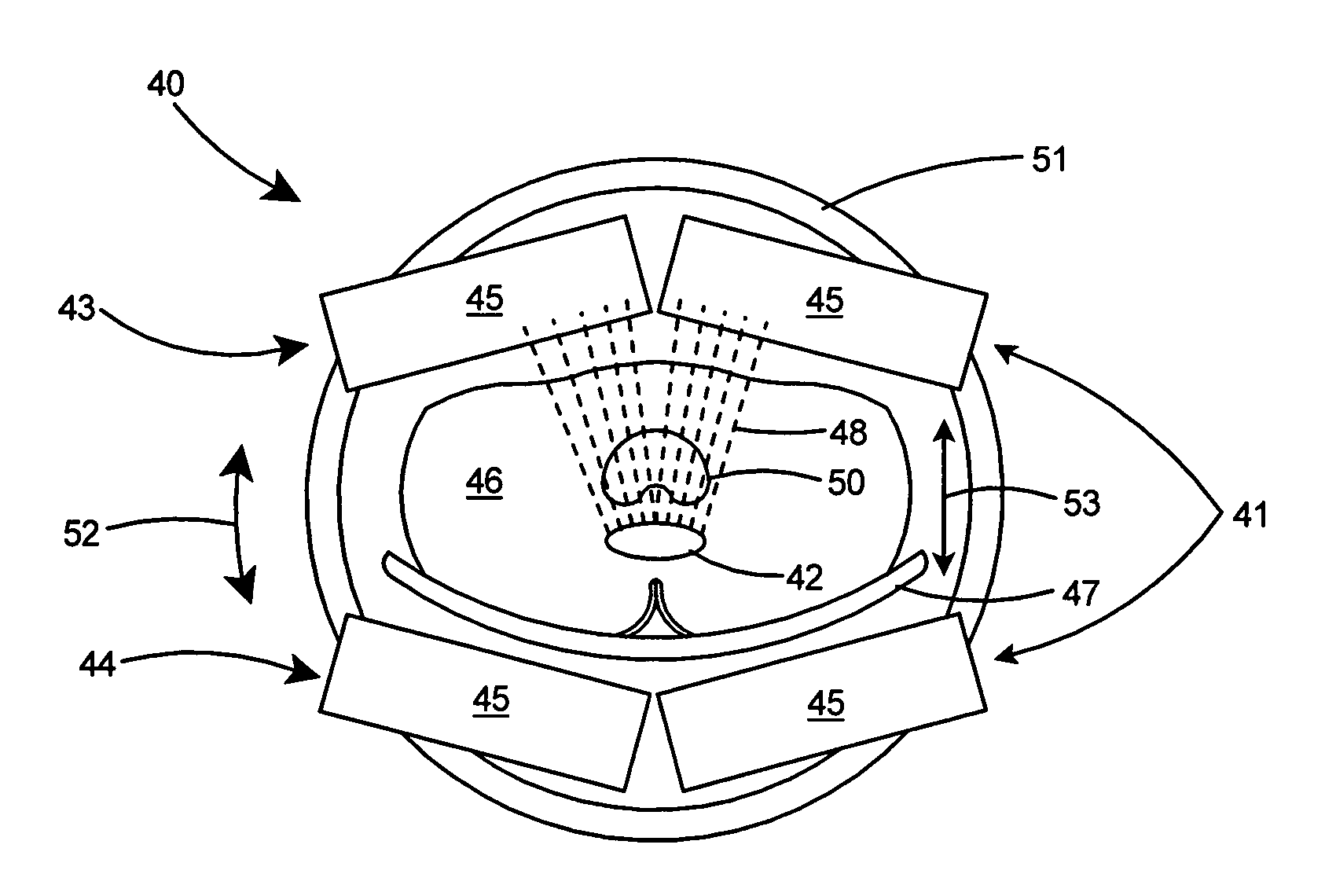
[0088]According to the present invention, a dedicated mobile high resolution PET imager to image the prostate and surrounding organs combines two major components including an outside high resolution dedicated PET imager placed close to the patient's torso with an insertable compact transrectal probe that is placed close to the prostate. The insertable probe operates in conjunction with the outside imager. The two detector systems are spatially co-registered to each other via electronic sensor positioning systems placed on all detector modules. The outside imager mounted on an open rotating gantry provides torso-wide 3D images of the prostate and surrounding tissue and organs. The insertable probe provides closer high sensitivity and very high resolution but limited, mostly 2D, view of the prostate and immediate surroundings. While the outside imager can operate separately, the critical focus of the present invention is the operation of the probe in conjunction with the outside imag...


PUM


Abstract

A dedicated mobile PET imaging system to image the prostate and surrounding organs. The imaging system includes an outside high resolution PET imager placed close to the patient's torso and an insertable and compact transrectal probe that is placed in close proximity to the prostate and operates in conjunction with the outside imager. The two detector systems are spatially co-registered to each other. The outside imager is mounted on an open rotating gantry to provide torso-wide 3D images of the prostate and surrounding tissue and organs. The insertable probe provides closer imaging, high sensitivity, and very high resolution predominately 2D view of the prostate and immediate surroundings. The probe is operated in conjunction with the outside imager and a fast data acquisition system to provide very high resolution reconstruction of the prostate and surrounding tissue and organs.

Description

[0001]The United States of America may have certain rights to this invention under Management and Operating contract No. DE-AC05-06OR23177 from the Department of Energy.FIELD OF THE INVENTION[0002]This invention relates to diagnostic imaging and more particularly to a high resolution PET imaging system for imaging the prostate gland and surrounding organs in a patient.BACKGROUND OF THE INVENTION[0003]In the current state of the art, standard PET imagers are bulky devices that are placed in dedicated imaging rooms and require the patient to be transported to the imager. These standard imagers have poor spatial resolution that is inadequate for accurate imaging of small organs such as the prostate. In addition, in some clinical situations, there would be an advantage to having a dedicated imager that can be, for example, assisting in the surgery suite where imaging can provide immediate biopsy guidance of (suspected) cancerous lesions, and with cancerous tissue removal both from prost...

Claims


Application Information

Patent Timeline
Patent Type & Authority Patents(United States)
IPC IPC(8): G01T1/164
CPCA61B6/037A61B6/425A61B6/4266A61B6/50G01T1/1603G01T1/2985A61B6/547
Inventor MAJEWSKI, STANISLAWPROFFITT, JAMES
Owner JEFFERSON SCI ASSOCS LLC
Why Eureka
Try Eureka