Ultrasonic diagnostic apparatus

a diagnostic apparatus and ultrasonic technology, applied in the field of ultrasonic diagnostic equipment, can solve the problems of unavoidable time and high time requirements, and achieve the effect of reducing the overall diagnostic tim

Inactive Publication Date: 2011-01-18
HITACHI LTD
View PDF10 Cites 3 Cited by
  • Summary
  • Abstract
  • Description
  • Claims
  • Application Information

AI Technical Summary

Benefits of technology

[0009]As described above, according to the ultrasonic diagnostic apparatus of the present invention, a plurality of elastic image acquisition modes are provided. Accordingly, by setting one acquisition mode suitable for a close examination mode and setting another acquisition mode suitable for a screening mode, not only the close examination mode, but also the screening mode can be adapted, and thus an elastic image suitable for each examination purpose can be obtained. As a result, the diagnosing time as a whole can be shortened.

Problems solved by technology

However, much time is needed to obtain an elastic image having high image quality, and thus much time is unavoidably taken if only an elastic image of high image quality is obtained.

Method used


Image

  • Ultrasonic diagnostic apparatus
  • Ultrasonic diagnostic apparatus
  • Ultrasonic diagnostic apparatus

Examples


first embodiment

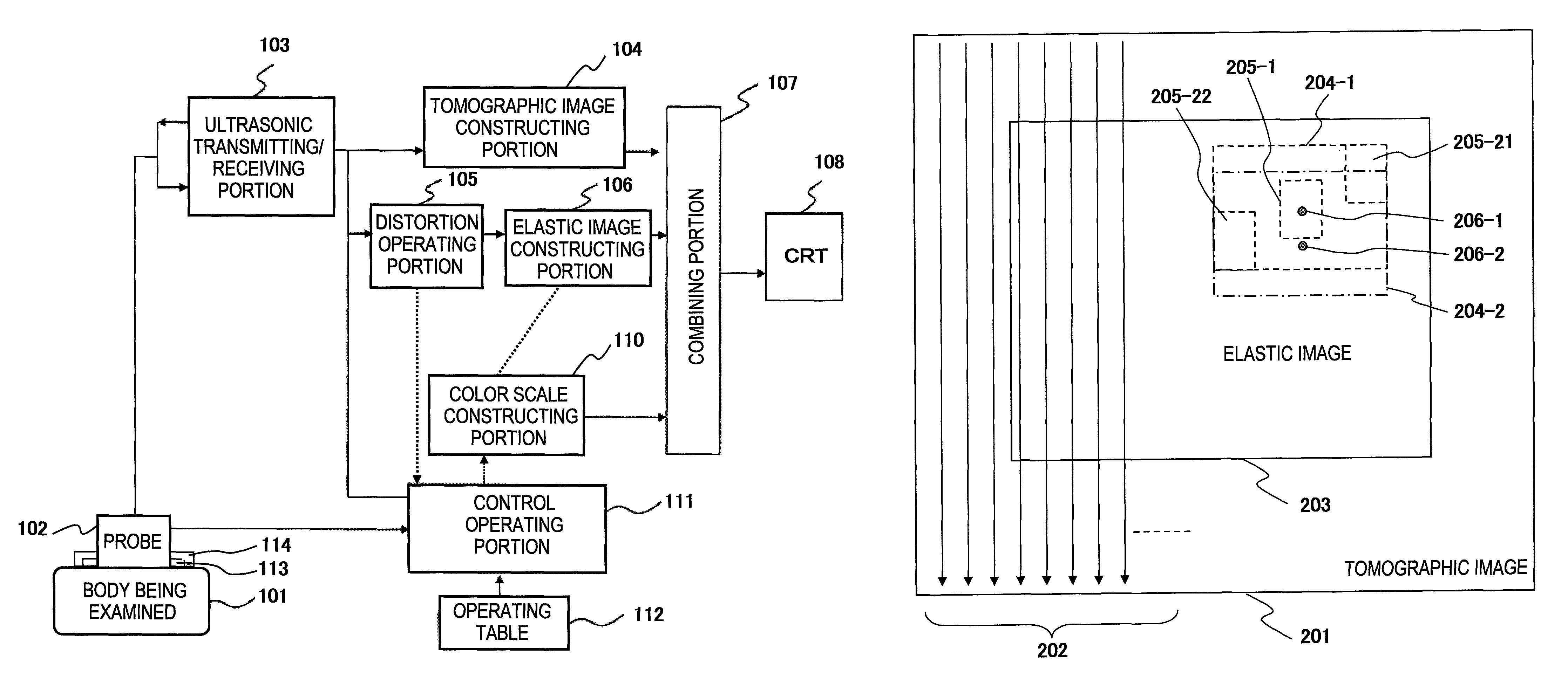
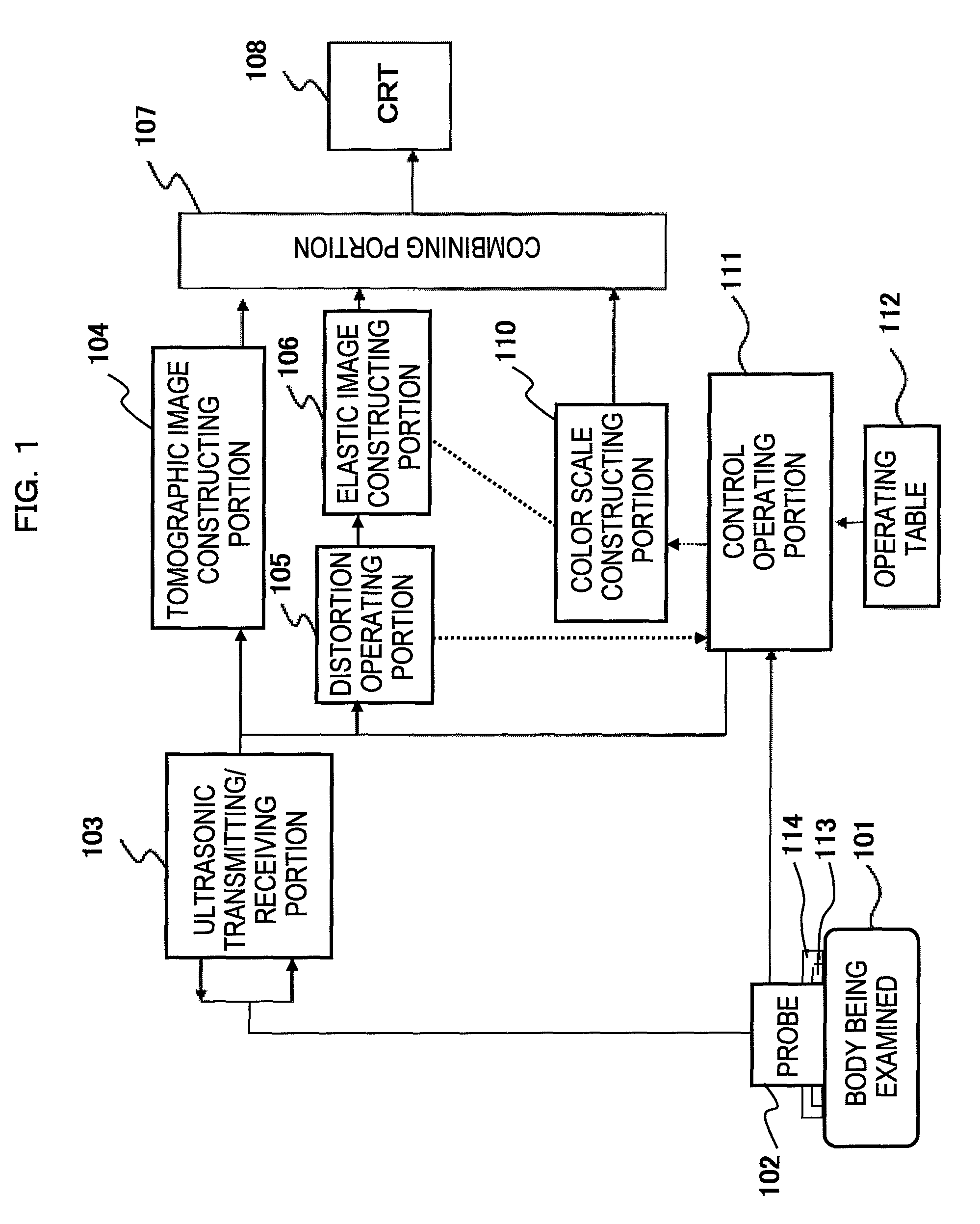
[0030]Next, a first embodiment of the ultrasonic diagnostic apparatus according to the present invention will be described. In this embodiment, the size of a correlation window for detecting the displacement amount of an ultrasonic signal between plural frames is controlled, thereby providing plural acquisition modes which are different at least in the precision of the elastic image.

[0031]In a general examination process, search and specification of a diagnostic site is carried out over a broad region in short time, and then the specified diagnostic site is subjected to close examination. According to this operating process, this embodiment is equipped with not only a close examination mode suitable for close examination of a diagnostic site, but also a screening mode suitable for search and specification of a diagnostic site over a broad region in short time as an acquisition mode for acquiring an elastic image. Furthermore, each acquisition mode may be segmentalized to provided th...

second embodiment

[0042]Next, a second embodiment of the ultrasonic diagnostic apparatus according to the present invention will be described. In this embodiment, the breadth of the search range for detecting the displacement amount of the ultrasonic signal is controlled among plural frames, whereby plural acquisition modes different at least in the image quality of the elastic image are provided.

[0043]As in the case of the first embodiment described above, this embodiment is also provided with plural acquisition modes containing not only the close examination mode, but also the screening mode as the acquisition mode for acquiring an elastic image, and the following description on this embodiment will be made on the basis of these two acquisition modes.

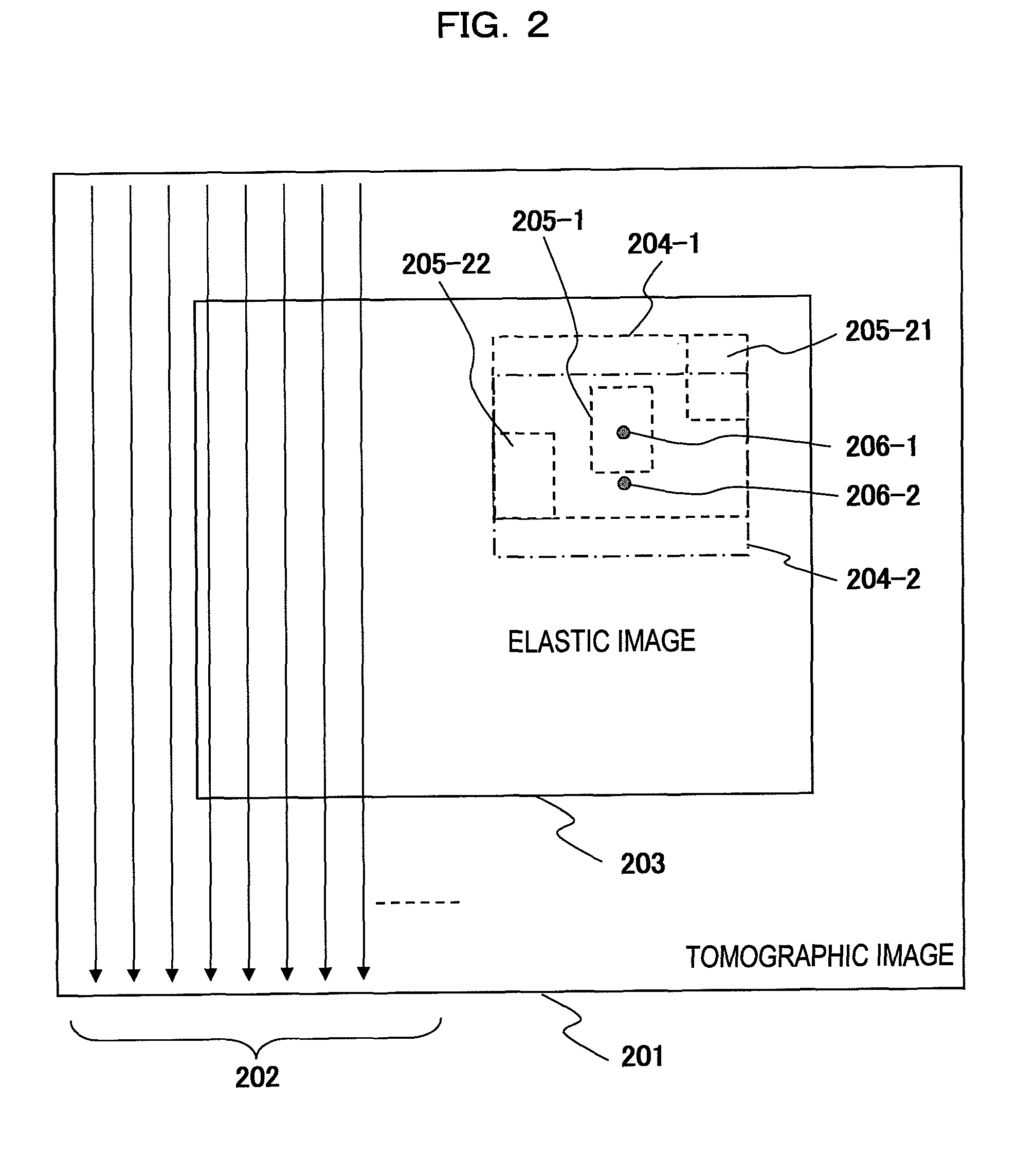
[0044]An example of this embodiment will be described with reference to FIG. 2 as in the case of the example of the first embodiment.

[0045]As described above, in order to detect the displacement amount of the ultrasonic signal at the point 206-1, the d...

third embodiment

[0053]Next, a third embodiment of the ultrasonic diagnostic apparatus according to the present invention will be described. According to this embodiment, plural acquisition modes different in at least one of the spatial resolution and the frame rate of the elastic image are provided by controlling the moving interval of the search range set to detect the displacement amount of an ultrasonic signal among plural frames, that is, controlling the thinning-out of the calculation of the tissue elasticity amount.

[0054]As in the case of the first embodiment, this embodiment is provided with plural acquisition modes containing not only the close examination mode, but also the screening mode as the acquisition mode for acquiring the elastic image, and the following description of this embodiment will be made on the basis of these two acquisition modes.

[0055]A first example of this embodiment will be described. In this example, at least one of the spatial resolution and the frame rate of the e...


PUM


Abstract

An ultrasonic diagnostic apparatus having a function of obtaining a tomographic image and an elastic image is adaptable to not only a close examination mode, but also a screening mode, and obtains an elastic image suitable for each examination purpose. Accordingly, the ultrasonic diagnostic apparatus of the present invention comprises tomographic image acquisition means for transmitting an ultrasonic wave from a probe to a body being examined, and receiving a reflection echo signal corresponding to the transmission of the ultrasonic wave to obtain a tomographic image, elastic image acquisition means having a first acquisition mode for determining a tissue elasticity amount of a biomedical tissue of the body being examined on the basis of the reflection echo signal to obtain an elastic image, and display means for displaying at least the elastic image. The elastic image acquisition means has a second acquisition mode different from the first acquisition mode.

Description

TECHNICAL FIELD[0001]The present invention relates to an ultrasonic diagnostic apparatus having a function of acquiring an ultrasonic reflectance tomographic image and an tissue elastic image for a diagnosis region in a body being examined by using ultrasonic waves, and particularly to control of a mode for acquiring a tissue elastic image.BACKGROUND ART[0002]According to an ultrasonic diagnostic apparatus, the ultrasonic reflectance of a biomedical tissue at a diagnosis site in a body being examined is measured by ultrasonic waves and the reflectance is converted to brightness to thereby display the measurement result as a reflectance tomographic image (hereinafter abbreviated as “tomographic image”) of the diagnosis site.[0003]Furthermore, according to the ultrasonic diagnostic apparatus, it has been recently implemented that the displacement of a biomedical tissue is measured on a tomographic image which is acquired by applying a pressure variation to the biomedical tissue with a...

Claims


Application Information

Patent Timeline
Patent Type & Authority Patents(United States)
IPC IPC(8): A61B8/00G06K9/00
CPCA61B8/08A61B8/14A61B8/463A61B8/485G01S7/52074G01S7/52042G01S7/52085
Inventor WAKI, KOJIMURAYAMA, NAOYUKI
Owner HITACHI LTD
Why Eureka
Try Eureka