
Robotic 5-dimensional ultrasound

a technology of ultrasound imaging and robotics, applied in the field of ultrasound imaging, can solve the problems of inability to effectively identify, monitor or track the target tissue, and facilitate planning or control, and achieve the effect of accurate visualization of the target area, and accurate identification and segmentation of the target area

Active Publication Date: 2011-03-08
THE JOHN HOPKINS UNIV SCHOOL OF MEDICINE
View PDF5 Cites 38 Cited by
  • Summary
  • Abstract
  • Description
  • Claims
  • Application Information

AI Technical Summary

Benefits of technology

Enables accurate identification and segmentation of isoechoic tissues, improves imaging in noisy environments, and provides precise control during ablation procedures by integrating strain and ultrasound data for enhanced visualization and planning.

Problems solved by technology

There are several problems associated with conventional thermal ablation techniques.
While ultrasound is commonly used for target imaging, in general, and ablation monitoring, problems still exist even with these systems, as they do not provide adequate imagery and, as a result, they cannot effectively identify, segment, monitor or track target tissue, nor do they facilitate planning or control prior to and during the ablation process.
One reason conventional robotic 3D ultrasound systems are inadequate is that ultrasound cannot detect all lesions and tumors.
Ultrasound cannot visualize lesions or tumors that are isoechoic.
Thus, conventional 3D ultrasound systems cannot effectively identify, segment, monitor or track target tissue, or facilitate planning and control prior to and during an ablation process where the target tissue is isoechoic.
Another reason conventional 3D ultrasound based systems are not always effective is that such systems do not necessarily provide adequate imaging in a noisy environment.
As one skilled in the art will appreciate, the operation of an ablation probe generates a significant amount of noise which otherwise interferes with the ultrasound signal, and the ability of the system to generate accurate imagery.

Method used


Image

  • Robotic 5-dimensional ultrasound
  • Robotic 5-dimensional ultrasound
  • Robotic 5-dimensional ultrasound

Examples


Embodiment Construction

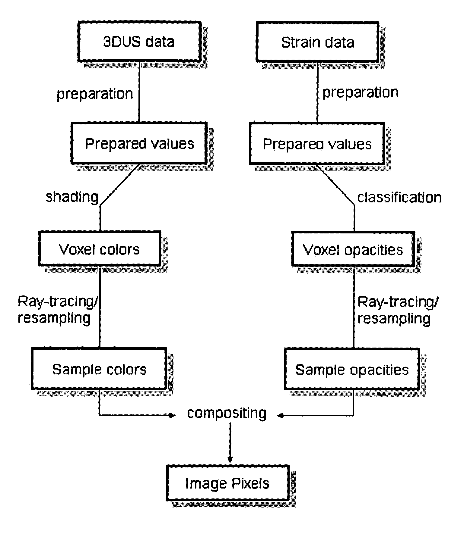
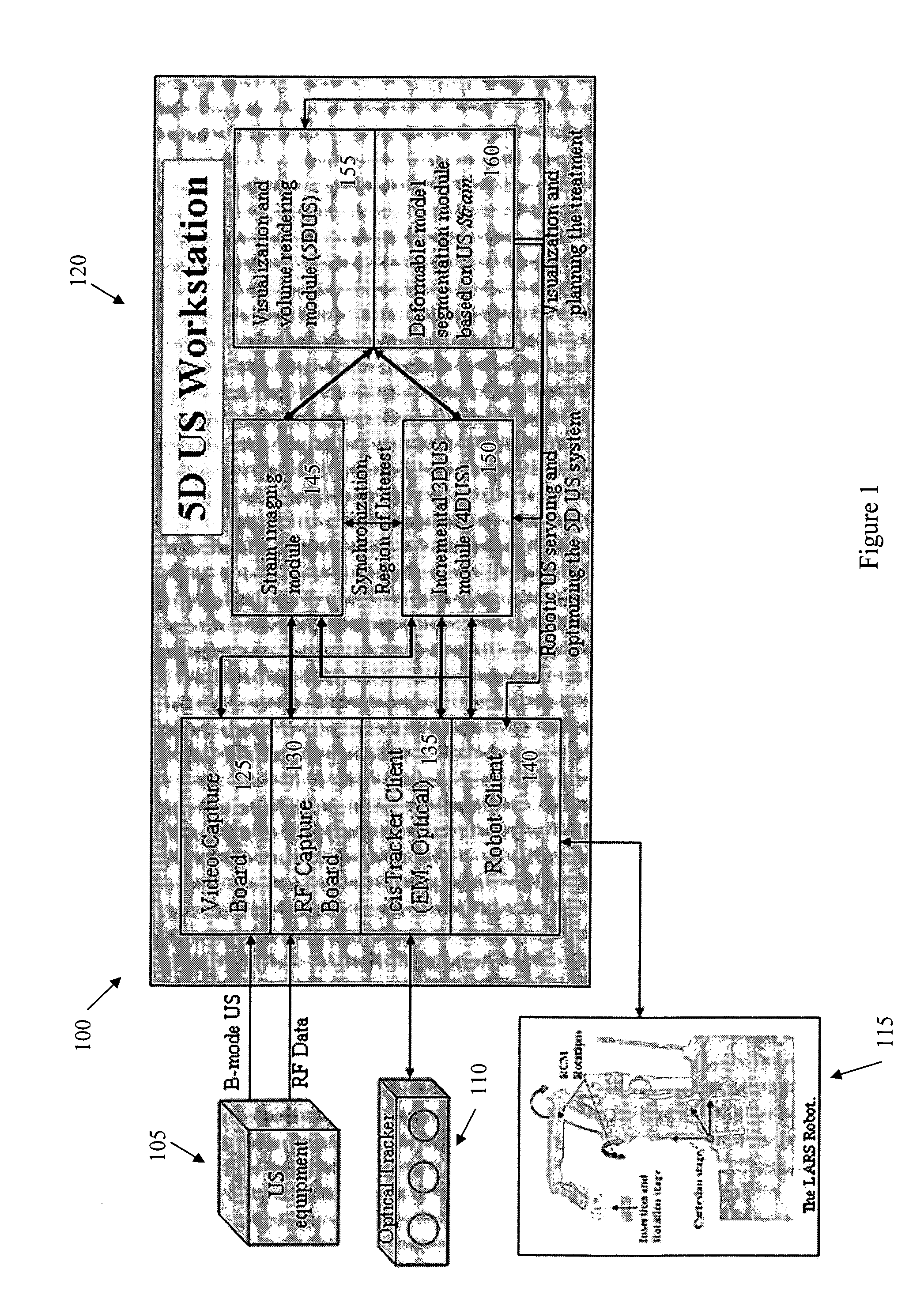
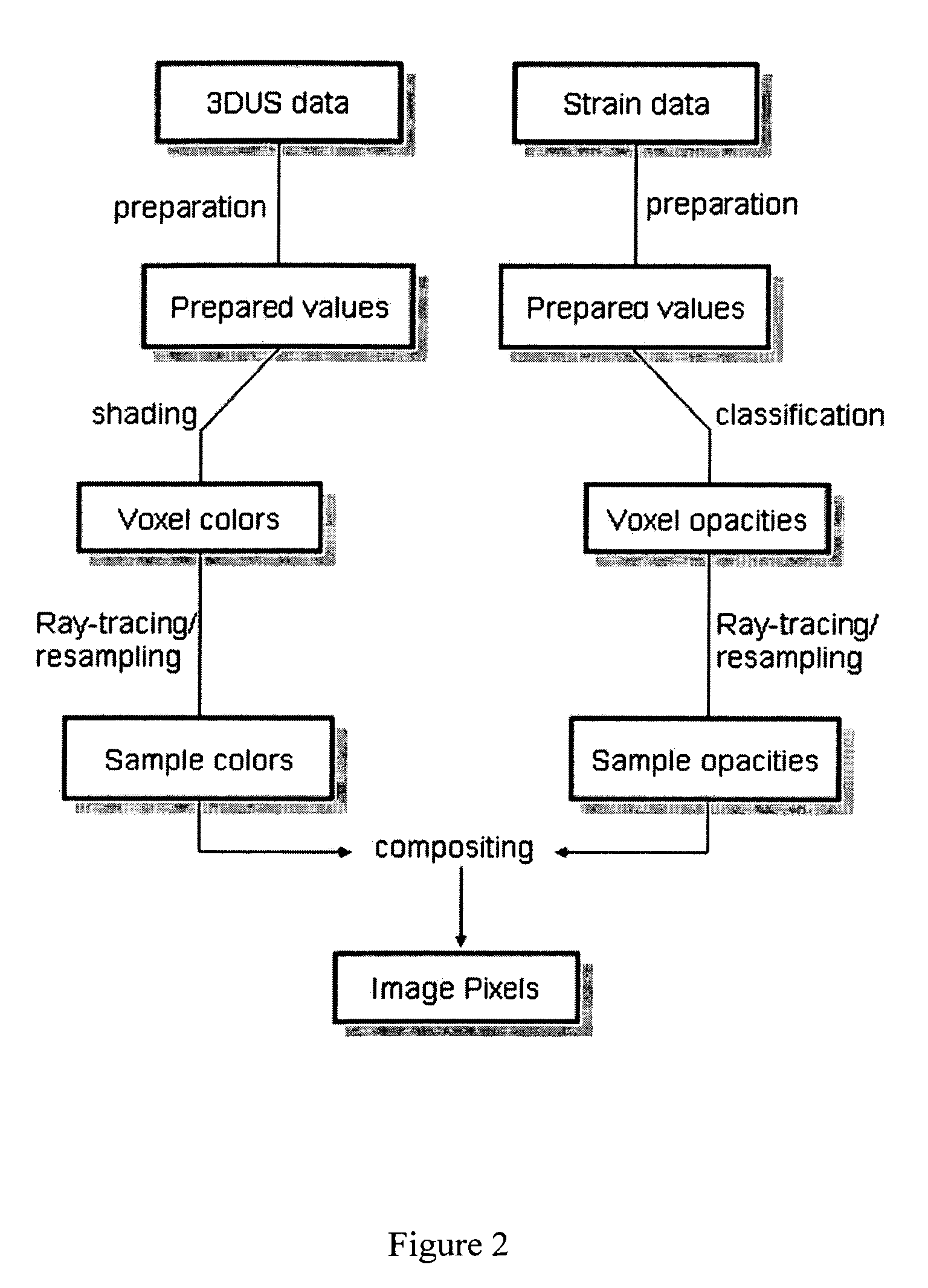
[0029]The focus of the present invention is a robotic 5D ultrasound system and method for use in the field of Image Guided Therapy (IGT) and, more generally, Computer Integrated Surgery (CIS). The robotic 5D ultrasound system and method, in accordance with exemplary embodiments of the present invention enhance “surgical CAD-CAM-TQM” systems by integrating over time 3D ultrasonic image data with strain (i.e., elasticity) image data.

[0030]“Surgical CAD” is analogous to computer-aided design (i.e., CAD) in manufacturing systems, wherein the CAD phase involves the design and / or planning of the intended treatment using an IGT system. In general, prior to such medical procedures, medical images, anatomical atlases and other information are combined for the purpose of generating a model of an individual patient. The model is then used for planning the treatment or intervention procedure. The present invention, however, offers new capabilities with respect to the planning based on more accu...


PUM


Abstract

A robotic 5D ultrasound system and method, for use in a computer integrated surgical system, wherein 3D ultrasonic image data is integrated over time with strain (i.e., elasticity) image data. By integrating the ultrasound image data and the strain image data, the present invention is capable of accurately identifying a target tissue in surrounding tissue; segmenting, monitoring and tracking the target tissue during the surgical procedure; and facilitating proper planning and execution of the surgical procedure, even where the surgical environment is noisy and the target tissue is isoechoic.

Description

CROSS-REFERENCE TO RELATED APPLICATIONS[0001]This application claims the benefit of U.S. Provisional Patent Application No. 60 / 488,941, filed on Jul. 21, 2003, which is hereby incorporated by reference for all purposes as if fully set forth herein.[0002]The research and development effort associated with the subject matter of this patent application was supported by the National Science Foundation under grant no. EEC9731478.BACKGROUND OF THE INVENTION[0003]1. Field of the Invention[0004]The present invention relates to ultrasound imaging, and more particularly, to robotic 5-dimensional (5D) ultrasound imaging.[0005]2. Discussion of the Related Art[0006]Ultrasound is widely used in the medical community for intervention therapy. Examples include thermal ablation, external beam radiation therapy (EBRT), Brachy therapy, and needle insertion procedures, including biopsy procedures.[0007]As stated, thermal ablation is but one procedure for which ultrasound is commonly used. Primary and m...

Claims


Application Information

Patent Timeline
Patent Type & Authority Patents(United States)
IPC IPC(8): A61B8/00A61B8/12A61B8/14G06F
CPCA61B8/08A61B8/4209A61B8/483A61B8/485A61B8/4245
Inventor BOCTOR, EMAD M.CHOTI, MICHAELFICHTINGER, GABORTAYLOR, RUSSELLPRINCE, JERRY L.
Owner THE JOHN HOPKINS UNIV SCHOOL OF MEDICINE
Learn More