Systems and methods for determining organ wall mass by three-dimensional ultrasound

Inactive Publication Date: 2011-09-13
VERATHON
View PDF2 Cites 14 Cited by
  • Summary
  • Abstract
  • Description
  • Claims
  • Application Information

AI Technical Summary

Problems solved by technology

Moreover, the existing 2D methods are manually based, utilizing leading edge-to-leading edge of opposing bladder walls laboriously executed upon a series of two-dimensional images, and are fraught with analytical inaccuracies (H. Miyashita, M. Kojima, and T. Miki, “Ultrasonic measurement of bladder weight as a possible predictor of acute urinary retention in men with lower urinary tract symptoms suggestive of benign prostate hyperplasia”, Ultrasound in Medicine and Biology 2002, 28(8): 985-990; M. Oelke, K. Hofner, B. Wiese, V. Gruneweld, and U. Jonas, “Increase in detrusor wall thickness indicates bladder outlet obstruction in men,”World J. of Urology, 2002, 19(6), 443-452; L. Muller, T. Bergstrom, M. Hellstrom, E. Svensson, and B. Jacobson, “Standardized ultrasound method for assessing detrusor muscle thickness in children,” J. Urol., 200, 164: 134-138; and Naya, M. Kojima, H. Honjyo, A. Ochiai, O. Ukimura, and H. Watanabe, “Intraobserver and interobserver variance in the measurement of ultrasound-estimated bladder weight,”Ultrasound in Med.

Method used


Image

  • Systems and methods for determining organ wall mass by three-dimensional ultrasound
  • Systems and methods for determining organ wall mass by three-dimensional ultrasound
  • Systems and methods for determining organ wall mass by three-dimensional ultrasound

Examples


Embodiment Construction

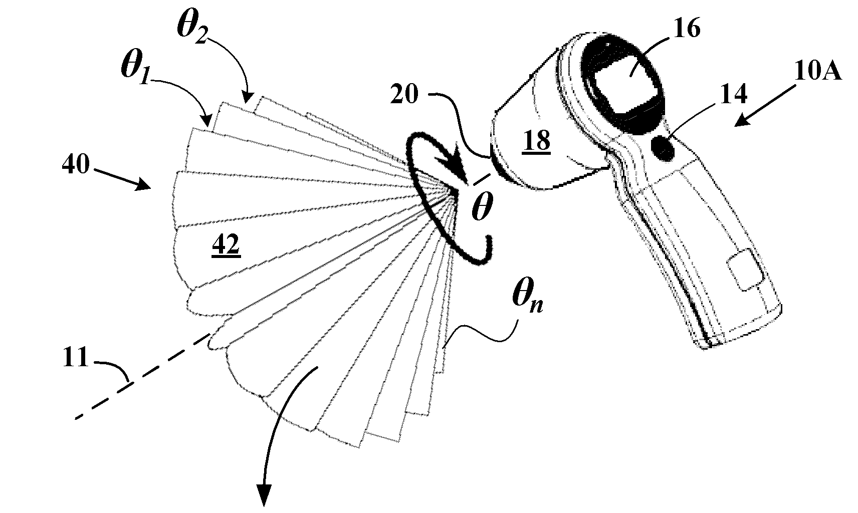
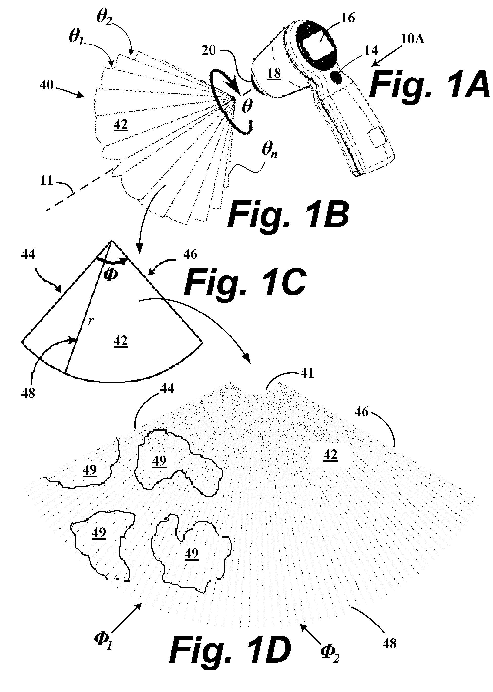
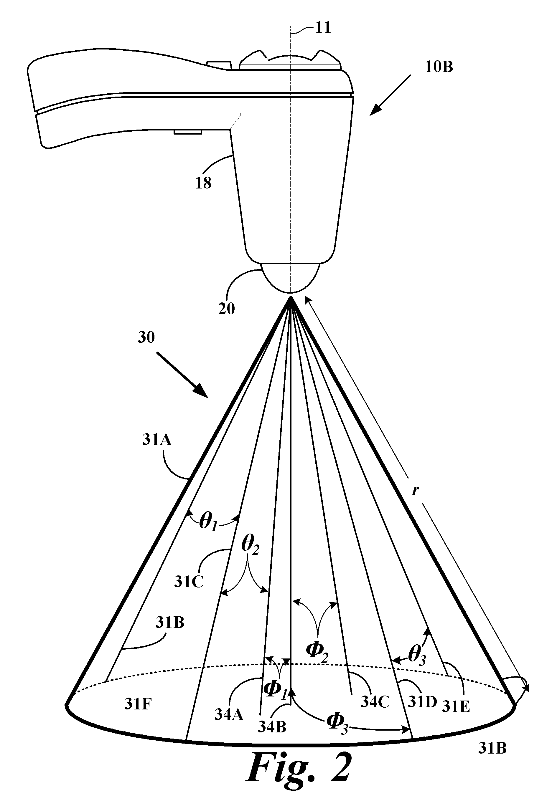
[0038]Methods and systems to acquire an ultrasound estimated organ wall mass and / or weight such as a bladder using three dimensional ultrasound echo information are described. The three-dimensional (3D) based ultrasound information is generated from a microprocessor-based system utilizing an ultrasound transceiver that properly targets the organ or other region of interest (ROI) and utilizes algorithms to delineate the inner (sub-mucosal) and outer (sub-serosal) wall boundaries of the organ wall as part of a process to determine the organ wall weight or mass. When the organ is a bladder, bladder wall algorithms operate without making geometric assumptions of the bladder so that the shape, area, and thickness between the sub-mucosal and subserosal layers of the bladder wall are more accurately determined to provide in a turn a more accurate determination of the bladder wall volume. Knowing the accurate bladder volume allows a more accurate determination of bladder wall weight or mass...


PUM


Abstract

An ultrasound system and method to measure an organ wall weight and mass. When the organ is a bladder, a bladder weight (UEBW) is determined using three-dimensional ultrasound imaging that is acquired using a hand-held or machine controlled ultrasound transceiver. The infravesical region of the bladder is delineated on this 3D data set to enable the calculation of urine volume and the bladder surface area. The outer anterior wall of the bladder is delineated to enable the calculation of the bladder wall thickness (BWT). The UEBW is calculated as a product of the bladder surface area, the bladder wall thickness, and the bladder wall specific gravity.

Description

CROSS REFERENCE TO RELATED APPLICATION[0001]This application is a continuation of U.S. patent application Ser. No. 11 / 295,043 filed Dec. 6, 2005 now U.S. Pat. No. 7,727,150, which application claims priority to U.S. provisional patent application Ser. No. 60 / 633,485 filed Dec. 6, 2004, each of which is incorporated by reference in its entirety as if fully set forth herein.FIELD OF THE INVENTION[0002]This invention relates generally to ultrasound-based diagnostic systems and procedures.BACKGROUND OF THE INVENTION[0003]Benign prostate hyperplasia (BPH) and other disorders can cause mechanical bladder outlet obstruction (BOO). A marker for predicting BOO is determining the weight of the bladder wall. Using probing ultrasound, an ultrasound estimated bladder wall weight (UEBW) might be obtained in a non-invasive way. Existing methods for acquiring UEBW assumes that the bladder is spherically shaped and that the thickness of the bladder wall is relatively constant in near empty to nearly...

Claims


Application Information

Patent Timeline
Patent Type & Authority Patents(United States)
IPC IPC(8): A61B8/00
CPCA61B8/0858A61B8/4254A61B8/483A61B5/204G01S15/8993
Inventor CHALANA, VIKRAMDUDYCHA, STEPHENYUK, JONGTAEMCMORROW, GERALD
Owner VERATHON
Why Eureka
Try Eureka