Method for imaging and spectroscopy of tumors and determination of the efficacy of anti-tumor drug therapies

a tumor and imaging method technology, applied in the field of tumor imaging and spectroscopy and determination of the efficacy of tumor drug therapy, can solve the problem of no single imaging method capable of quantitatively analyzing tumor angiogenesis

Active Publication Date: 2011-12-13
THE JOHN HOPKINS UNIV SCHOOL OF MEDICINE
View PDF26 Cites 13 Cited by
  • Summary
  • Abstract
  • Description
  • Claims
  • Application Information

AI Technical Summary

Benefits of technology

[0011]The invention also provides a method for determining the extent of vascularization of a mass using infrared imaging, comprising obtaining an infrared image of th

Problems solved by technology

At present there is no single imaging method capable of providing quantitative characterization o

Method used


Image

  • Method for imaging and spectroscopy of tumors and determination of the efficacy of anti-tumor drug therapies
  • Method for imaging and spectroscopy of tumors and determination of the efficacy of anti-tumor drug therapies
  • Method for imaging and spectroscopy of tumors and determination of the efficacy of anti-tumor drug therapies

Examples


Embodiment Construction

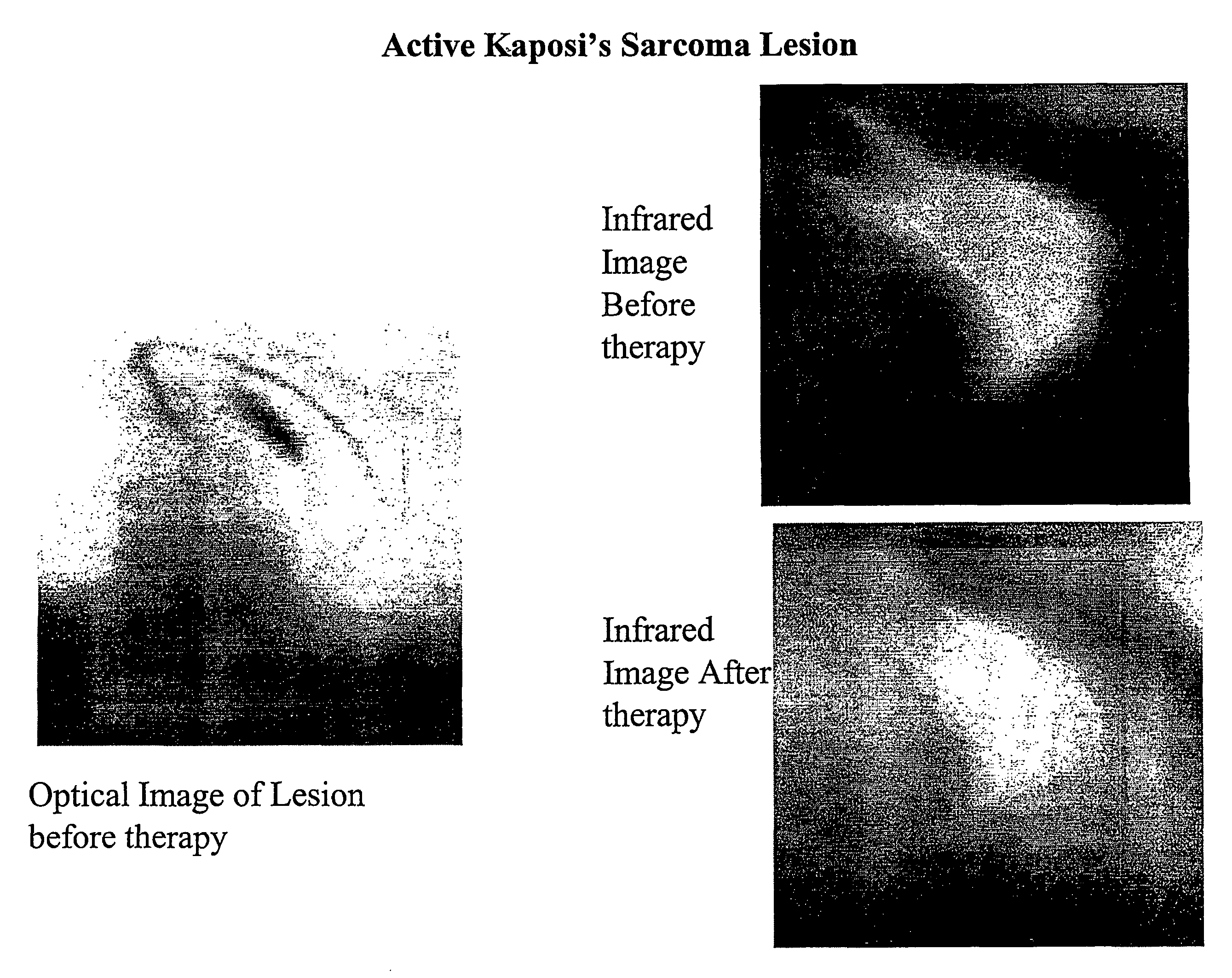
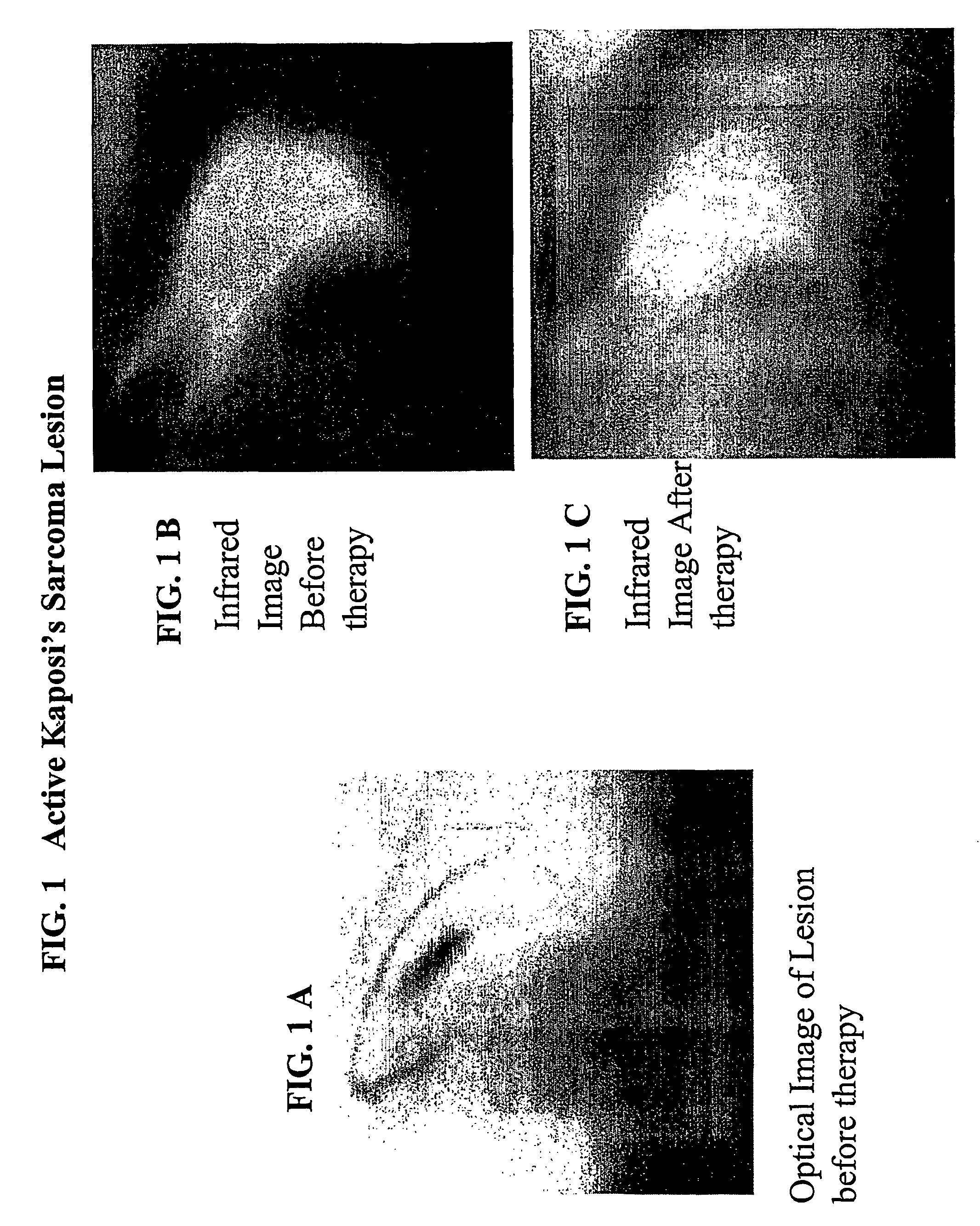
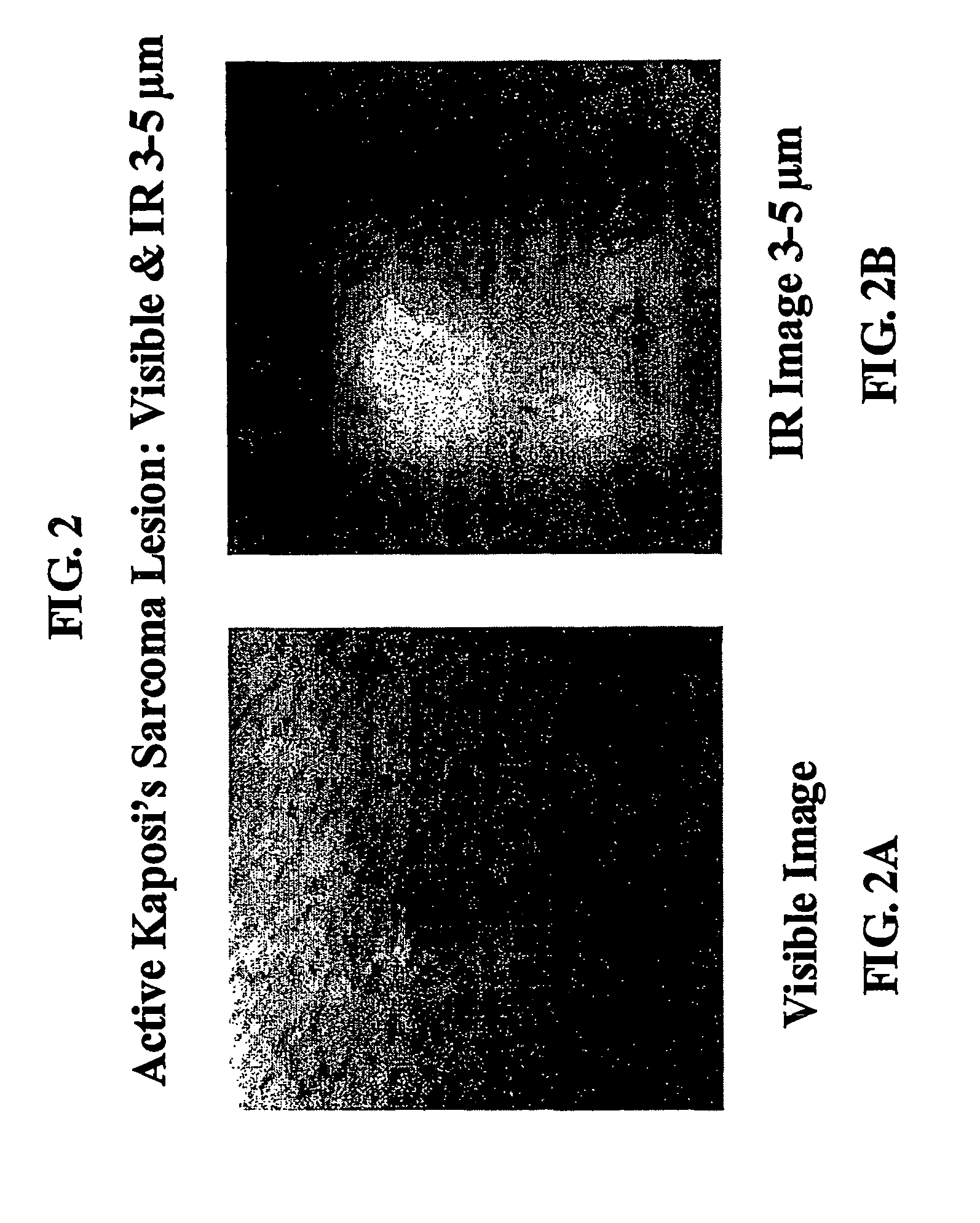
[0019]The present invention is directed to a method of determining regions of vascularity and higher than average metabolic activity of a tissue, tumor or lesion using infrared (“IR”) radiometric thermal imaging. Information on the extent of vascularization and the amount of metabolic activity obtained from IR images taken before and at one or more times after the onset of drug therapy, can be used to assess the efficacy of anticancer or angiogenic disease treatments with drugs or radiation. Because tumor angiogenesis is vital to tumor survival and growth, information on changes in tumor vascularization is useful to monitor efficacy of any treatment, including treatment with antiangiogenesis factors or other regimen that directly or indirectly affects tumor growth.

[0020]The invention is described in part with reference to specific embodiments thereof. It will, however, be evident that various modifications and changes may be made to the inventions without departing from the broader ...


PUM


Abstract

The present invention relates to methods of passive infrared imaging and dynamic infrared imaging of a tumor or lesion (“DIR”) where multiple images are taken before, during and after heating or cooling the tumor or lesion. Patterns of intensity of infrared emission in the infrared image(s) are related to the level of vascularity or metabolic activity of the tumor or lesion, and are used to identify size and shape of the tumor or lesion. The patterns can also be used to determine whether Kaposi's sarcoma lesions, skin cancer lesions and melanomas are active or inactive. Changes in the patterns of infrared emission determined by comparing images taken before, during and after the onset of drug or radiation therapy, are used to assess efficacy of the therapy. The methods can also be used to study tissue affected by angiogenic diseases.

Description

CROSS-REFERENCE TO RELATED APPLICATIONSBACKGROUND[0001]1. Field of the Invention[0002]The present invention relates to methods of passive and active dynamic infrared imaging of a tumor or lesion (“DIRI”) that heats or cools the tumor or lesion. Patterns of intensity of infrared emission in the infrared image are related to the level of vascularity or metabolic activity of the tumor or lesion, and are used to identify the size and shape of the tumor or lesion. Changes in the patterns of infrared emission determined by comparing images taken before, during and after the onset of drug or radiation therapy, are used to assess the efficacy of the therapy. The methods can also be used to study tissue that has been affected by angiogenic diseases.[0003]2. Description of the Related Art[0004]Tumor angiogenesis, the recruitment of new blood vessels by a growing tumor from existing neighboring vessels, is essential for the growth and persistence of solid tumors and their metastases (Folkman, ...

Claims


Application Information

Patent Timeline
IPC IPC(8): A61B6/00A61B5/00
CPCA61B5/015A61B5/415A61B5/416A61B5/418
Inventor MURPHY, JOHN CWILLIAMS, JERRY R.OSIANDER, ROBERT
Owner THE JOHN HOPKINS UNIV SCHOOL OF MEDICINE
Why Eureka
Try Eureka