

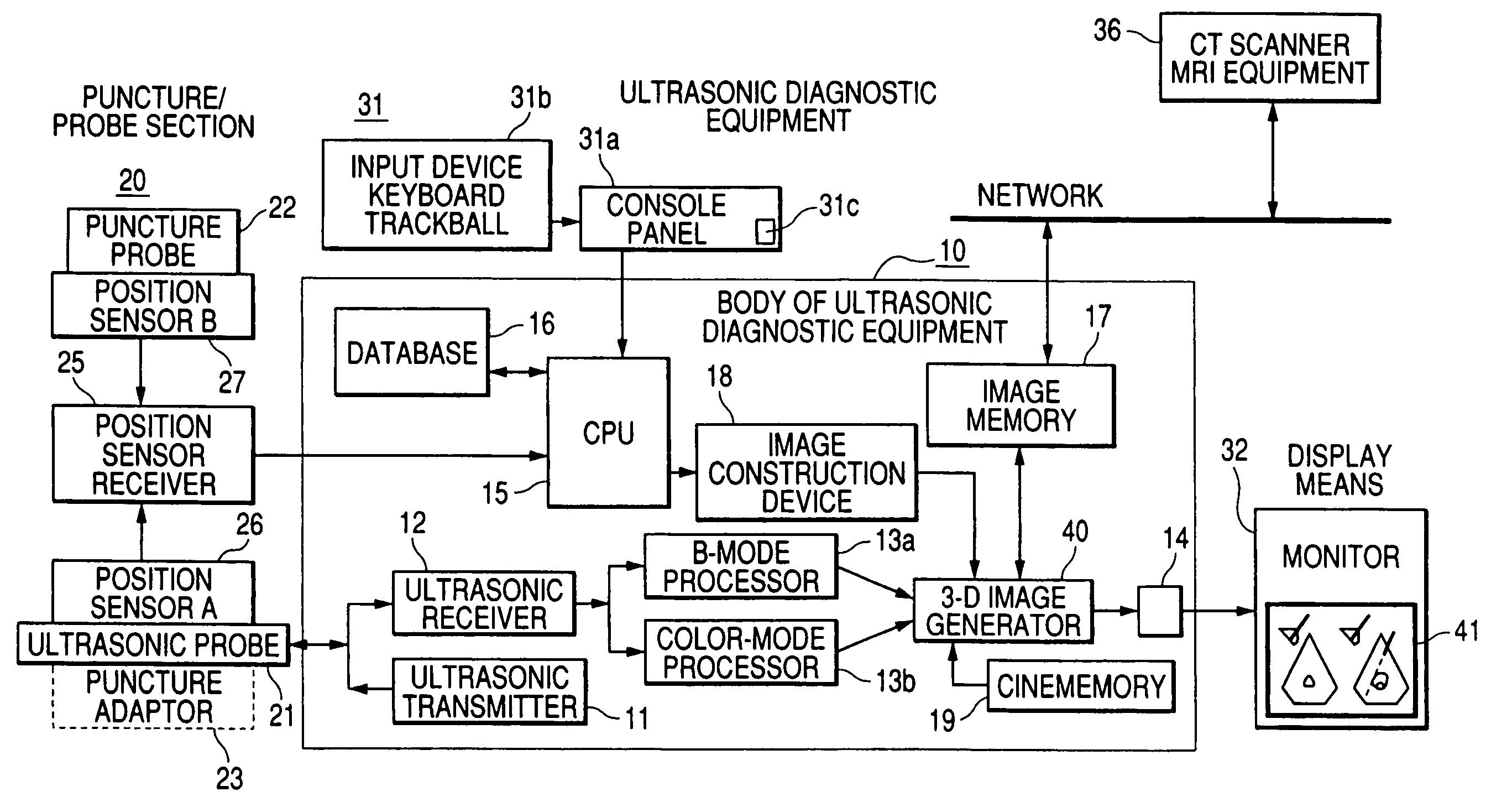
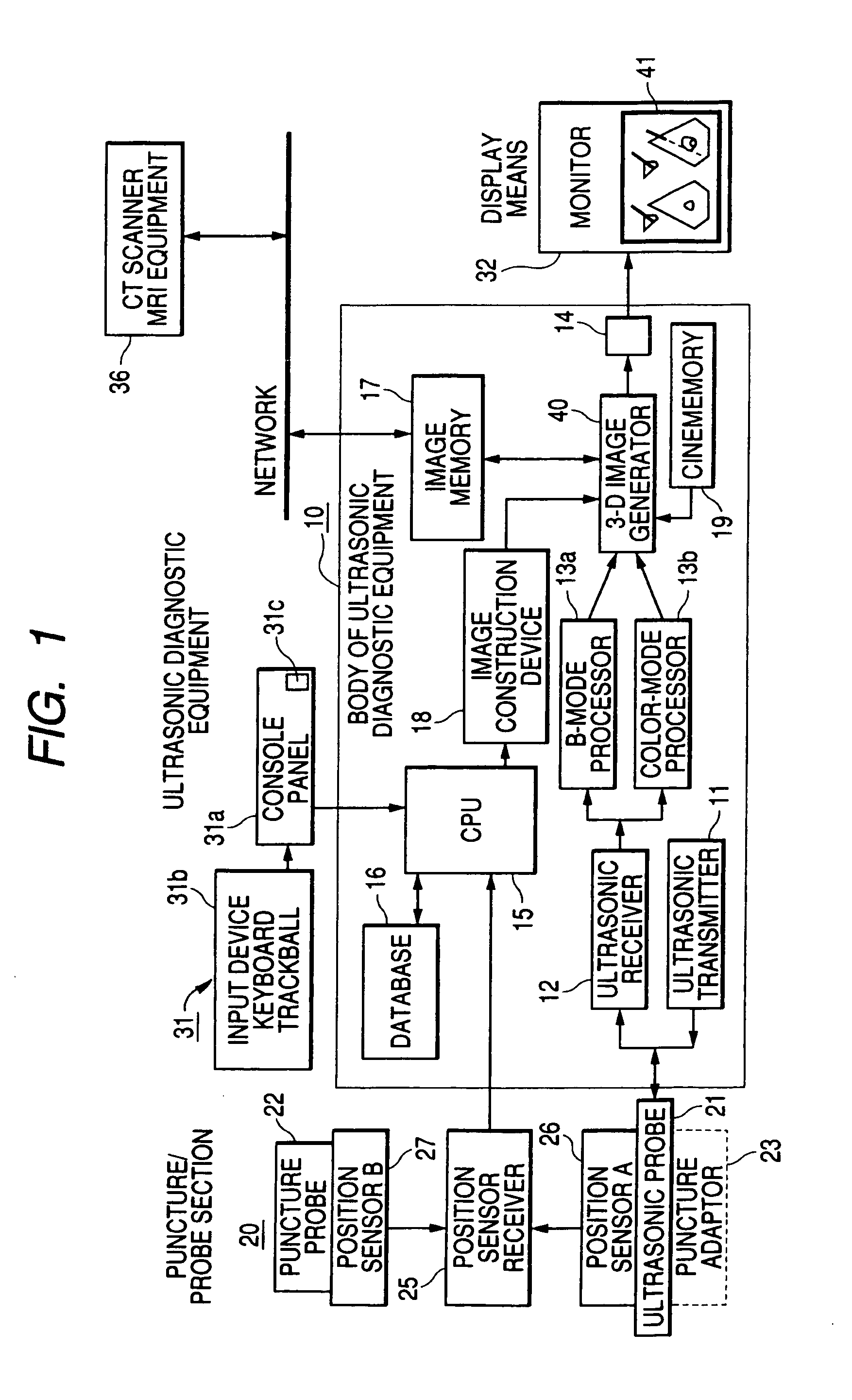
Ultrasonic diagnostic apparatus for fixedly displaying a puncture probe during 2D imaging
a technology of ultrasonic diagnostic equipment and probes, which is applied in ultrasonic/sonic/infrasonic image/data processing, applications, and catheters. it can solve the problems of inability to observe a cauterized part at the back of a cauterized part from the position of a probe over a treated part, and achieve the effect of easy detection
- Summary
- Abstract
- Description
- Claims
- Application Information
AI Technical Summary
Benefits of technology
Problems solved by technology
Method used
Image

Examples
4 embodiment
1-4 Embodiment
[0105]Referring to FIGS. 5 and 6A to 6F, a 1-4 embodiment will be described below.
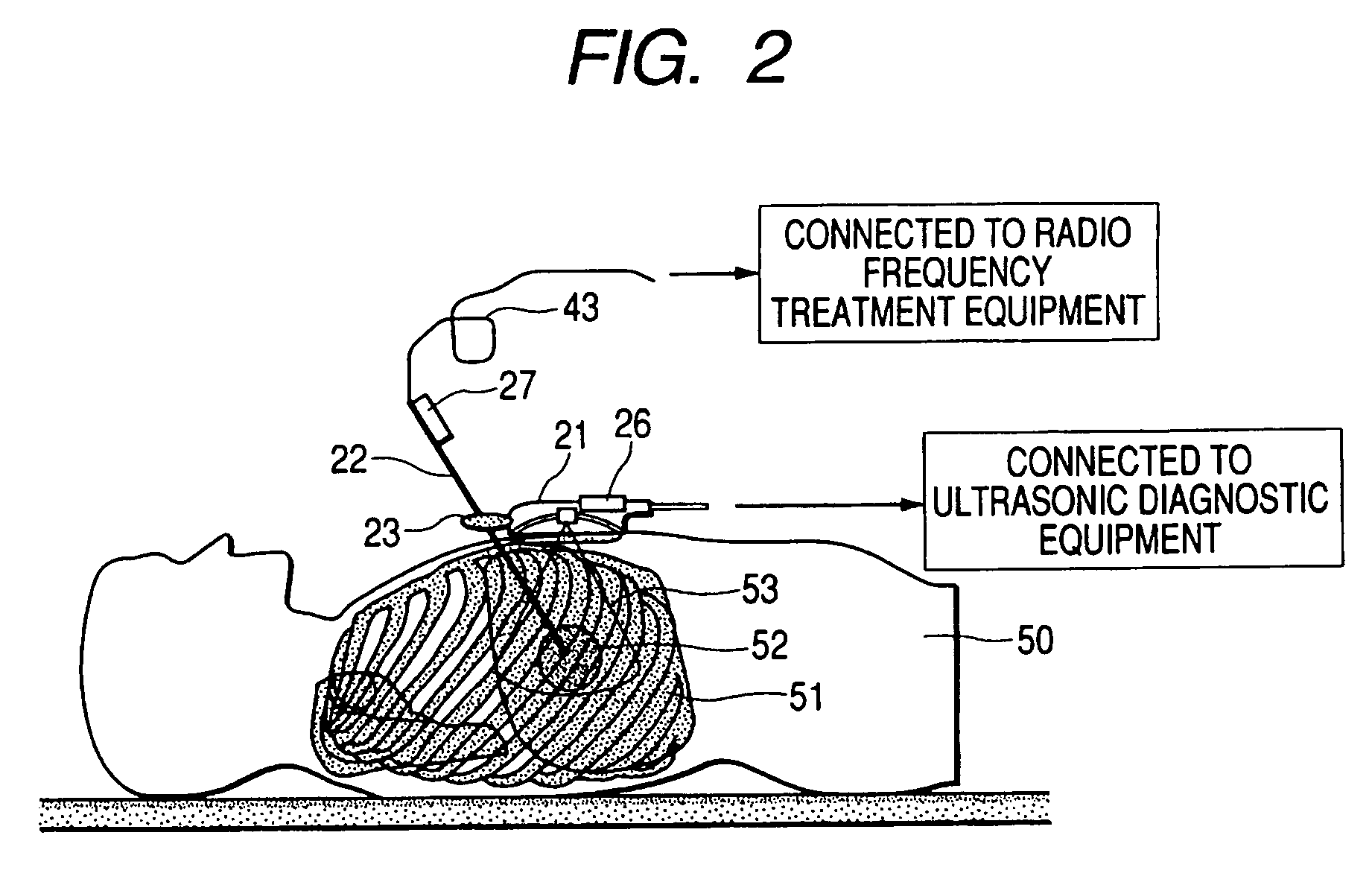
[0106]In this embodiment, first an ultrasonic probe 21 is independently placed on the surface of the body of a patient as shown in FIG. 5 without utilizing a puncture adaptor, a tumor of a target internal organ, its ambient internal organs and its ambient blood vessels are scanned, an inserted position and a puncture path are observed and determined. A position in which the tumor can be caught in a field of view of an ultrasonotomographic image and a clear tomographic image of a target part is acquired is a therapeutic observation position.
[0107]Next, the ultrasonic probe 21 is held in the therapeutic observation position, the end of a puncture probe 22 provided with a position sensor B 27 is located in the vicinity of the surface of the body of a patient 50, and the puncture probe is set in its inserted direction. FIG. 6B is a schematic drawing in which the tomographic image 71 in the th...
5 embodiment
1-5 Embodiment
[0117]FIGS. 7, 8, 9 and 10 are schematic drawings showing an embodiment of monitor display by the ultrasonic diagnostic equipment according to the invention. In this embodiment, the last tomographic image acquired before cautery or in the last cautery and a real-time tomographic image showing an inserted puncture probe during cautery are simultaneously displayed on two screens in various display modes of a monitor.
[0118]In a first display example shown in FIG. 7, a tomographic image 81 formerly acquired and recorded is displayed including an image of a tumor 82 verified as a therapeutic object on the left side for example of the monitor 32. On the right side, a real-time tomographic image 83 observed by an ultrasonic probe 21 is displayed including an image of the tumor 84 treated by the puncture probe 22.
[0119]Next, when the puncture probe 22 provided with a position sensor B 27 is set in the vicinity of the surface of the body over a target internal organ in an inser...
6 embodiment
1-6 Embodiment
[0132]Referring to a schematic drawing shown in FIGS. 11A and 11B, a 1-6 embodiment of the invention will be described below.
[0133]A three-dimensional ultrasonographic image by the ultrasonic diagnostic equipment according to the invention is not limited to an image three-dimensionally scanned by manually operating an ultrasonic probe and acquired by reconstructing three-dimensional tomographic image data based upon a two-dimensional tomographic image. A three-dimensional tomographic image by a mechanical three-dimensional ultrasonic probe for three-dimensionally scanning by the mechanical parallel motion or the mechanical rotational motion of the ultrasonic probe or a three-dimensional tomographic image by an electronic three-dimensional ultrasonic probe for electronically scanning three-dimensionally based upon a two-dimensional array probe can be similarly applied for a three-dimensional tomographic image by the ultrasonic diagnostic equipment according to the inven...
PUM
 Abstract
Description
Claims
Application Information