Method and apparatus for positioning a device in a tubular organ

a tubular organ and positioning technology, applied in the field of medical systems, can solve the problems of unoptimized deployment of therapeutic devices such as stents, difficult navigation of guide wires in anatomies such as bifurcations and branches, and difficulty in insertion of guide wires, so as to improve the resemblance of the operator and improve the resemblance of the vessel

Inactive Publication Date: 2012-02-28
PAIEON INC
View PDF87 Cites 94 Cited by
  • Summary
  • Abstract
  • Description
  • Claims
  • Application Information

AI Technical Summary

Problems solved by technology

In cases of difficult morphology of the vessel the guide wire insertion becomes a challenging task even for skilled physicians.
Furthermore, navigation of the guide-wire in anatomies such as bifurcations and branches is always a challenging task.
It is known that in some of cases, deployment of a therapeutic device such as a stent is not optimal, an indication to that is a necessity to deliver an additional stent to cover the stenotic segment.
The problem is to identify each checked location and provide a method of automatic navigation to the chosen one.
Additional equipment increases the cost of procedure, makes it more complicated, and requires accurate calibration.
These methods serve specific types of treatments and therefore are not universal.
However, ultrasound modality cannot be applied for some organs, such as coronary arteries.
Thus, the main difficulty of the registration, which is an essential step in data fusion, is solved by using additional equipment—the organs are imaged by ultrasound throughout the insertion.
The last solution is not valid for the case of X-ray angiography, for example.
However, as mentioned above, ultrasound technology is not applicable to all organs, and especially to coronary arteries, since the technology does not compensate for changes in imaging conditions and for movement of organs.
Additionally, the suggested method is not applicable to moving organs, such as arteries whose shape changes with the heart beat cycle.
Yet another drawback of the method is that the catheter is identified in low quality fluoroscopic images by usage of special intensity modulated catheter.
Thus, the method requires complete information of the imaging conditions, a static scene and a special catheter, making it inapplicable for standard coronary angioplasty.

Method used


Image

  • Method and apparatus for positioning a device in a tubular organ
  • Method and apparatus for positioning a device in a tubular organ
  • Method and apparatus for positioning a device in a tubular organ

Examples


Embodiment Construction

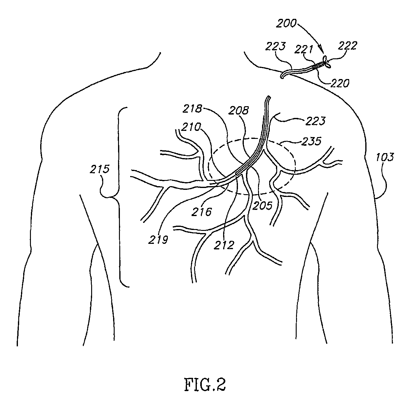
[0022]The disclosed invention presents an apparatus and method for automatic detecting and tracking of a device within a tubular organ, such as an artery, a blood vessel, or a urethra of a subject. In the context of the disclosed invention, “device” relates either to a guide wire tip, a catheter tip, or a therapeutic device. When the tubular organ under discussion is an artery, the therapeutic device is usually an intravascular therapeutic device, such as a stent, a balloon, a pacing lead, or the like.

[0023]The present invention can be implemented, but is not restricted to in the following procedures: catheterization procedure, bi-ventricular pacing, coronary angiography, coronary angioplasty, peripheral angiography, peripheral angioplasty, carotid angiography, carotid angioplasty, neuro angiography / plasty, biliary imaging or intervention, and the like.

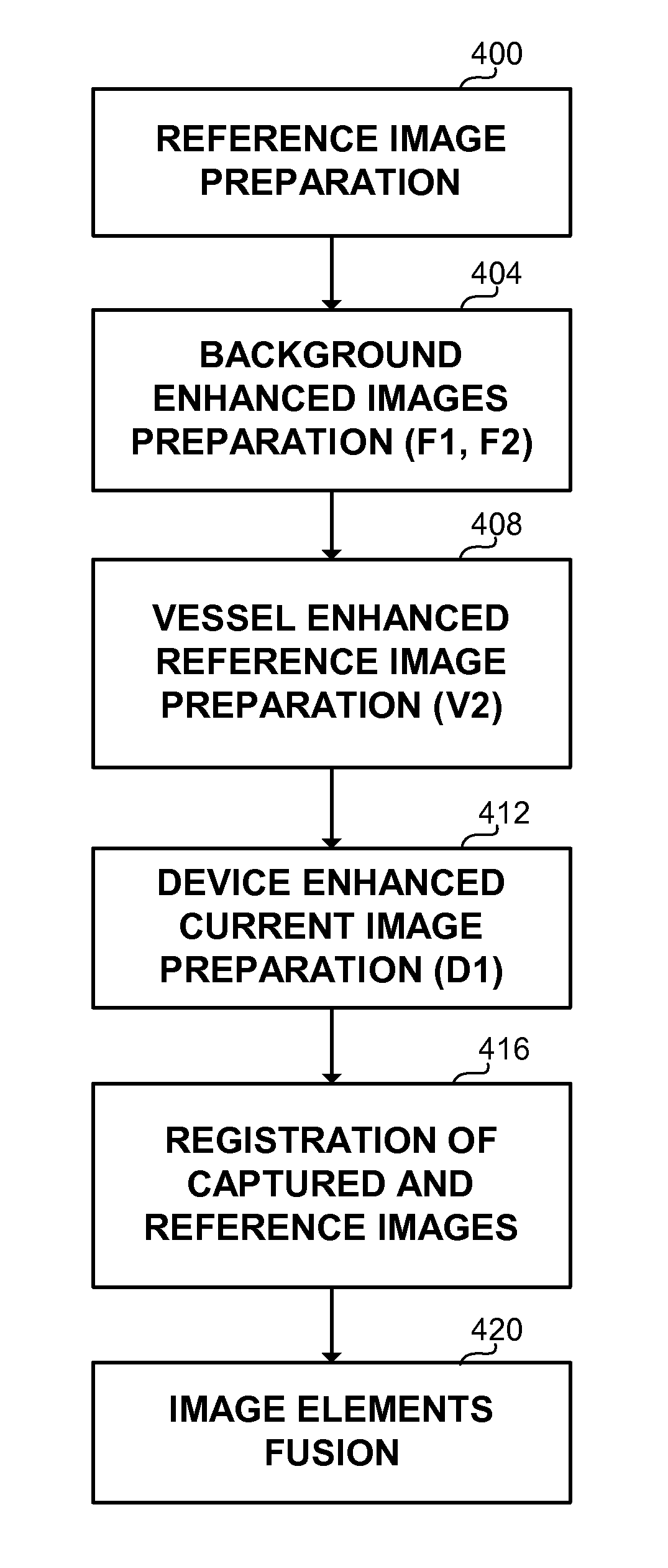
[0024]In a preferred embodiment, the method of the present invention employs a diagnostic stage and a therapeutic stage. During the ...


PUM


Abstract

An apparatus and method for detecting, tracking and registering a device within a tubular organ of a subject. The devices include guide wire tip or therapeutic devices, and the detection and tracking uses fluoroscopic images taken prior to or during a catheterization operation. The devices are fused with images or projections of models depicting the tubular organs.

Description

RELATED APPLICATIONS[0001]The present invention relates to international patent application serial number PCT / IL01 / 00955 titled “METHOD AND SYSTEM FOR POSITIONING A DEVICE IN A TUBULAR ORGAN” filed on Oct. 15, 2001.BACKGROUND OF THE INVENTION[0002]1. Field of the Invention[0003]The present invention relates to medical systems in general, and to a method and apparatus for positioning and presenting a device in tubular organs, in particular.[0004]2. Discussion of the Related Art[0005]Interventional cardiology procedures are becoming increasingly important in the treatment of physiological abnormalities such as lumen stenosis or aneurysm. For example, in order to treat a stenotic coronary artery, it is often required to inflate a balloon, apply an atherectomy or thrombectomy device and place a stent (prosthesis) at a diseased artery site. In this example, the devices are either a guide wire or a therapeutic intravascular device, such as a balloon, a stent, an atherectomy, or a thrombec...

Claims


Application Information

Patent Timeline
Patent Type & Authority Patents(United States)
IPC IPC(8): A61B6/03
CPCA61B6/12A61B6/481A61B6/504G06K9/6203G06T7/0026G06T7/0038A61B6/541G06T2207/30101G06T7/32G06T7/38G06V10/7515
Inventor ZARKH, MICHAELKLAIMAN, MOSHEEVRON, RAMI
Owner PAIEON INC
Why Eureka
Try Eureka