Method for assisting in diagnosis of cerebral diseases and apparatus thereof

a technology for diagnosing cerebral diseases and assisting in diagnosis, applied in the field of assisting in diagnosis of cerebral diseases and an apparatus thereof, can solve the problems of many people needed in evaluating the processing, the operation error of overlooking processing errors, and the complexity of image processing

Active Publication Date: 2012-07-10
DAI NIPPON PRINTING CO LTD
View PDF5 Cites 15 Cited by
  • Summary
  • Abstract
  • Description
  • Claims
  • Application Information

AI Technical Summary

Benefits of technology

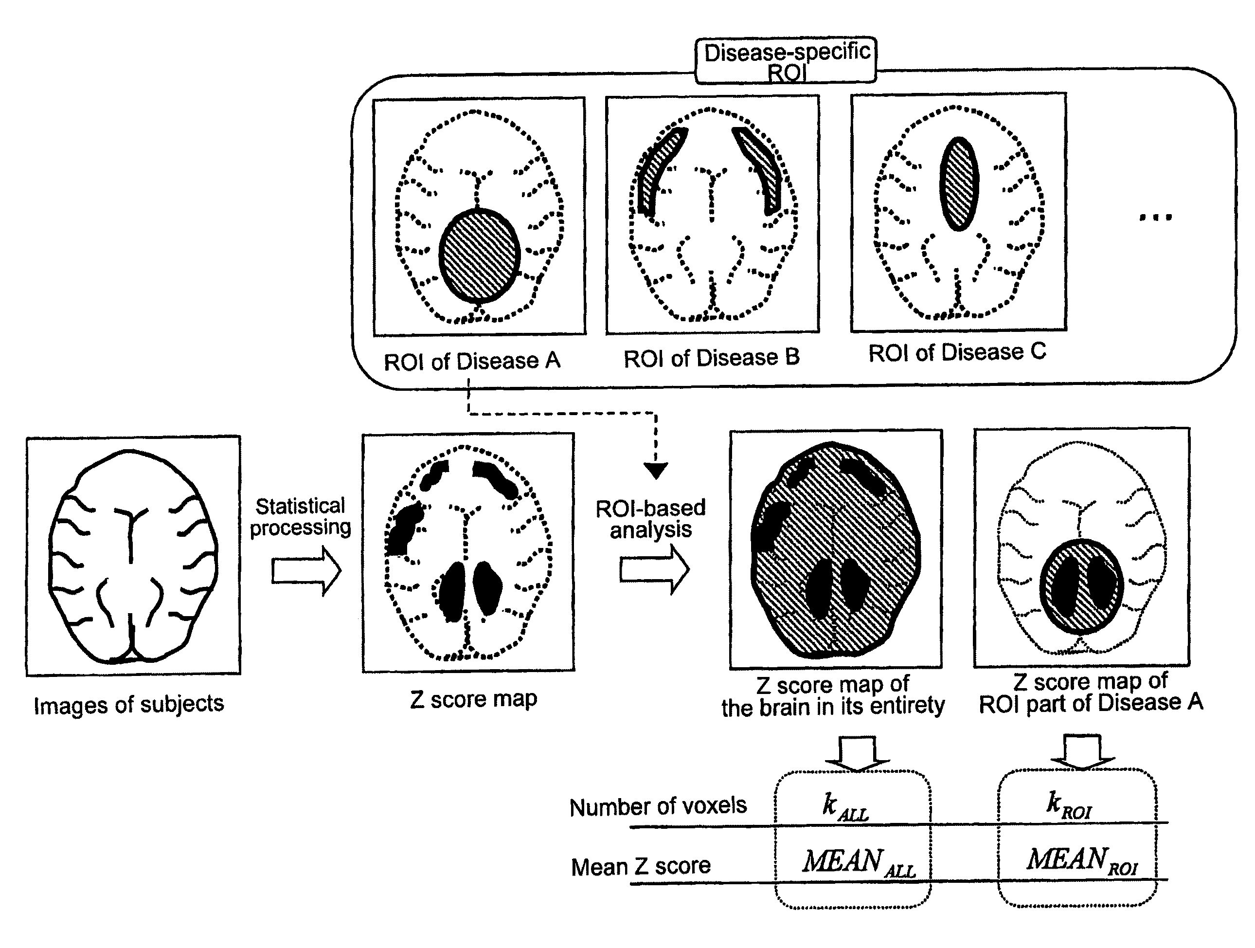
[0013]An object of the present invention is to provide a method for assisting in the diagnosis of cerebral diseases in which an operator is able to establish an ROI for inputted brain images (data) of a subject without manual procedures such as manual depiction, thereby offering objective diagnosis results and an apparatus thereof.
[0014]The present invention is a method for assisting in the diagnosis of cerebral diseases in which brain images of a subject are inputted and subjected to image processing to exhibit the diagnosis result, thereby assisting in the diagnosis. More specifically, a statistical method is used to determine in advance a disease-specific region of interest (ROI) in the brain images, and the thus input brain images of the subject are statistically compared with previously prepared brain images of normal cases to exhibit the diagnosis result, in which the ROI is applied to solve the above-described object.
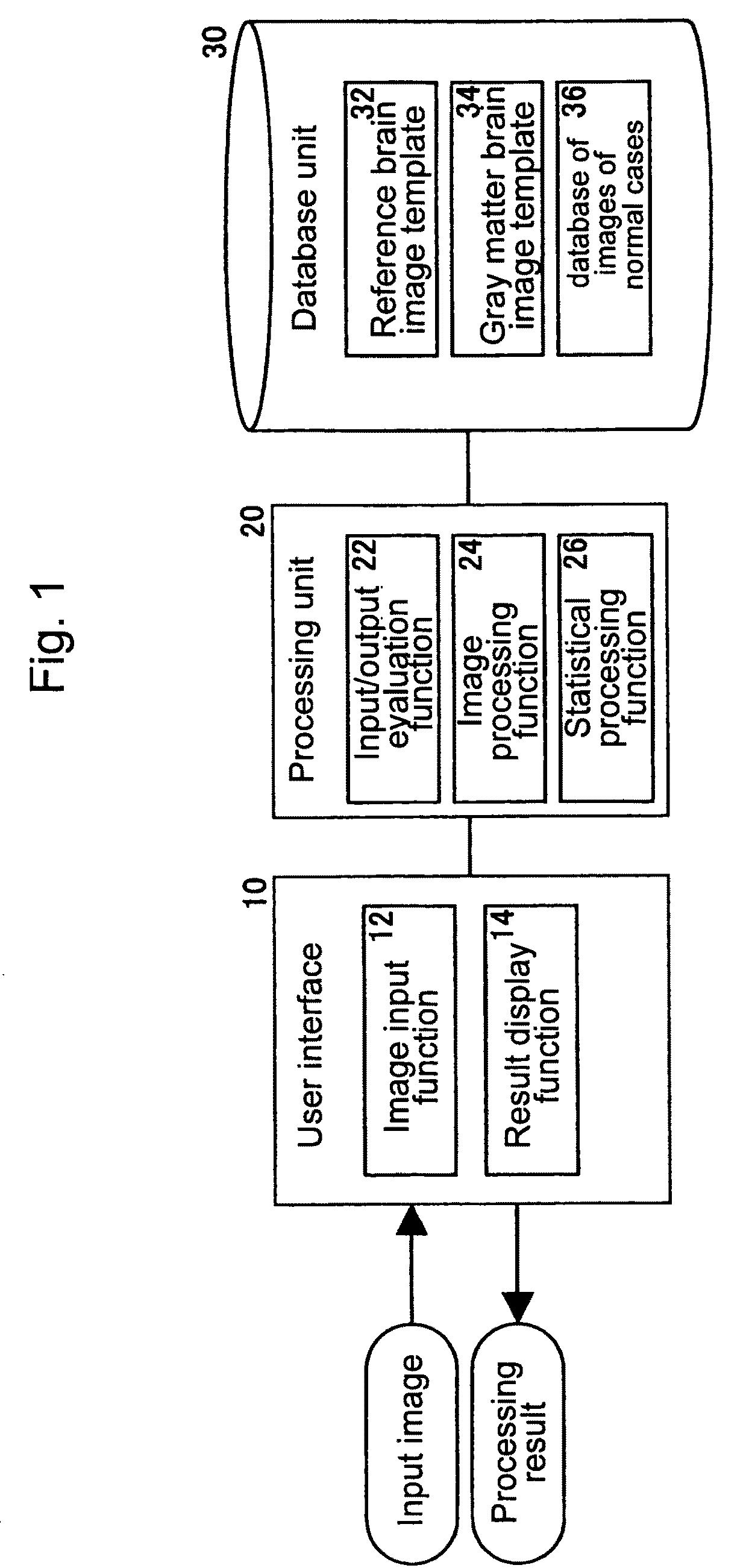
[0015]The present invention is also an apparatus for assisting in the diagnosis of cerebral diseases in which brain images of a subject are inputted and subjected to image processing to output the diagnosis result, thereby assisting in the diagnosis. The apparatus is provided with a retention means for determining in advance a disease-specific region of interest (ROI) in the brain images by a statistical method and retaining them and an image / statistical processing means for statistically comparing the thus input brain images of the subject with the previously prepared brain images of normal cases, also having the function to apply the ROI when the image / statistical processing means makes a statistical comparison to provide the diagnosis result, thereby also solving the above-described object.
[0022]According to the present invention, a previously prepared ROI on the basis of statistical processing is established for inputted brain images of a subject so as to apply the ROI-based analysis, thus making it possible to provide objective diagnosis results without manual procedures.

Problems solved by technology

In particular, a highly accurate technique such as the VBM method targeting MRI images requires complicated processing of images.
Consequently, in the above judgment, the results may include subjective elements or there may be operational mistakes that overlook errors in processing.
In this case, there is a problem that many people are needed in evaluating the processing results.
Further, in the conventional ROI method in which an operator establishes an ROI by manually depicting the contour of a corresponding site on an image, the accuracy is likely to be influenced by accidental errors resulted from difference in visual perception or difference in operator's experience.
Therefore, the conventional ROI method fails in providing diagnosis assistance on the basis of objective data, which is another problem.

Method used


Image

  • Method for assisting in diagnosis of cerebral diseases and apparatus thereof
  • Method for assisting in diagnosis of cerebral diseases and apparatus thereof
  • Method for assisting in diagnosis of cerebral diseases and apparatus thereof

Examples


example

[0282]In order to make a diagnosis of Alzheimer's dementia (AD), MRI is used to take T1-weighted images of the brain in subjects and normal cases, and these images are retained in the DICOM format. The DICOM format is an imaging format commonly used in medical images having a header part and an image data part in one file and able to retain parameters at the time of taking images and diagnosis information. In most cases, one file of the DICOM images has information on one piece of slice image, and a plurality of the DICOM images are used to express a three-dimensional brain image. DICOM images are stored at a DICOM server and can be called up whenever necessary.

[0283]A DICOM image file expresses three-dimensional information on the brain in its entirety by using a plurality of images, with only the header part and image data part of the DICOM file being converted into the Analyze format, which is a concatenated format. The Analyze format is able to constitute an image of the head in...


PUM


Abstract

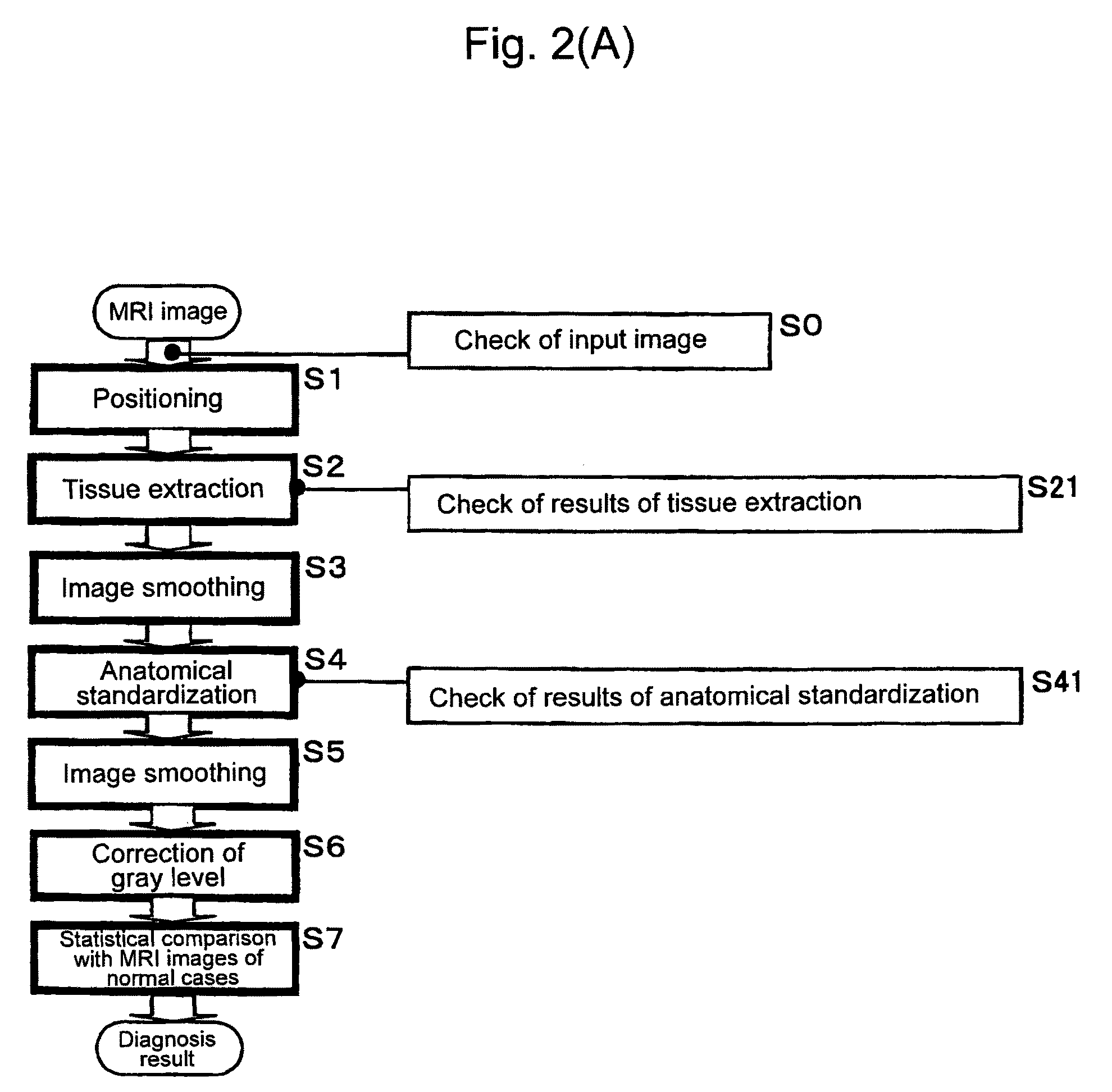
Input MRI brain images are positioned so as to correct a spatial deviation, gray matter tissues are extracted from these images to effect a first image smoothing, the thus-obtained images are subjected to anatomical standardization, a second image smoothing is effected, the gray level is corrected, brain images after correction are statistically compared with MRI brain images of normal cases, thereby providing the diagnosis result. In this instance, the brain images are automatically checked for input images regarding the resolution dot density and the like, the result of gray matter tissue extraction and the result of anatomical standardization, by which specifications of input images and the like can be confirmed objectively and automatically to make a diagnosis automatically by image processing. Further, an ROI-based analysis is made to provide the analysis result as the diagnosis result.

Description

TECHNICAL FIELD[0001]The present invention relates to a method for assisting in the diagnosis of cerebral diseases and an apparatus thereof and, in particular, to a method for favorably assisting in the diagnosis of cerebral diseases in which an MRI (Magnetic Resonance Imaging) and the like are used to input brain images and process the images, thereby providing disease-specific diagnosis assistance and to an apparatus thereof.BACKGROUND ART[0002]With the advent of an aging society, there is found a yearly increase in the number of patients with dementia-related diseases. There are several types of dementia-related diseases, thus necessitating an appropriate treatment depending on diseased conditions on the basis of differential diagnosis for these types.[0003]On the other hand, in recent years, in order to cope with the above-described situation, radioactive medical examinations such as SPECT (Single Photon Emission Computed Tomography) and PET (Positron Emission Tomography) or oth...

Claims


Application Information

Patent Timeline
Patent Type & Authority Patents(United States)
IPC IPC(8): G06K9/00G06K9/62G06K9/74G16H20/70G16H30/40G16H50/20
CPCA61B5/055G06F19/321G06T7/0012G06F19/345G06T2207/10072G06T2207/30016A61B5/4088G16H50/20G16H20/70G16H30/40
Inventor MATSUDA, HIROSHIONO, TETSUTAROMATSUBA, SEIJI
Owner DAI NIPPON PRINTING CO LTD
Why Eureka
Try Eureka