X-ray imaging device, method for detecting deviation of flat panel detector, and program for the same
a flat panel detector and imaging surface technology, applied in the field of xray imaging devices, can solve the problems of deviation of the imaging surface of the fpd from the x-ray field, undetectable center of the x-ray field, etc., and achieve the effect of precisely detecting the deviation amoun
- Summary
- Abstract
- Description
- Claims
- Application Information
AI Technical Summary
Benefits of technology
Problems solved by technology
Method used
Image
Examples
first embodiment
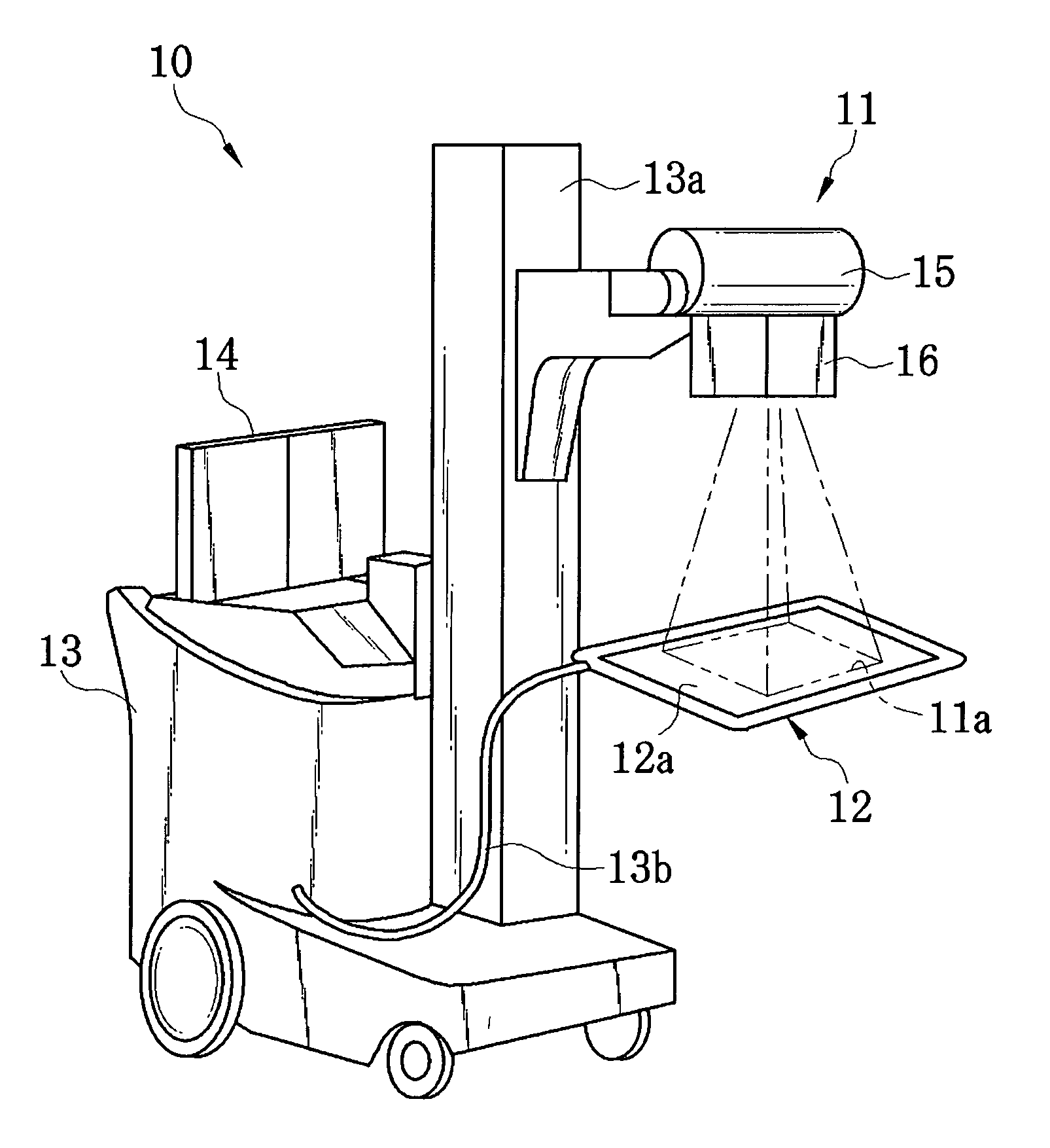
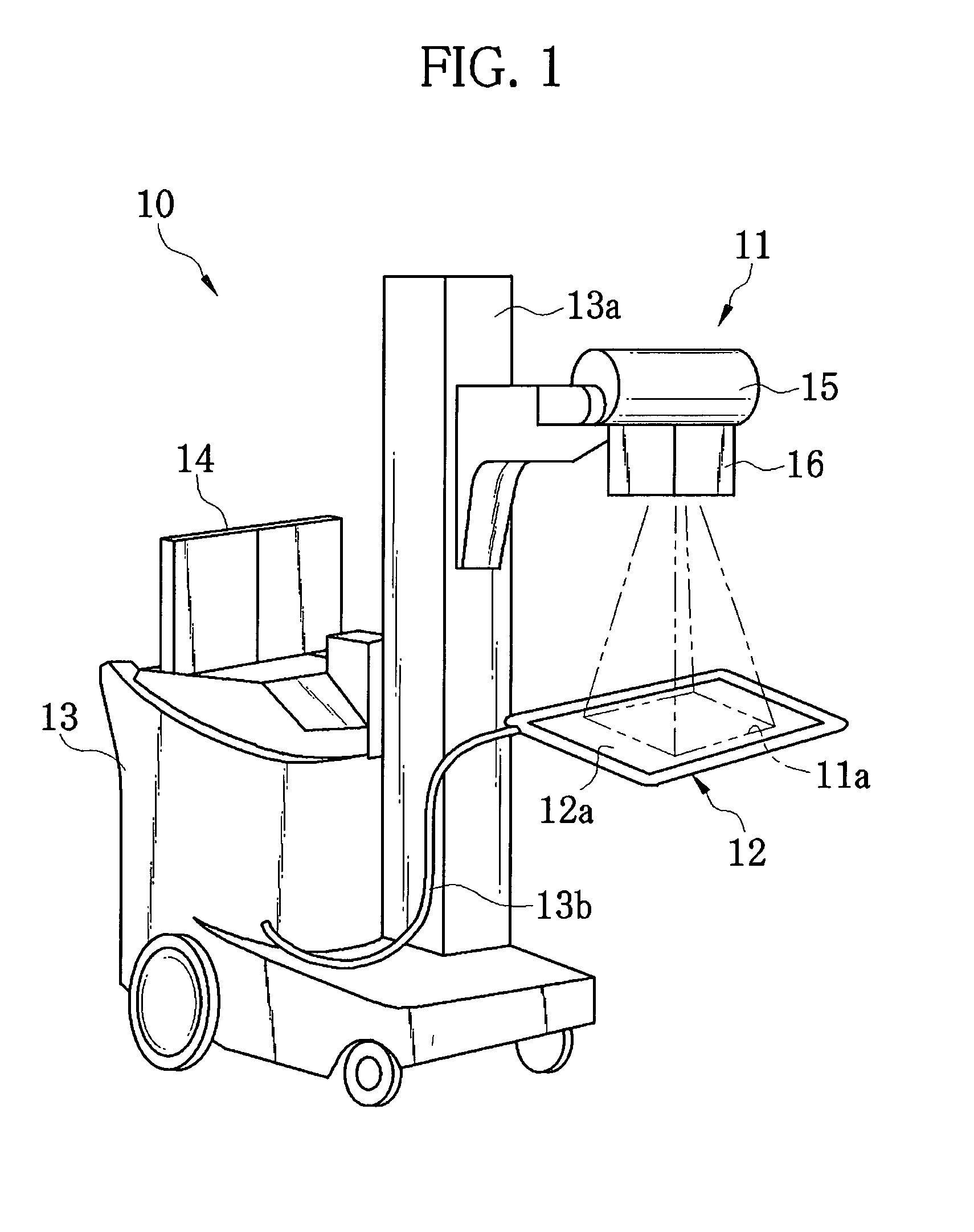
[0044]As shown in FIG. 1, an X-ray imaging device 10 is formed integrally with a cart or a wagon in a movable manner. The X-ray imaging device 10 is constituted of an X-ray generator 11, a portable flat panel detector (FPD) 12 called as an electronic cassette, a main body 13 integrally attached to the cart, and a monitor 14 attached to the main body 13. The X-ray generator 11 is slidably attached to a column 13a, and emits an X-ray radiation beam as shown by chain double-dashed lines of FIG. 1. The emitted X-ray radiation beam passes through patient's body, and then the FPD 12 receives the X-ray radiation beam to produce a radiographic image. The FPD 12 is connected to the main body 13 through a cable 13b.
[0045]On the monitor 14, which is a liquid crystal display, the radiographic image outputted from the FPD 12, a GUI (graphical user interface) image, and the like are displayed.
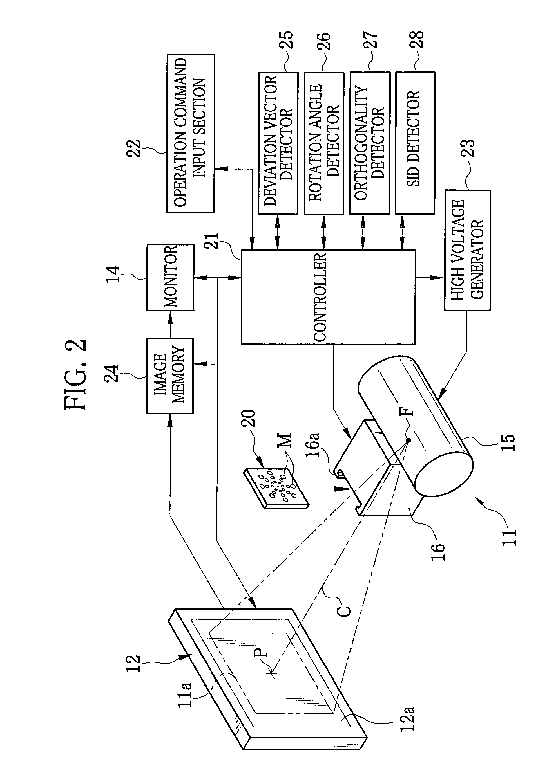
[0046]The main body 13 contains a controller 21 (see FIG. 2). The controller 21 controls the operation o...
second embodiment
[0086]In an X-ray imaging device according to a second embodiment, the markers M0 to M3 are formed in the collimator leaves, in order to produce the preliminary radiographic image without using the filter plate 20 or 30. The X-ray imaging device according to the second embodiment has the same structure as that of the X-ray imaging device 10 according to the first embodiment, except for the collimator leaves.
[0087]As shown in FIG. 13, a collimator unit 40 according to the second embodiment is provided with first and second collimator leaves 41a and 41b that are openable and closable in the X direction, and third and fourth collimator leaves 41c and 41d that are openable and closable in the Y direction. In the first and second collimator leaves 41a and 41b, circular holes are formed as the markers M0 to M3. By closing the first and second collimator leaves 41a and 41b, the markers M0 to M3, which are in the same layout as those of the filter plate 20 shown in FIG. 3, appear in the fir...
third embodiment
[0091]In the X-ray imaging devices according to the first and second embodiments, the holes of the markers M0 and M3 are formed in the filter plate or the collimator leaves, and the preliminary radiographic image is produced from the X-ray radiation beam that has passed through the markers M0 to M3. In contrast, an X-ray imaging device according to a third embodiment produces the preliminary radiographic image in which X-ray density is reduced in stages from center to edge, with use of a filter plate that has different X-ray transmittance from area to area.
[0092]In FIG. 14, a filter plate 50 of the third embodiment is divided into first to fourth areas 50a to 50d that have different X-ray transmittance. The square first area 50a disposed at the center of the filter plate 50 has transmittance of approximately 90%. The square frame-shaped second area 50b surrounding the first area 50a has transmittance of approximately 70%. The square frame-shaped third area 50c surrounding the second...
PUM
 Abstract
Description
Claims
Application Information
 - R&D
- Intellectual Property
- Life Sciences
- Materials
- Tech Scout
- Unparalleled Data Quality
- Higher Quality Content
- 60% Fewer Hallucinations
Browse by: Latest US Patents, China's latest patents, Technical Efficacy Thesaurus, Application Domain, Technology Topic, Popular Technical Reports.
© 2025 PatSnap. All rights reserved.Legal|Privacy policy|Modern Slavery Act Transparency Statement|Sitemap|About US| Contact US: help@patsnap.com


