Application of image-based dynamic ultrasound spectrography (IDUS) in detection and localization of breast microcalcifcation

a dynamic ultrasound and image-based technology, applied in the field of medical conditions, can solve the problems of more difficult the inability to identify a discrete mass with sufficient clarity, and the inability to image the structure of the breast with sufficient clarity

Inactive Publication Date: 2013-02-19
BIOQUANTETICS
View PDF15 Cites 13 Cited by
  • Summary
  • Abstract
  • Description
  • Claims
  • Application Information

AI Technical Summary

Benefits of technology

[0041]It is an object of the present invention to provide a method for the detection and localization of breast microcalcification through a precise method of ultrasound impacting and ultrasound frequency wave detection which will enable the system to detect not only if there is a microcalcification in the breast, but the precise location of the microcalcification and the mass of the microcalcification.
[0044]It is also an object of the present invention to provide a methodology to locate microcalcification and determine the nature of the microcalcification without providing the drawbacks of other imaging and screening modalities such as being invasive, having no ionization radiation, being safe, reliable, cost-effective, not Doppler based, not angular-dependent and being portable, suitable for field applications and suitable for operating room conditions.

Problems solved by technology

When the breast is glandular it is more difficult to image its architecture than when the breast contains large amounts of fat.
The breast might be so radiographically dense that breast structure cannot be imaged with sufficient clarity to identify a discrete mass.
Although the results for women younger than age 50 are still controversial.
However, the reported positive predictive value (PPV) which includes the prevalence of detecting the disease is quite poor, varying from 10% to 50%.
Ultrasound imaging of the breast may also help radiologists to evaluate some lumps that can be felt but that are difficult to see on X-ray mammogram, especially in dense breast or implants.
3-Dimensional ultrasound imaging is seldom used in breast screening due to very limited added information.
However, traditional ultrasound has poor calcification detectability.
However, neither the technique nor the interpretive criteria are standardized as of to date, leading to variability in performance and in results interpretation.
In addition, MRI can only be performed in a setting in which it is possible to perform biopsy of lesions detected solely by MRI.
Despite its high accuracy in detecting malignancies in breast, MRI is not recommended as a routine examination for the differentiation of benign and malignant lesions / tumors.
MRI is a prohibitively expensive modality and it is unsuitable for large-scale screening programs.
The major drawback associated with optical imaging remains light propagation in biological tissue, which is highly scattered, resulting in poor resolution.
Optical mammography has yet to demonstrate its potential to be a stand-alone imaging modality, mainly because of its poor specificity and sensitivity.
Secondly, all information about optical absorption inhomogeneities is carried to the breast surface by ultrasound waves which have low attenuation and scattering in soft tissue and thus, resulting in poor sensitivity.
One of the major disadvantages of these techniques is the difficulties in displaying and analyzing the 3-dimensional information retained from the targeted area.
Therefore, the time and cost required for image retrieval and analysis of the thermo / photo-acoustic techniques are potentially greater when compared with that of X-ray mammography and ultrasound.
Moreover, these techniques have yet to demonstrate reproducibility, adequate sensitivity, specificity and practicality.

Method used


Image

  • Application of image-based dynamic ultrasound spectrography (IDUS) in detection and localization of breast microcalcifcation
  • Application of image-based dynamic ultrasound spectrography (IDUS) in detection and localization of breast microcalcifcation
  • Application of image-based dynamic ultrasound spectrography (IDUS) in detection and localization of breast microcalcifcation

Examples


Embodiment Construction

[0064]Although specific embodiments of the present invention will now be described with reference to the drawings, it should be understood that such embodiments are by way of example only and merely illustrative of but a small number of the many possible specific embodiments which can represent applications of the principles of the present invention. Various changes and modifications obvious to one skilled in the art to which the present invention pertains are deemed to be within the spirit, scope and contemplation of the present invention as further defined in the appended claims.

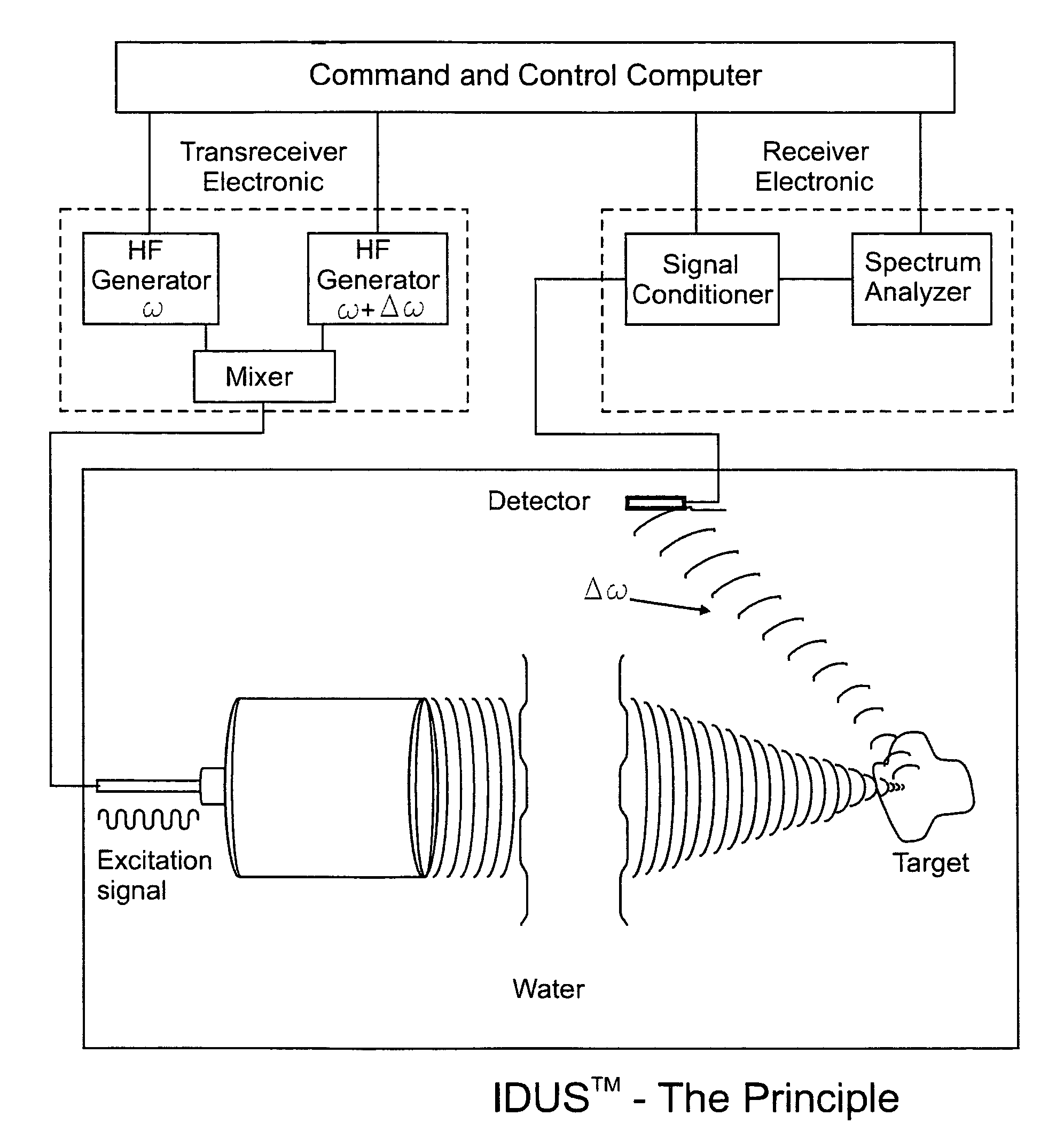
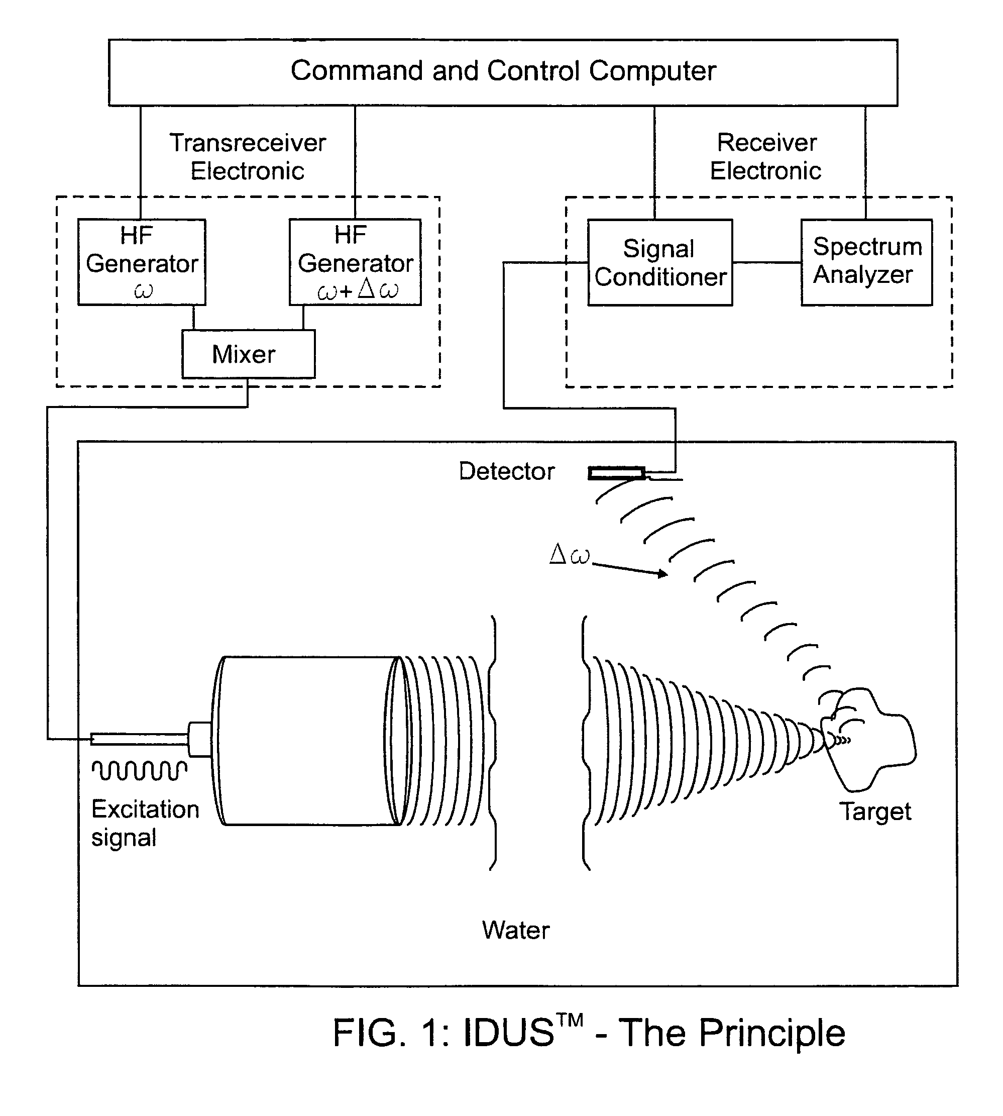
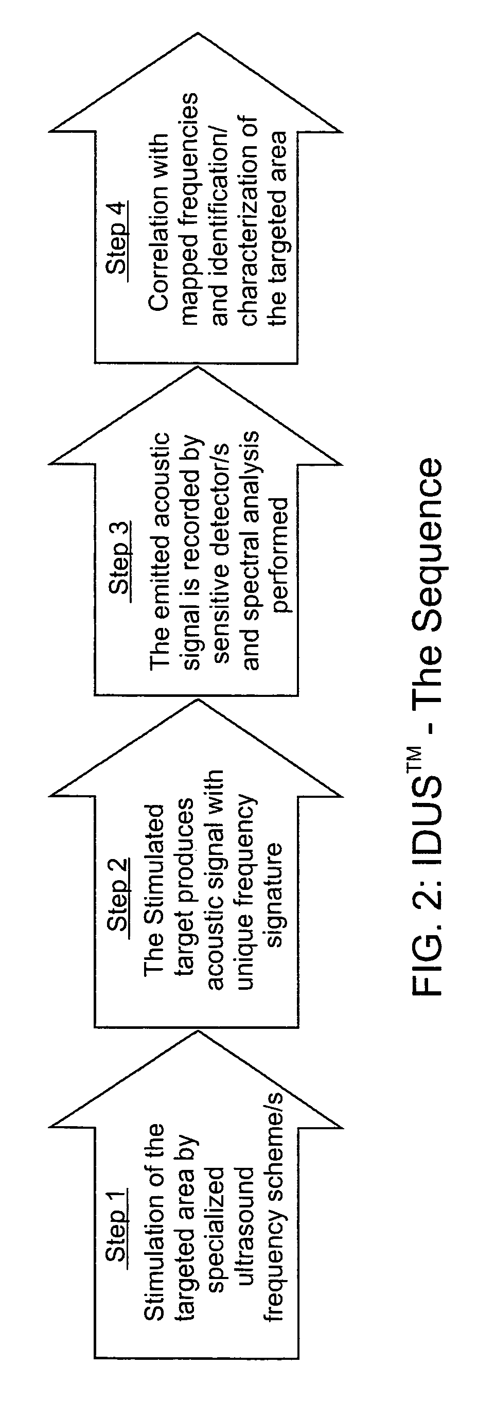
[0065]The fundamental concept of the present invention is depicted in the enclosed FIG. 1 which is a schematic representation of the present invention as described below.

[0066]The invention for the noninvasive remote ultrasound detection and localization of the targeted breast microcalcification masses by sensing their emanating acoustic response is based on the insonification of the breast areas with micr...


PUM


Abstract

A method for the detection and localization of breast microcalcification through a precise method of ultrasound impacting and ultrasound frequency wave detection which will enable the system to detect not only if there is a microcalcification in the breast, but the precise location of the microcalcification and the mass of the microcalcification. The invention is also a method and system that provides bi-modal guided stimulation of a targeted mass within the breast to determine the location and nature of the microcalcification within the breast.

Description

BACKGROUND OF THE INVENTION[0001]1. Field of the Invention[0002]The present invention relates to the diagnosis of medical conditions and in particular to medical diagnosis of abnormalities within a female breast which is performed through the use of non-invasive ultrasound techniques to determine whether or not there is any microcalcification in a female breast and further, to determine the location where the microcalcification is located as well as the size of the microcalcification.[0003]2. Description of the Prior Art[0004]In general, breast screening for cancer and microcalcification detection have been performed using different imaging modalities in the prior art and there are several techniques that are currently in active use. The techniques are as follows:X-Ray Mammography[0005]To date, X-ray mammography is the method of choice and the “gold standard” for breast screening and diagnosis with which other technologies are compared. In order to perform this test a breast is expo...

Claims


Application Information

Patent Timeline
Patent Type & Authority Patents(United States)
IPC IPC(8): A61B8/00
CPCA61B8/0825A61B8/483G01S7/52022G01S7/52036A61B8/4477A61B8/485A61B8/0833A61B8/5215G01S15/8977G01S15/8993G01S15/8952
Inventor RAMBOD, EDMONDITZCHAK, YACOVSHAMISS, ARIWEIHS, DANIEL
Owner BIOQUANTETICS
Why Eureka
Try Eureka