Vertebra segmentation apparatus, vertebra segmentation method, and recording medium with program for vertebra segmentation

a segmentation apparatus and recording medium technology, applied in the field of vertebral segmentation methods, vertebral segmentation apparatuses, recording media with program for vertebral segmentation, can solve the problems of reducing image rendering capability, particularly difficult to render intervertebral discs and vertebral body endplates separately, etc., to reduce matching errors, high spatial frequency noise, and high accuracy

Active Publication Date: 2013-05-14
FUJIFILM CORP
View PDF10 Cites 1 Cited by
  • Summary
  • Abstract
  • Description
  • Claims
  • Application Information

AI Technical Summary

Benefits of technology

[0013]The third feature quantity is calculated in view of cross-sectional shapes that are transverse and parallel to the central axis of each of the vertebrae. Accordingly, it is possible to perform a quantitative density analysis based on cross-sectional shapes from two different directions, thereby allowing the vertebrae to be partitioned and recognized in a three-dimensional medical image of the subject whose spinal shape is deformed entirely or locally. This process is particularly effective in analyzing a medical image of a spine, which is deformed in its entirety due to spinal curvature.
[0016]Preferably, the vertebra segmentation apparatus further comprises a function matcher for matching the third feature quantity calculated for each of the respective points on the central axis to a prescribed periodic function or a quasi-periodic function, wherein the vertebral position estimator estimates the positions of the vertebrae on the central axis based on the prescribed periodic function or the quasi-periodic function matched by the function matcher. Since the third feature quantity representative of the regularity of the array of the vertebrae is matched to a prescribed regular function form, high spatial frequency noise which is possessed by the third feature quantity can be reduced, for thereby estimating positions of the vertebrae with high accuracy.
[0017]Preferably, the prescribed periodic function or the quasi-periodic function includes a phase as a variable. In this case, the vertebra segmentation apparatus further comprises a phase determiner for determining the phase successively for the points on the central axis. It is thus possible to reduce an error in matching the third feature quantity to the matching function, for thereby estimating the position of each vertebra with high accuracy. This process is particularly effective in analyzing a medical image of a spine, which is deformed locally due to a bone fracture.
[0024]With the vertebra segmentation apparatus, the vertebra segmentation method, and the recording medium storing the program according to the present invention, tomographic images are generated at intervals along the central axis of each of respective vertebrae, and based on the generated tomographic images, a first feature quantity representative of a clearness of sectional shapes crossing the central axis at the vertebrae is calculated for each of respective points on the central axis. Further, based on the generated tomographic images, a second feature quantity representative of a clearness of sectional shapes parallel to the central axis at the vertebrae is calculated for each of the respective points on the central axis. A third feature quantity representative of a regularity of an array of the vertebrae is calculated for each of the respective points on the central axis based on the calculated first feature quantity and the calculated second feature quantity. Positions of the vertebrae on the central axis are calculated based on the third feature quantity, which is calculated for each of the respective points on the central axis. Accordingly, it is possible to perform a quantitative density analysis based on cross-sectional shapes as viewed from two different directions, thereby allowing the vertebrae to be partitioned and recognized as a three-dimensional medical image of a subject whose spinal shape is deformed entirely or locally. Such a process is particularly effective in analyzing a medical image of a spine, which is deformed in its entirety due to spinal curvature.

Problems solved by technology

It is generally known that if slices are thick in the scanning direction, i.e., the direction of the body axis, then the spatial resolution along the direction of the body axis becomes insufficient due to a partial volume effect, resulting in a reduction in the image rendering capability.
It is particularly difficult to render intervertebral discs and vertebral body endplates separately on tomographic images within a range from the cervical vertebrae to the upper thoracic vertebrae, where each of the vertebrae is relatively short.

Method used


Image

  • Vertebra segmentation apparatus, vertebra segmentation method, and recording medium with program for vertebra segmentation
  • Vertebra segmentation apparatus, vertebra segmentation method, and recording medium with program for vertebra segmentation
  • Vertebra segmentation apparatus, vertebra segmentation method, and recording medium with program for vertebra segmentation

Examples


Embodiment Construction

[0040]A vertebra segmentation method according to a preferred embodiment of the present invention, and a vertebra segmentation apparatus for carrying out the vertebra segmentation method will be described in detail below with reference to the drawings.

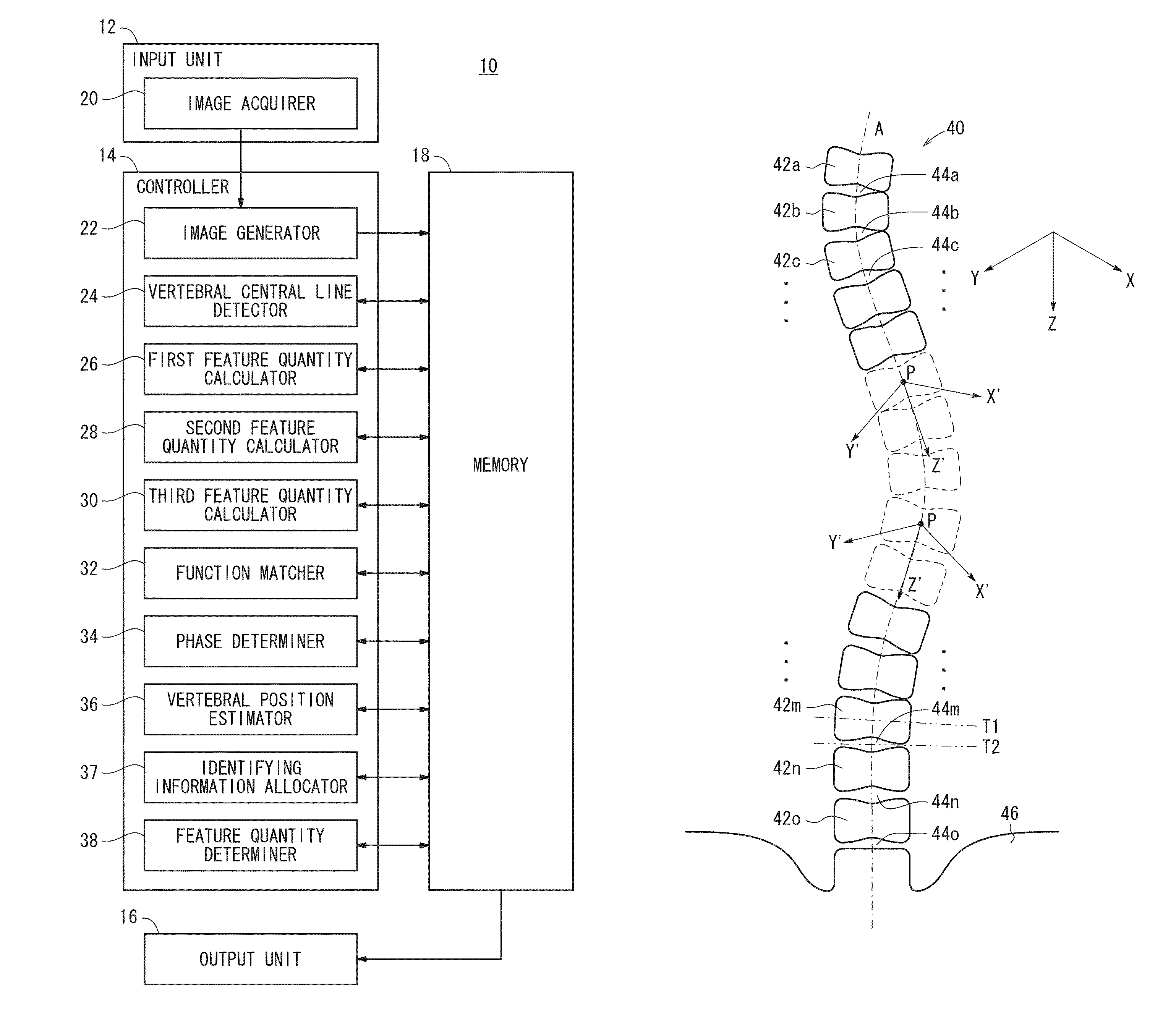
[0041]FIG. 1 is an electric block diagram of a vertebra segmentation apparatus 10 according to an embodiment of the present invention. As shown in FIG. 1, the vertebra segmentation apparatus 10 comprises an input unit 12, a controller 14 that serves as an information processor such as a CPU or the like, an output unit 16, and a memory 18 that serves as a recording medium. The memory 18 stores a program for carrying out functions of the vertebra segmentation apparatus 10. The input unit 12 receives signals from external circuits. The input unit 12 includes an image acquirer 20 for acquiring a three-dimensional medical image, which includes a plurality of vertebrae. The image acquirer 20 may acquire not only a medical image, such as a CT...


PUM


Abstract

Based on generated tomographic images, a first feature quantity representative of a clearness of sectional shapes crossing the Z′ axis at the vertebrae is calculated for each of respective points on the Z′ axis. Further, based on the generated tomographic images, a second feature quantity representative of a clearness of sectional shapes parallel to the Z′ axis at the vertebrae is calculated for each of the respective points on the Z′ axis. A third feature quantity representative of a regularity of an array of the vertebrae is calculated for each of the respective points on the Z′ axis, based on the calculated first feature quantity and the calculated second feature quantity. Positions of the vertebrae on the Z′ axis are calculated based on the third feature quantity, which is calculated for each of respective points on the Z′ axis.

Description

CROSS-REFERENCE TO RELATED APPLICATION[0001]This application is based upon and claims the benefit of priority from Japanese Patent Applications No. 2009-270837 filed on Nov. 27, 2009 and No. 2010-194795 filed on Aug. 31, 2010, of which the contents are incorporated herein by reference.BACKGROUND OF THE INVENTION[0002]1. Field of the Invention[0003]The present invention relates to a vertebra segmentation apparatus, a vertebra segmentation method, and a recording medium storing a program for partitioning and recognizing a plurality of vertebrae from a three-dimensional medical image that includes such vertebrae.[0004]2. Description of the Related Art[0005]Heretofore, there have been proposed in the medical field a variety of image processing algorithms for partitioning and recognizing a plurality of vertebrae based on tomographic images generated by scanning a subject from a chest region to the legs at a plurality of sectional positions. Such an image processing sequence is referred t...

Claims


Application Information

Patent Timeline
Patent Type & Authority Patents(United States)
IPC IPC(8): A61B5/00G16Z99/00
CPCA61B6/505G06F19/3437G06T7/004G06T7/0085G06T2207/10072G06T2207/30012G06T2207/30172G06T7/70G06T7/13G16H50/50G16Z99/00
Inventor WANG, CAIHUA
Owner FUJIFILM CORP
Why Eureka
Try Eureka