Medical surgical navigation sensor mounting system

a sensor mounting and medical technology, applied in the field of surgical devices employed, can solve the problems of high failure rate during manufacturing, high cost, and high precision of similar medical sensors, and achieve the effect of facilitating wider deployment of such systems and accurate registration of engagemen

Active Publication Date: 2013-10-15
VERAN MEDICAL TECH
View PDF10 Cites 5 Cited by
  • Summary
  • Abstract
  • Description
  • Claims
  • Application Information

AI Technical Summary

Benefits of technology

[0019]Employing polymeric material formed as a tube adapted to shrink, or in a sheet adapted to thermoform around the formed sensor device, the sensing component may be electrically engaged to the lead wire, without the employment of solder, heating, crimping, welding or other methods which have proved unreliable and destructive. This provides a much more reliable manner to manufacture the sensors with much less chance of damage thereto during the process.
[0020]During thermoforming or heating, the polymeric material will form a tube or jacket about the already electrically engaged sensor and instrument. The resulting engagement of the shrunken polymeric material about the circumference of the engaged wire and sensing component will provide a slight to moderate compressive force thereto, along the aggregate length of both of the assembled components. This force as well as any adhesive aspect of the polymeric material, provides a means to maintain both the sensing component and lead wire, in their pre-positioned electrical communicative engagement. The formed engagement thereafter ensures the electrical connection between the sensing component and lead electrical wire is maintained during shipping, storage, and eventually its use within the body of a patient.
[0021]The method and structure provides an advantage over conventional mounting techniques, such as adhesives, or solder, or welding, or crimping. Such conventional methods and apperatuses use mechanical or other methods to join the sensing component to the lead wire and frequently destroy or yield an unreliable resulting device.
[0022]Heat shrinking material forming the jacket is formed of polymeric material such as a thermoplastic material such as polyolefin, fluoropolymer (such as FEP, PTFE or Kynar), PVC, neoprene, silicone elastomer or Viton. Depending on the material to be employed, there are generally two ways that heat shrink polymeric material may work. If the polymeric material contains many monomers, then when the tubing is heated the monomers polymerise. This increases the density of the material as the monomers become bonded together, therefore taking up less space. Accordingly, the volume of the material shrinks when heated.

Problems solved by technology

Because of their small size and delicate nature of the wire-wrapped EM sensor components, and similar delicate electronic devices, problems have arisen in their manufacturing and their engagement to surgical devices resulting in a higher than preferred failure rate during manufacturing which drives up costs.
This and similar such medical sensors requiring high precision, are extremely fragile.
Consequently, while maintaining a secured electrical connection to a lead wire is a must, conventional engagement methods noted above frequently result in a destroyed sensor during manufacture.
Even if not damaged during manufacturing, current mounting techniques frequently result in a poorly constructed sensor due to damage to delicate windings or to wire engagements which may fail during use.

Method used


Image

  • Medical surgical navigation sensor mounting system
  • Medical surgical navigation sensor mounting system
  • Medical surgical navigation sensor mounting system

Examples


Embodiment Construction

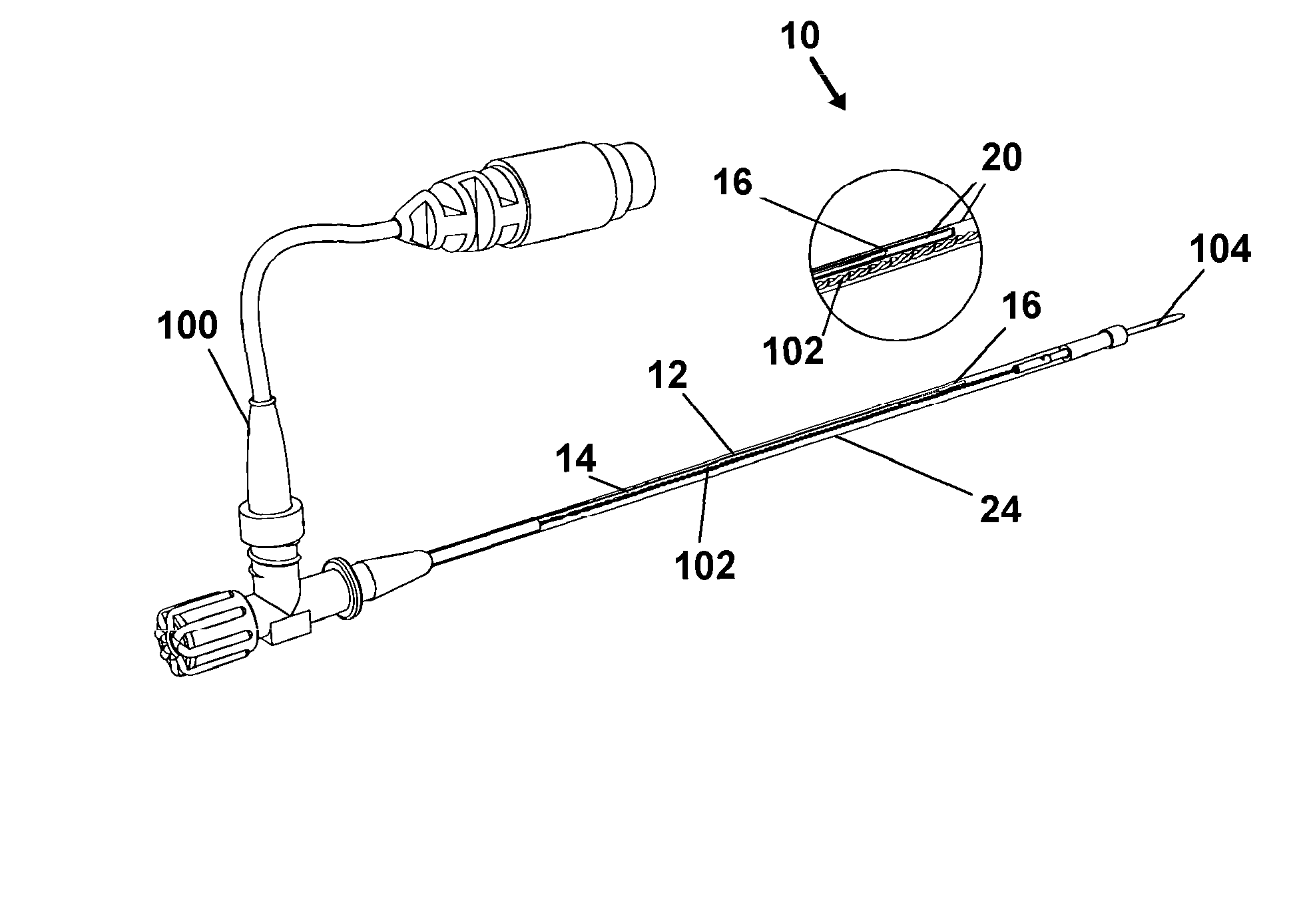
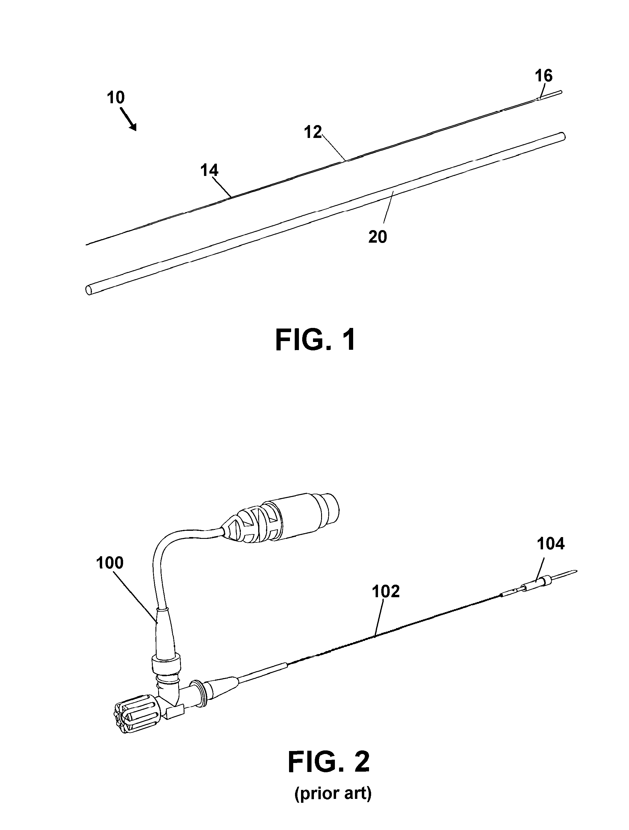
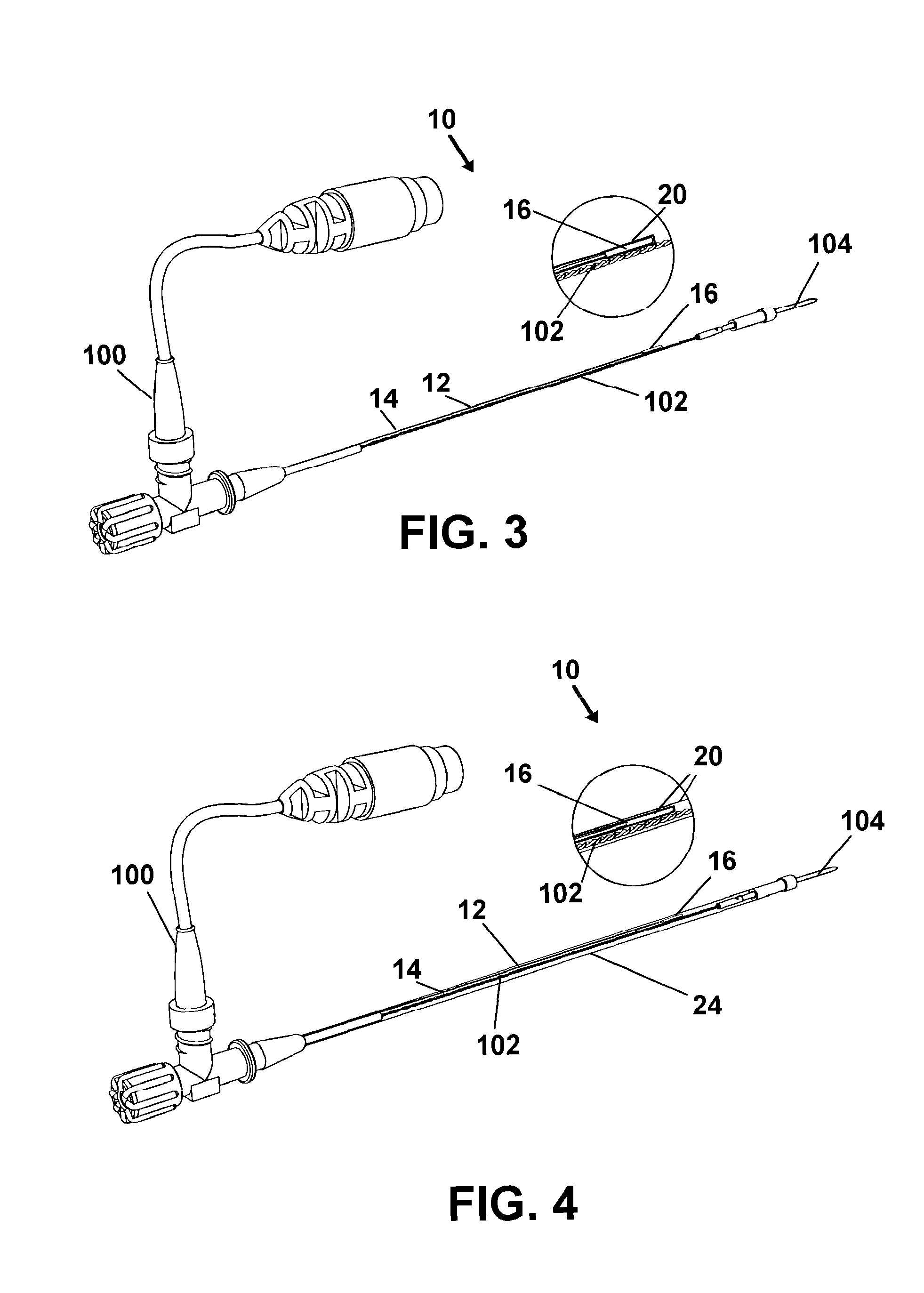
[0052]Now referring to drawings in FIGS. 1-16, wherein similar components are identified by like reference numerals, there is seen in FIG. 1 the sensor 12 and the heat shrinking or polymeric material shown as a shrink tube 20 which optionally may include an interior thermoplastic adhesive surface to provided increased adhesion of the formed polymeric jacket to the underlying assembly and / or catheter or wire. These components are employed in all particularly preferred modes of the medical sensor operative engagement and mounting system 10 herein. The assembled sensor 12 generally consists of an electrically conductive lead wire 14 and the sensing component 16 engaged thereon.

[0053]Again, the sensing component 16 can be any type of sensor or marker employed in the art of surgical navigation for example electromagnetic sensors which broadcast when provided electrical current from the lead wire 14 which forms the sensor assembly of an aggregate length of the two operationally and electr...


PUM


Abstract

An engagement system for a surgical sensor assembly, formed of a sensing component in a fixed position and a lead wire in an electrical communication therewith. The engagement system employs a jacket of heat shrink polymeric material to hold the sensing component to the lead wire and eliminates the employment of soldering and welding and crimping which can damage such sensing components. The system is employed in combination with catheters having a lumen for control wires for surgical instruments.

Description

BACKGROUND OF THE INVENTION[0001]1. Field of the Invention[0002]The present invention relates to surgical devices employed for minimally invasive surgery. More specifically the invention relates to a sensor mounting device and system which provides secured non-damaging electronically communicative engagement of a medical sensor to a lead wire and surgical instrument. The invention is most suitably adapted for engagement with needles, guide wires, catheters, cytology brushes, biopsy forceps, and other devices employed using electronic navigation to determine positioning in the body of a patient.[0003]2. Prior Art[0004]The medical industry is a field which is constantly seeing technological changes, advancements, and improvements. Minimally invasive surgery and computer assisted surgery (CAS) is a recent development which allows physicians to perform procedures with the aid of computer technology which provides a means to visually determine instrument positioning in the body of a pati...

Claims


Application Information

Patent Timeline
Patent Type & Authority Patents(United States)
IPC IPC(8): A61M25/098
CPCA61B5/065A61B5/6852A61B10/06A61B2010/0216A61B2562/12A61B5/062A61B2019/5251A61B2019/5483A61B2090/3983A61B2034/2051
Inventor SALEH, RAFIC
Owner VERAN MEDICAL TECH
Why Eureka
Try Eureka